
Research article · 2021 · Full text
International Journal of Biology SciencesIsolation of pathogens from leaf, root and rhizosphere water samples of aquatic weeds, Eichhornia crassipes and Pistia stratiotes present in the backwaters of YMCA canal, Alappuzha, Kerala, India
Volume 3(2) · Pages 24–29 · DOI: 10.33545/26649926.2021.v3.i2a.131 ↗
Research areasBiological Control of Invasive Species
Abstract
Author-writtenThe presence of pathogenic microorganisms in backwaters is of a great concern and the unsafe water is responsible for 1.2 million deaths each year. In the present study, water samples beneath the aquatic weeds, leaf and root samples of E. crassipes and P. stratiotes were collected from YMCA canal, Alappuzha, Kerala state, India, and subjected to physicochemical and microbiological analysis. The pH of the water samples were in the range of 6.6 to 6.9, salinity was nil, electrical conductivity was 5.327 mS for Eichhornia crassipes sample and 882.2 mS for Pistia stratiotes rhizosphere water sample. Total dissolved solids (TDS) were 2.777 ppm and 448 ppm for E. crassipes and P. stratiotes respectively. Heterophilic plate count of E. crassipes is 11 x 103 and 3 x 10² CFU/ml for bacteria and fungi respectively. Similarly the rhizosphere water sample of P. stratiotes showed a bacterial count of 75 × 103 CFU/ml and the fungal count was 8 x 10² CFU/ml. Human pathogens such as Klebsiella. pneumoniae, Salmonella typhi, Enterococcus faecalis, E. coli, Vibrio cholerae, Salmonella enteritidis, Enterobacter aerogenes, Vibrio vulnificus etc. were isolated from leaf, root and rhizosphere water samples. Antibiotic sensitivity studies revealed that Klebsiella pneumoniae was resistant to antibiotics like Azithromycin, Co-Trimoxazole, Cefuroxime and Cefalexin. Most of the pathogens were resistant to Cefalexin.
Summary
Source-checkedPlain-language reading of the full paper, prepared with AI and checked against the text. Each point links to the authors' own words.
Why it matters
- Pathogenic microorganisms in backwaters are a major public health concern, contributing to approximately 1.2 million deaths annually. View evidence ↗
- Backwater pollution is driven by several factors, including untreated waste discharge, agricultural runoff, and the accumulation of aquatic weeds. View evidence ↗
- Aquatic weeds form dense mats that block sunlight and deplete dissolved oxygen, which negatively impacts aquatic life and water chemistry. View evidence ↗
The question
- The study aimed to analyze the physicochemical and microbiological properties of E. crassipes and P. stratiotes from the YMCA canal in Kerala, specifically isolating human pathogens and testing their antibiotic sensitivity. View evidence ↗
What they did
- Researchers collected whole plants of E. crassipes and P. stratiotes, along with water samples from beneath the weeds in 250 ml sterile flasks, from the YMCA canal. View evidence ↗
- Physicochemical analysis included measuring pH, temperature, electrical conductivity, Total Dissolved Solids (TDS), salinity, and specific gravity using standardized meters and a refractometer. View evidence ↗
- Antibiotic sensitivity was evaluated using the disc diffusion method and agar dilution method following NCCLS standards. View evidence ↗
What they found
- Rhizosphere water for E. crassipes had a pH of 6.6, TDS of 2.777 ppm, and conductivity of 5.327 mS, while P. stratiotes had a pH of 6.9, TDS of 448.0 ppm, and conductivity of 882.2 mS. View evidence ↗
- Bacterial counts in rhizosphere water were higher for P. stratiotes (75 x 10³ CFU/ml) than E. crassipes (11 x 10³ CFU/ml), and fungal counts were also higher for P. stratiotes (8 x 10² CFU/ml) than E. crassipes (3 x 10² CFU/ml). View evidence ↗
- Most isolated pathogens were resistant to Cefalexin, including all pathogens from P. stratiotes and all from E. crassipes except V. cholerae. View evidence ↗
- Five key pathogens (K. pneumoniae, S. typhi, E. faecalis, E. coli, and V. cholerae) were susceptible to Ciprofloxacin, Chloramphenicol, Doxycycline hydrochloride, and Gatifloxacin. View evidence ↗
What it means
- The authors conclude that aquatic weeds act as reservoirs for human pathogens, including antibiotic-resistant strains, posing a threat to human health. View evidence ↗
- The authors recommend prioritizing the removal of aquatic weeds and implementing regular monitoring to prevent sewage from mixing with water bodies. View evidence ↗
Visual summary
2 visualsIllustrations explain the paper; charts are replotted from the paper's own table values.
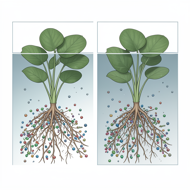
Aquatic Weeds as Pathogen Reservoirs
AI-generated conceptual illustration (Water hyacinth (Eichhornia crassipes) and Water lettuce (Pistia stratiotes) plants in canal water). It explains the idea and does not show study data.
View source evidence ↗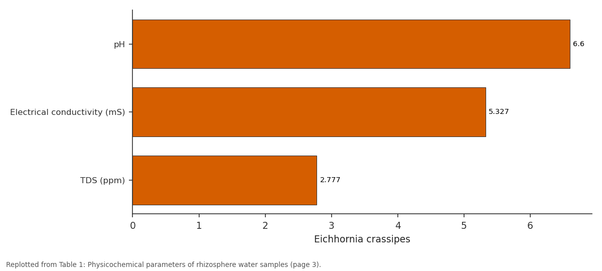
Eichhornia crassipes: pH vs Electrical conductivity... vs TDS (ppm)
Replotted from Table 1 (page 3) of the original paper. Every bar is the paper's own table value.
View source evidence ↗Figures & tables
From the original paper Table 1
 Table 2
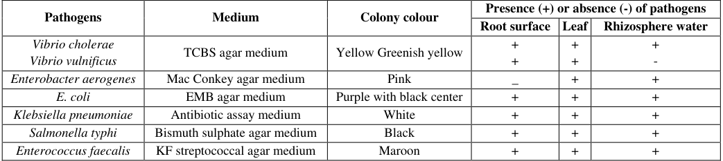 Table 3
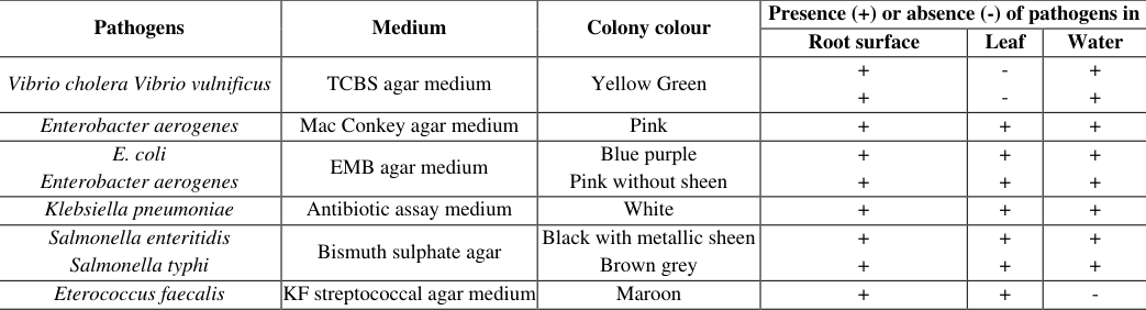 Table 4
 Table 5
 Table 6
Introduction
Water has been of great importance to human beings and other organisms of the environment for sustenance of life and maintaining the balance of the nature, hence water is called as the "life blood of the earth" (Ram et al., 2009) [10] . Dead leaf, woody debris, animal remains etc. constitute the main sources of organic matter in backwater environment. Alappuzha, known as the Venice of the East, boasts of shimmering waterways and a rich biodiversity. A common sight in Alappuzha is the mesmerizing houseboats. But because of the lack of proper regulation and hygiene they give adverse effects to the backwaters. Oil spillages from the boats, CO 2 emissions, discharge of untreated wastes, disposal of organic waste, fertilizer residues and run offs from agricultural fields and accumulation of aquatic weeds play a major role in backwater pollution. As a result of this water bodies have become a congenial breeding ground for water borne vectors like mosquitoes and pathogenic microorganisms (Chandran 2014) [8] . Aquatic weeds are unwanted and undesirable plants that are floating on the surface of water and form a dense mat there by reducing the penetration of sunlight into the water and reduce the photosynthetic efficiency of microscopic algae and results in the depletion of dissolved oxygen. Reduced levels of dissolved oxygen seriously affect the aquatic life and also alter the equilibrium of physicochemical properties of water. The microbial quality of water have an important influence on public health. Poor microbiological quality is likely to lead water borne disease outbreaks. Opportunistic pathogens are naturally present in the environment and normally present no risk to human health. They are able to cause disease in people with impaired local or general immune defences (Sreedevi and Sebastian 2020) [21] . Anthropogenic activities result in a significant decrease in surface water quality of aquatic systems in watersheds (Massoud et al., 2006) [16] . River inflows contribute many pollutants, thereby tending to induce ecological and hygienic problems (Wang et al., 2007) [24] . Escalating water pollution causes not only the deterioration of water quality but it also compromises human wellbeing and the permanence of aquatic ecosystems, economic growth and community affluent (Milovanovic, 2007) [17] . Eichhornia crassipes and Pistia stratiotes are the two major aquatic weeds found in the canals of Alappuzha. Water hyacinth (E. crassipes) is an aquatic plant which can flourish and reproduce floating freely on the surface of water or it can also be anchored in mud. It is a perennial weed, form dense rafts in the water and mud. Some of the principal problems are its interference with navigation, water flow, and the recreational use of aquatic systems, as well as the risk it poses of mechanical damage to hydroelectric systems. It is also responsible for drastic changes in the plant and animal communities of freshwater environments and acts as an agent for the spread of serious diseases in tropical countries (Chandran, 2011) [6] . Pistia is a genus of aquatic plant in the arum family, Araceae. The single species it comprises, Pistia stratiotes, is often called water cabbage, water lettuce, Nile cabbage, or shell flower. Microbial assemblage as a biofilm commonly occurs on the leaves of submerged plants, rhizosphere, especially on rhizoplane and on the solid surfaces of sediments.
Aquatic weeds are also responsible for lowering quantity as well as quality of water. These weeds cause taste and odour problems and also increases biological oxygen demand because of organic loading (Gopal and Sharma, 1979) [12] . In the present study aquatic weeds E. crassipes and P. stratiotes were collected from YMCA canal, Alappuzha, Kerala state, India and subjected to physicochemical and microbiological analysis. The human pathogenic microorganisms were also isolated using selective media and performed antibiotic sensitivity studies.
Materials and methods
Materials and methods
Collection of samples
Water samples beneath the aquatic weeds of E. crassipes and P. stratiotes were collected from YMCA canal, Alappuzha were collected separately in sterile 250 ml Erlenmeyer flasks at normal atmospheric temperature and transported to the laboratory for further analysis (APHA 1998) [3] . Both the weeds as whole plants were also collected in buckets with water and brought to the laboratory for further studies.
Chemicals and media used
Glucose, tryptone yeast extract agar, potato dextrose agar, Thiosulphate Citrate Bile salt Sucrose (TCBS) agar, MacConkey agar, Eosin Methylene Blue (EMB) agar, antibiotic assay medium, Bismuth sulphate (BS) agar, KF streptococcal agar, Lauryl sulphate broth, antibiotic discs such as Co-Trimoxazole (25 mcg), Gatifloxacin (5 mcg), Cefuroxime (30 mcg), Cefalexin (30 mcg), Chloramphenicol (30 mcg), Doxycycline hydrochloride (30 mcg), Azithromycin (15 mcg), Ciprofloxacin (5 mcg) were procured from HiMedia Laboratories Private Limited, Mumbai, India.
Physicochemical parameters Physical parameters
For physicochemical analysis water samples were collected from the rhizosphere regions of E. crassipes and P. stratiotes. The pH of water samples were checked using pH meter (Systronics 361, India). The temperature was measured using standard mercury filled centigrade thermometer. The electrical conductivity and Total Dissolved Solids (TDS) were measured using pre calibrated conductivity TDS meter (Systronics 308, India). Salinity and specific gravity of the water samples were estimated using a handheld refractometer (Erma, ERS10, Tokyo Japan).
Microbiological analysis Hetrophilic plate count (HPC)
Water samples collected from YMCA canal, Alappuzha, Kerala state, India, were serially diluted and plated on Tryptone Glucose Extract Agar (tryptone 5g, yeast extract 3g, glucose 1 g, and agar 15 g and final pH (at 25°C) 7.0±0.2) and the plates were kept for 24 hours of incubation at 37 °C and the number of colonies were counted after incubation (APHA 1998) [3] . For fungal analysis, the serially diluted water samples were plated on potato dextrose agar and the plates were kept for incubation at room temperature for 48-72 hours. The fungal colonies developed were noted after incubation.
Isolation of microorganism from root, leaf and rhizosphere water sample
Rhizosphere water samples collected from E. crassipes and P. stratiotes were processed and were serially diluted and the dilution 10 -2 was spread on sterile specific media such as TCBS agar (for Vibrio cholerae), Bismuth sulphite agar (for Salmonella typhi), Eosin Methylene Blue agar (for E.coli), Antibiotic assay medium D (for Klebsiella pneumonia), Mac Conkey agar (for Enterobacter aerogenes), KF streptoccocal agar (for Enterococcus faecalis) for the isolation of pathogenic microorganisms. Pathogenic microorganisms found in the root, leaf and in the water sample near the weeds were isolated by using spread plate method after enriching the samples in respective enrichment media.
Enrichment of water samples Isolation of E. coli
For the enrichment of the E. coli strains, all samples were cultured in Lauryl sulphate broth overnight at 37 °C and subsequently were streaked onto Eosin Methylene Blue (EMB) agar plates and incubated at 37 °C for 24 h.
Isolation of Vibrio sp.
Water samples suspected to contain Vibrio spp. and V. parahaemolyticus were enriched by adding 100 ml water sample in 200 ml of double strength alkaline peptone water (pH 8.6) at 37 o C for 24 hours and swabs from the alkaline peptone water was streaked on to Thiosulphate Citrate Bile salt Sucrose (TCBS) agar and further incubated at 37 o C for 24 -48 hours.
Isolation of Salmonella typhi
Salmonella typhi was isolated by inoculating one ml of water sample into 10 ml of selenite enrichment broth and incubated at 37 o C for 12-18 hours. Swabs from the selenite broth were streaked on to Bismuth Sulfite (BS) Agar plates, and further incubated at 37 o C for 24 -48 hours.
Antibiotic sensitivity assay
The antibacterial sensitivity assay was carried out by disc diffusion method (Chandran 2013, 2015) [7,9] . Antibiotic susceptibility of the isolates was tested according to National Committee for Clinical Laboratory Standards (NCCLS) by disc diffusion method with an inoculum of 10 8 cfu, and agar dilution method with 10 4 fu/spot (Deb Mandal et al, 2011) [10] . The test bacterial cultures were evenly spread over Mueller Hinton agar plates using a sterile cotton swab. The antibiotic discs (6 mm in diameter) were placed in the inoculated agar. The plates were then incubated at 37 °C for 24 to 48 hours. After incubation, the zone of inhibition thus developed were measured with the scale to the nearest in centimeters (CM). The antibiotics (content per disc) used in the study were Co-Trimoxazole (25 mcg), Gatifloxacin (5 mcg), Cefuroxime (30 mcg), Cefalexin (30 mcg), Chloramphenicol (30 mcg), Doxycycline hydrochloride (30 mcg), Azithromycin (15 mcg), Ciprofloxacin (5 mcg).
Results and Discussion
Results and Discussion
Physicochemical parameters
The physicochemical parameters of the rhizosphere water sample collected from the YMCA canal were analysed and the results are given in table 1. The pH of the water samples ranged from 6.5 to 6.9. Temperature ranged from 32 to 32.3 o C, TDS ranged from 2.777 to 448 ppm and electrical conductivity ranged from 5.327 to 882.2 ms. Salinity and specific gravity is nil for the rhizosphere water sample. The pH (6.6 and 6.9) and temperature (32 o C) also favoured the bacterial growth. The variables such as pH, temperature, salinity, nutrient availability, and geographic locations influence the growth of microorganisms in the natural habitat (Amend et al., 2013) [1] . Over 1.6 million people directly or indirectly depend on water for various purposes such as agriculture, fishing, transportation and recreation. So there is a possibility of spread of waterborne diseases from these aquatic bodies will pose a great threat.
| Aquatic weeds | pH | Salinity | Electrical conductivity (mS) | TDS (ppm) | Temperature (oC) | Specific gravity |
|---|---|---|---|---|---|---|
| Eichhornia crassipes | 6.6 | Nil | 5.327 | 2.777 | 32.3 | 0 |
| Pistia stratiotes | 6.9 | Nil | 882.2 | 448.0 | 32 | 0 |
Table text extracted from the original PDF; superscript letters are shown inline. The original table image is below.
View original table image
Microbiological analysis Heterophilic plate count
The bacteriological examination of water has an important relevance in pollution studies and it can provide a direct measure of deleterious effect of pollution on human health. HPC of rhizosphere water sample of E. crassipes, i.e. 11 x 10 3 and 3 x 10 2 CFU/ml for bacteria and fungi respectively. The rhizosphere water sample of P. stratiotes showed a bacterial count of 75 × 10 3 CFU/ml and the fungal count was 8 x 10 2 CFU/ml (Table 2). In the present study the heterophilic bacterial population was high in the rhozsphere region of E. crassipes and P. stratiotes. The heavy heterophilic bacterial count in the rhizosphere region was mainly due to the chemotaxis of microbes towards rhizosphere region of aquatic weeds. This chemotaxis was attributed to the secretion of different amino acids found in the root exudates secreted by E. crassipes (Chandran 2014) [8] . The abundance of pathogen in water depends on actors such as the contamination level, pathogen persistence in water bodies, biological reservoirs including aquatic plants and sediments (Dechense et al., 2006) [11] . Sediments and submerged aquatic vegetation are the important reservoirs of microorganisms (Badgley et al., 2010) [5] .
| Microorganisms | Culture media | Eichhornia crassipes | Pistia stratiotes |
|---|---|---|---|
| Bacteria | Tryptone glucose extract agar | 11 × 103 | 75 × 103 |
| Fungi | Potato dextrose agar | 3 x 10² | 8 x 10² |
Table text extracted from the original PDF; superscript letters are shown inline. The original table image is below.
View original table image
Isolation of pathogens
Human pathogens isolated form root, leaf and rhizosphere water samples of E. crassipes, such as V. cholerae and V. vulnificus were cultured using TCBS agar medium. Enterobacter species such as Enterobacter aerogenes and Enterococcus faecalis were cultured using Mac Conkey agar medium produced pink and pale pink colonies. The increased number of coliform and faecal coliforms in water bodies is mainly due to the higher sewage contamination. In the EMB agar medium, E. coli formed purple with black centered colonies and K. pneumoniae formed white colonies in antibiotic assay medium. Species of S. enteritidis and S. typhi formed brown and black (with metallic sheen) colonies in BS agar medium. Maroon colonies of E. faecalis were observed in KF streptococcal agar medium. The presence or absence of microorganisms found in the root, leaf and rhizosphere water samples of aquatic weeds are given in table 3 and 4. Faecal contamination of water introduces a variety of pathogens into water ways, including bacteria, viruses, protozoa and parasitic worms. E. coli, Salmonella and Vibrio spp. forms the most important pathogen that spread through water. E. coli is the only member of the total coli form group that is found exclusively in faeces, other members of the group are found naturally in water, soil, and vegetation, as well as in faeces. S. typhi is one of the major causes of food and water borne gastroenteritis in human (Tsen et al., 2000) [23] and remains an important health problem worldwide (Athira et al., 2019) [4] . The presence of E. coli, Klebsiella, and Enterobacter species in water is a likely indicator of the presence of pathogenic organisms such as Clostridium perfringens, Salmonella, and Protozoa (Anyamene and Ojiagu 2014) [2] . These pathogens cause diarrhea, giardiasis, dysentery, and gastroenteritis, which are common among the rural dwellers of developing nations (Thliza et al., 2015, Oludairo and Aiyedun 2016, Isikwue and Chikezie 2014) [22,18,13] .
| Pathogens | Medium | Colony colour | Presence (+) or absence (-) of pathogens | — | — |
|---|---|---|---|---|---|
| Root surface | Leaf | Rhizosphere water | |||
| Vibrio cholerae Vibrio vulnificus | TCBS agar medium | Yellow Greenish yellow | + + | + + | + - |
| Enterobacter aerogenes | Mac Conkey agar medium | Pink | _ | + | + |
| E. coli | EMB agar medium | Purple with black center | + | + | + |
| Klebsiella pneumoniae | Antibiotic assay medium | White | + | + | + |
| Salmonella typhi | Bismuth sulphate agar medium | Black | + | + | + |
| Enterococcus faecalis | KF streptococcal agar medium | Maroon | + | + | + |
Table text extracted from the original PDF; superscript letters are shown inline. The original table image is below.
View original table image
Antibiotic sensitivity assay
The antibiotic sensitivity assay revealed that all the organisms isolated from E. crassipes except V. cholerae were found to be resistant to antibiotic Cefalexin. V. cholerae showed a zone of inhibition towards the antibiotic, Celalexin with a zone of inhibition of 1.1 cm. All the five organisms K. pneumoniae, S. typhi, E. faecalis, E. coli and V. cholerae showed a zone of inhibition towards the antibiotics Ciprofloxacin, Chloramphenicol, Doxycycline hydrochloride and Gatifloxacin. The overall result of the antibiotic sensitivity test is given in table 5. Based on the antibiotic sensitivity assay, it was found that all the pathogens present in P. stratiotes were also found to be resistant to the antibiotic Cefalexin. All the five organisms (Klebsiella pneumoniae, S. typhi, E. faecalis, E. coli and V. cholerae showed a zone of inhibition towards the antibiotics Ciprofloxacin, Chloramphenicol, Doxycycline hydrochloride and Gatifloxacin. The overall results of the antibiotic sensitivity assay are given in table 6. Development and spread of antimicrobial resistance have become a global public health problem, impacted by both human and nonhuman antimicrobial usage (Rigos et al., 2010) [20] . Aquatic environments, rivers and streams are considered as an ideal reservoir for antibiotic resistance dissemination because antimicrobials and antimicrobial resistant bacteria are often directly released in the environment (Lupo et al., 2012) [15] .
| Pathogens isolated from Pistia stratiotes | Antibiotics/ diameter of zone of inhibition in cm | — | — | — | — | — | — | — |
|---|---|---|---|---|---|---|---|---|
| Ciprofloxacin | Azithromycin | Chloramphenicol | Co- Trimoxazole | Cefuroxime | Cefalexin | Doxycycline hydrochloride | Gatifloxacin | |
| Klebsiella pneumoniae | 1.1 | 0.7 | 0.4 | 0.4 | _ | _ | 1 | 1 |
| Salmonella typhi | 1.6 | 1.7 | 1.6 | 0.5 | 0.4 | _ | 1.1 | 1.7 |
| Enterobacter faecalis | 1.2 | 1 | 0.5 | 0.5 | 0.5 | _ | 0.7 | 1.2 |
| E. coli | 1.7 | _ | 0.3 | 1 | 0.5 | _ | 0.8 | 1.7 |
| Vibrio cholerae | 1.6 | 1.3 | 0.8 | _ | 0.3 | _ | 1.5 | 1.6 |
Table text extracted from the original PDF; superscript letters are shown inline. The original table image is below.
View original table image
| Pathogens isolated from Eichhornia crassipes | Antibiotics/ Diameter of zone of inhibition in cm | — | — | — | — | — | — | — |
|---|---|---|---|---|---|---|---|---|
| Ciprofloxacin | Azithromycin | Chloramphenicol | Co- Trimoxazole | Cefuroxime | Cefalexin | Doxycycline hydrochloride | Gatifloxacin | |
| Klebsiella pneumoniae | 1.5 | _ | 0.7 | _ | _ | _ | 1 | 1.5 |
| Salmonella typhi | 1.5 | _ | 1 | 0.5 | 0.7 | _ | 1.1 | 1.6 |
| Enterobacter faecalis | 1.2 | 1 | 0.9 | 0.3 | 0.4 | _ | 0.7 | 1.3 |
| E. coli | 1.5 | _ | 0.4 | 0.4 | 0.4 | _ | 0.8 | 1.5 |
| Vibrio cholerae | 1.4 | 0.6 | 1.4 | _ | 0.4 | 1.1 | 1.5 | 1.2 |
Table text extracted from the original PDF; superscript letters are shown inline. The original table image is below.
View original table image
Conclusion
The aquatic weeds harbor the growth of human pathogenic microorganisms, even some of them are antibiotic resistant and the water bodies can act as a reservoir of pathogens which pose a threat to human population. The isolation of human pathogenic microorganisms such as K. pneumoniae, S. typhi, E. faecalis, E. coli, V. cholerae, S. enteritidis, E. aerogenes, V. vulnificus etc. clearly indicates the mixing of sewage and other pollutants with the water body. Regular monitoring and controlling the mixing of sewage with water body has to be prevented. Removal of aquatic weeds from the backwaters should also be given priority.
Figures and tables from the paper
| Pathogens | Medium | Colony colour | Presence (+) or absence (-) of pathogens in | — | — |
|---|---|---|---|---|---|
| Root surface | Leaf | Water | |||
| Vibrio cholera Vibrio vulnificus | TCBS agar medium | Yellow Green | + + | - - | + + |
| Enterobacter aerogenes | Mac Conkey agar medium | Pink | + | + | + |
| E. coli Enterobacter aerogenes | EMB agar medium | Blue purple Pink without sheen | + + | + + | + + |
| Klebsiella pneumoniae | Antibiotic assay medium | White | + | + | + |
| Salmonella enteritidis Salmonella typhi | Bismuth sulphate agar | Black with metallic sheen Brown grey | + + | + + | + + |
| Eterococcus faecalis | KF streptococcal agar medium | Maroon | + | + | - |
Table text extracted from the original PDF; superscript letters are shown inline. The original table image is below.
View original table image
References
24 in the original paper- Amend AS, Oliver TA, Amaral-Zettler LA, Boetius A, Fuhrman JA, Horner-Devine MC. et al. Macroecological patterns of marine bacteria on a global scale. J Biogeogr. 2013;40:800-811.
- Anyamene NC, Ojiagu DK. Bacteriological Analysis of sachet water sold in Akwa Metropolis, Nigeria. Int. J Agric. Biosci. 2014;3:120-122.
- APHA. Standards methods for the examination of water and wastewater. 20 th edition, American Public Health Association, Washington, D.C. 1998.
- Athira TS, Vijayan B, Gopika G, George T, Sudhakaran R, Franklin N, et al. Screening and Identification of pathogenic microorganisms in backwaters of Alappuzha District, Kerala State, India. Int. J Adv. Res. Biol. Sci. 2019;6(4):62-69.
- Badgley BD, Nayak BS, Harwood VJ. The importance of sediments and submerged aquatic vegetation as potential habitats for persistant strains of Enterococci in a subtropical watershed. Water Res. 2010;44:5857-5866.
- Chandran PR, Kiran K, Divakaran D, Prajisha PK. Analysis of bacteriological quality of drinking water samples from Cherthala taluk, Kerala, India. Asian J Water Environ. Pollut. 2011;8:61-68.
- Chandran PR, Manju S, Vysakhi MV, Shaji PK, Nair GA. In vitro antimicrobial activities of Hygrophila schulli (Buch.-Ham) leaf and root extracts against clinically important human pathogens. Biomed. Pharmacol. J. 2013;6:421-428.
- Chandran PR. Harbouring of pathogenic microorganisms by aquatic weed, Eichhornia crassipes in its rhizosphere. Int. J ChemTech Res. 2014;(2):1413-1417.
- Chandran PR. Antimycobacterial activity of Glycyrrhiza glabra root extract against Mycobacterium smegmatis. Int. J PharmTech Res. 2015;7(3):458-463.
- DebMandal M, Mandal S, Pal NK. Antibiotic Resistance Prevalence and Pattern in Environmental Bacterial Isolates. The Open Antimicrobial Agents J. 2011;3:45-52.
- Dechense M, Soyeux E, Loret JF, Westrell T, Stenstorm TA, Gornik V, et al. Pathogens in source water, Microbiological Risk Assessment: A Scientific basis for managing drinking water safety from source to tap; Microrisk European Project: Nieuwegein, The Netherland, 2006, 1-42.
- Gopal B, Sharma KP. Aquatic weed control versus utilisation. Econ. Bot. 1979;33(3):340-346.
- Isikwue MO, Chikezie A. Quality assessment of various sachet water brands marketed in Bauchi metropolis of Nigeria. Int. J Adv. Eng. Technol. 2014;6:2489-2495.
- Hemanta Pokhrel, Sangipran Baishya, Bipul Phukan, Devika Pillai and Mohd Ashraf Rather. Occurrence and distribution of multiple antibiotic resistance bacteria of public health significance in backwaters and aquaculture farm. Int. J Curr. Microbiol. App. Sci. 2018;7(02):975-987.
- Lupo A, Coyne S, Berendonk TU. Origin and evolution of antibiotic resistance: the common mechanisms of emergence and spread in water bodies. Front. Microbiol. 2012;3:18.
- Massoud MA, El-Fadel M, Scrimshaw MD, Lester JN. Factors influencing development of management strategies for the Abou Ali River in Lebanon: i. Spatial variation and land use. Sci. Total Environ. 2006;362:15-30.
- Milovanovic M. Water quality assessment and determination of pollution sources along the Axios/Vardar River, Southeastern Europe. Desalination 2007; 213:159173.
- Oludairo VO, Aiyedun J. Contamination of commercially packaged sachet water and the public health implications: an overview. Bangladesh J Vet. Med. 2016;13(2):73-81.
- Ram HK, Mohan R, Shivabasavaiah. Water Quality Status of Fresh Water Lake (Thalli Lake) Krishnagiri, Tamil Nadu. Indian J Environ. Ecoplan. 2009;16:103-112.
- Rigos G, Bitchava K. Antibacterial drugs in products originating from aquaculture: assessing the risks to public welfare. Mediterr. Mar. Sci. 2010;11(1):33-42.
- Sreedevi NK, Sebastian CD. Assessment on Bacteriological Contamination of River Water in the Urban Areas of Kozhikode, Kerala. Int. J Sci. Res. 2020;9(4):1638-1642.
- Thliza LA, Khan AU, Dangora DB, Yahaya A. Study of some bacterial load of some brands of sachet water sold in Ahmadu Bello University (Main Campus), Zaria, Nigeria. Int. J curr. Sci. 2015;14:91-97.
- Tsen HY, Hu HH, Lin JS, Huang CH, Wang TK. Analysis of Salmonella typhimurium isolates from food poisoning cases by molecular sub typing methods. Food Microbiol. 2000;17:143-152.
- Wang X, Lu Y, Han J, He G, Wang T. Identification of anthropogenic influences on water quality of rivers in Taihu watershed. J Environ. Sci. 2007;19:475-481.
Cited by 5
From OpenAlex, OpenCitations and reference lists read by Scinovex
- Statistical modeling of electronic waste (e-waste) generated in India
G.B. Singh, Jitendra Singh Bhadauria · International Journal of Statistics and Applied Mathematics · 2023
Electronic Waste (e-waste) is one of the fastest growing waste streams in the country. Growth of Information and Communication Technology sector has enhanced the usage of the electronic equipment exponentially. Developing countries are facing enormous challenges related to the generation and management of E-waste which are either internally generated or imported illegally; India is no exception to it. Electronic wastes can cause widespread environmental damage due to the use of toxic materials in the manufacture of electronic goods. Hazardous materials such as lead, mercury and hexavalent chro
- Occurrence, distribution, characteristics, toxicity, human health effects and removal techniques of Microplastic: A Review
Rakhi Dwivedi · International Journal of Advanced Chemistry Research · 2023
Since the inception of commercial plastic production around 1950, modern society's reliance on plastics has surged dramatically. This growing dependence is attributed to the remarkable advantages of plastics, including their versatility, stability, lightweight nature, and cost-effectiveness, all of which have propelled their global demand to unprecedented levels. These materials find applications in packaging, construction, electronics, textiles, medical devices, and more, owing to their capacity to be moulded into a wide array of shapes and sizes. Regrettably, the prevalent pattern involves u
- Interactions, processes and biotechnological potential of microbiomes of freshwater macrophytes ↗
Farheen Mushtaq, Sumira Tyub, Aabroo Fatima, Safeera Akbar, Uzma Ali, Syed Nasir Ahmad · Discover Plants. · 2026
Freshwater macrophytes harbour microbiomes in their rhizosphere, phyllosphere and endosphere, creating dynamic plant-microbe interactions modulating ecological processes. These interactions drive biogeochemical cycling (such as nitrogen and phosphorus), organic matter degradation and breakdown of emergent contaminants and thus promote water quality and ecosystem health. Recent research demonstrates that macrophyte microbiomes promote plant performance, stress resistance and phytoremediation in natural and constructed…
❝ 0PDF - Assessment of impact of nickel stress on the accumulation, pigments and protein content of water hyacinth (Pontederia crassipes L.) ↗
Manas Ranjan Satpathy, Sagarika Samantaray · South Asian Journal of Agricultural Sciences · 2023
Pontederia crassipes L. plants were collected and exposed to different concentrations (Control, 0.01, 0.1, 1.0 and 10.0 ppm) of Ni by using NiSO4 in medium in the laboratory. The effect of Ni2+ concentrations on visible symptoms such as toxicity, pigments (Chlorophyll- a, b and total) contents were estimated. Plants exposed to high nickel (1.0 and 10.0 ppm) showed visible toxicity…
- Study on diversity and therapeutic potential of aquatic macrophytes in the freshwater bodies of Canchipur, Manipur (India)
Chongtham Umabati Devi, Khumukcham Susheela Devi, Akoijam Nirmala Devi · International Journal of Horticulture and Food Science · 2023
The state of Manipur in North- east India is endowed with a rich repository of medicinal and aromatic plants and more over well-known for its diverse culture of human races. This could be due to its climatic condition, perennial rivers, fertile soil and dense forests. Despite of such wide availability of aquatic vegetation, the literature pertaining to the therapeutic utility of these plants is meagre because of their unproductive and impractical nature. Hence, keeping these facts in mind, the present investigation was formulated to study the diversity of aquatic macrophytes of some freshwater