
Research article · 2025 · Full text
International Journal of Advanced Chemistry ResearchEncapsulation of probiotics in alginate-chitosan beads for enhanced survival in gastric conditions
Volume 7(10) · Pages 120–123 · DOI: 10.33545/26646781.2025.v7.i10b.483 ↗
Research areasMicroencapsulation and Drying ProcessesProbiotics and Fermented FoodsAdvanced Drug Delivery Systems
Abstract
Author-writtenDelivering viable probiotic bacteria through the harsh acidic environment of the human stomach to the intestine—where they exert their health benefits—remains one of the most persistent challenges in functional food science. Lactobacillus rhamnosus GG, among the best-characterised probiotic strains, loses more than 99.9% of its viability within 90 minutes of exposure to simulated gastric fluid at pH 2.0. This research encapsulated L. rhamnosus GG in alginate beads, alginate-chitosan beads, and alginate-chitosan-resistant starch beads prepared by extrusion into calcium chloride hardening baths. Bead morphology, size distribution, encapsulation efficiency, and survival under simulated gastric (SGF, pH 2.0, pepsin) and intestinal (SIF, pH 7.4, pancreatin) conditions were evaluated. The triple-layer alginate-chitosan-starch beads offered the best protection: after 180 minutes in SGF, viable counts dropped by only 1.41 log CFU/g (from 9.09 to 7.68), compared to a complete loss for free cells. In SIF, the same beads released 87.3% of entrapped bacteria within 120 minutes. Mean bead diameter was 1.14 ± 0.28 mm with encapsulation efficiency of 93.7%. These results indicate that combining chitosan's pH-responsive cation barrier with resistant starch's prebiotic matrix provides a practical dual-protection strategy for oral probiotic delivery.
Summary
Source-checkedPlain-language reading of the full paper, prepared with AI and checked against the text. Each point links to the authors' own words.
Why it matters
- A major challenge in functional food science is ensuring probiotic bacteria survive the stomach's acidic environment to reach the intestine and provide health benefits. View evidence ↗
- Lactobacillus rhamnosus GG is highly sensitive to gastric conditions, losing over 99.9% of viability within 90 minutes at pH 2.0. View evidence ↗
- Microencapsulation is the primary strategy used to protect probiotics by trapping them in a polymer matrix that resists stomach acid and releases them in the intestine. View evidence ↗
The question
- The study aimed to compare the protective ability and intestinal release of three different bead formulations: alginate, alginate-chitosan, and alginate-chitosan-resistant starch. View evidence ↗
What they did
- The research was conducted between March and October 2024 at Acharya Jagadish Chandra Bose University in West Bengal, India. View evidence ↗
- Probiotics were encapsulated by mixing cell suspensions with 2% sodium alginate (and 2% resistant starch for specific beads) and extruding the mixture into a 0.1 M calcium chloride solution. View evidence ↗
- Beads were coated with chitosan by immersion in a 0.4% chitosan solution for 40 minutes. View evidence ↗
- The study used simulated gastric fluid (SGF) at pH 2.0 containing pepsin and simulated intestinal fluid (SIF) at pH 7.4 containing pancreatin. View evidence ↗
What they found
- Encapsulation efficiency increased with complexity, reaching 93.7% for the alginate-chitosan-starch beads compared to 89.4% for plain alginate. View evidence ↗
- The alginate-chitosan-starch beads provided the best protection, maintaining 7.68 log CFU/g after 180 minutes in SGF, representing a loss of only 1.41 log. View evidence ↗
- The triple-layer beads successfully released 87.3% of the entrapped bacteria within 120 minutes when transferred to simulated intestinal fluid. View evidence ↗
- Approximately 72.9% of the alginate-chitosan beads had a diameter between 0.5 and 1.5 mm. View evidence ↗
What it means
- The authors conclude that combining chitosan and resistant starch creates a synergistic effect that provides superior gastric protection. View evidence ↗
- The triple-layer system is recommended for use in nutraceutical capsules and functional foods to deliver probiotics to the lower GI tract. View evidence ↗
- The bead size of approximately 1.1 mm is a limitation that may make them unsuitable for clear beverages. View evidence ↗
- Future work is needed to validate the results in a murine model and to test shelf-life stability during refrigeration. View evidence ↗
Visual summary
2 visualsIllustrations explain the paper; charts are replotted from the paper's own table values.
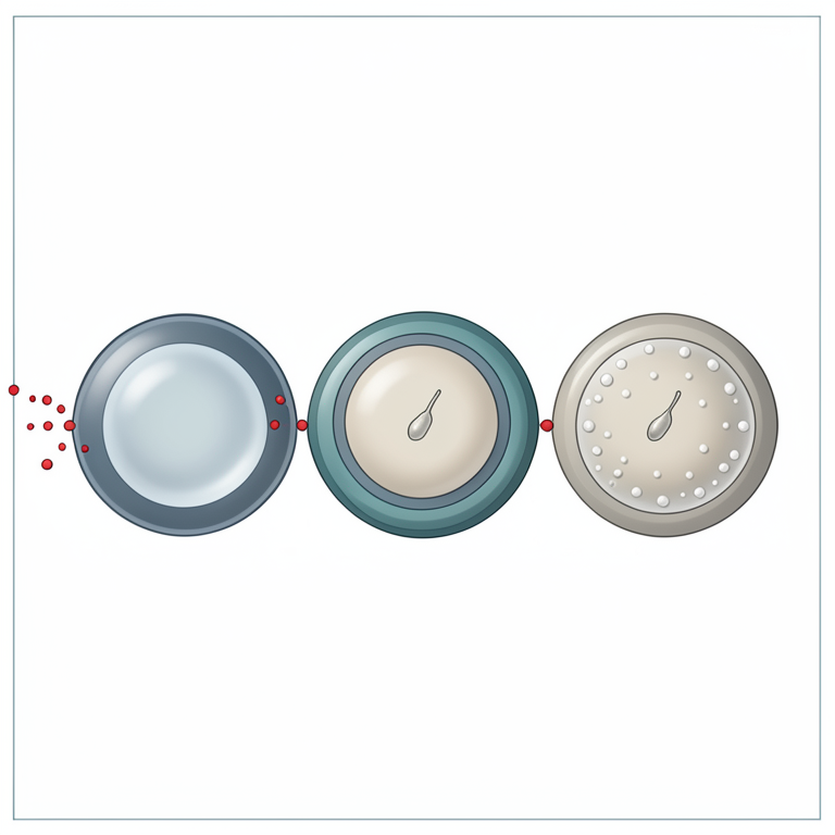
Triple-Layer Protection Concept
AI-generated conceptual illustration (probiotic bacteria encapsulated in alginate-chitosan polymer beads). It explains the idea and does not show study data.
View source evidence ↗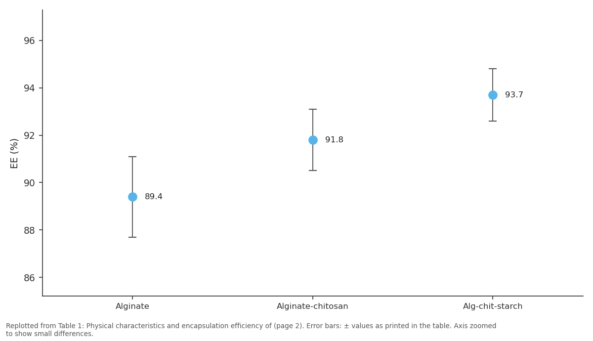
EE (%): Alginate vs Alginate-chitosan vs Alg-chit-starch
Replotted from Table 1 (page 2) of the original paper. Every bar is the paper's own table value.
View source evidence ↗Figures & tables
From the original paper Table 1
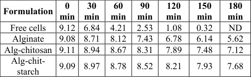 Table 2
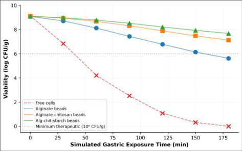 Figure 1
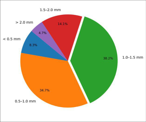 Figure 2
Introduction
The central challenge facing probiotic product developers is straightforward to state and difficult to solve: how can billions of living bacterial cells survive stomach acid, bile salts, and digestive enzymes long enough to colonise the intestinal mucosa and deliver measurable health outcomes [1] ? International guidelines recommend a minimum viable count of 10⁶-10⁷ CFU per gram at the point of consumption, yet unprotected cells of even robust strains like Lactobacillus and Bifidobacterium can drop by 4-6 log orders within minutes at gastric pH [2] . Microencapsulation-trapping cells within a polymeric matrix that resists gastric breakdown and releases its payload in the intestine-has emerged as the leading protective strategy [3] . Sodium alginate, a natural anionic polysaccharide extracted from brown seaweed, forms gel beads instantly upon contact with divalent cations (Ca²⁺) under mild, cell-compatible conditions [4] . But plain alginate beads are porous at low pH, allowing proton diffusion that kills entrapped cells. Coating these beads with chitosan, a cationic polysaccharide, plugs surface pores through electrostatic cross-linking and introduces a pH-responsive barrier that shrinks in acid and swells in neutral-to-alkaline environments [5] . Adding a third component-resistant starch-may further improve survival by acting both as a physical filler that slows acid penetration and as a prebiotic substrate that supports probiotic metabolism during transit [6] . Resistant starch granules resist digestion in the upper GI tract and are fermented by colonic bacteria, potentially creating a synbiotic effect when co-encapsulated with probiotics [7] . This research compared three bead formulations (alginate, alginate-chitosan, and alginatechitosan-resistant starch) for their ability to protect L. rhamnosus GG during simulated gastrointestinal transit and to release viable cells under intestinal conditions, with the goal of identifying a practical formulation for functional food and nutraceutical applications.
Material and Methods Material
All experiments were performed between March and October 2024 at the Department of Food Chemistry and Nutrition, Acharya Jagadish Chandra Bose University, Barrackpore, West Bengal, India. Lactobacillus rhamnosus GG (ATCC 53103) was procured from HiMedia Laboratories (Mumbai, India). Sodium alginate (food grade, viscosity 200-400 cP), low-molecular-weight chitosan (75-85% deacetylated), calcium chloride dihydrate, pepsin from porcine gastric mucosa (≥ 250 U/mg), and pancreatin from porcine pancreas (8× USP) were purchased from Sigma-Aldrich (Bengaluru, India). High-amylose maize resistant starch (Hi-Maize 260, ~60% RS) was obtained from Ingredion (Westchester, IL, USA). MRS agar and broth were from HiMedia.
Extraction Protocol
L. rhamnosus GG was cultured in MRS broth at 37°C for 18 h under anaerobic conditions (AnaeroGen sachets, Oxoid). Cells were harvested by centrifugation (4000 × g, 10 min, 4°C), washed twice with sterile 0.85% NaCl, and resuspended to approximately 10¹⁰ CFU/mL. For encapsulation, cell suspension was mixed with 2% (w/v) sodium alginate solution (1:1 v/v). For starch-containing beads, resistant starch (2% w/v) was dispersed in the alginate solution before mixing with cells. The cell-polymer mixture was extruded through a 21-gauge needle into 0.1 M CaCl₂ solution under gentle stirring (100 rpm) and allowed to harden for 30 min. Chitosan coating was applied by immersing hardened beads in 0.4% (w/v) chitosan solution (in 1% acetic acid, pH 5.5) for 40 min, followed by rinsing with sterile water.
Methods
Bead diameter was measured on 100 randomly selected beads per formulation using a digital calliper (Mitutoyo, Japan). Morphology was observed by optical microscopy (Olympus CX23) and scanning electron microscopy (JEOL JSM-6390LV). Encapsulation efficiency (EE%) was determined by dissolving 1 g of beads in 0.1 M sodium citrate buffer (pH 6.0) for 15 min, serial diluting, plating on MRS agar, incubating 48 h at 37°C anaerobically, and calculating EE% = (log CFU_encapsulated / log CFU_initial) × 100.
In-vitro Protocol
Simulated gastric fluid (SGF) was prepared with NaCl (2 g/L) and pepsin (3.2 g/L) adjusted to pH 2.0 with HCl. Simulated intestinal fluid (SIF) contained KH₂PO₄ (6.8 g/L) and pancreatin (10 g/L) at pH 7.4. One gram of beads or equivalent free cells was incubated in 10 mL SGF at 37°C with shaking (100 rpm). At 0, 30, 60, 90, 120, 150, and 180 min, beads were removed, dissolved in citrate buffer, and viable counts enumerated on MRS agar. After 180 min in SGF, surviving beads were transferred to SIF for a further 120 min to assess intestinal release. Experiments were performed in triplicate on three independent bead batches.
Results
Optical microscopy showed that alginate beads were spherical with smooth surfaces, alginate-chitosan beads had a slightly rough texture, and starch-containing beads displayed an opaque, granular appearance. Mean diameters were 1.42 ± 0.31 mm (alginate), 1.28 ± 0.25 mm (alginatechitosan), and 1.14 ± 0.28 mm (alginate-chitosan-starch). The shrinkage with each additional coating or filler reflected tighter polymer cross-linking. Encapsulation efficiencies were 89.4%, 91.8%, and 93.7%, respectively.
Comprehensive Interpretation
The triple-layer beads maintained viability above 7.68 log CFU/g even after 180 min of gastric exposure (Table 2, Figure 1), a loss of just 1.41 log-well within the range considered acceptable for commercial probiotic supplements. Upon transfer to SIF, these beads released 87.3% of entrapped bacteria within 120 min, confirming intestinal-triggered disintegration. The bead size distribution (Figure 2) shows that 72.9% of alginate-chitosan beads fell within 0.5-1.5 mm, a range suitable for incorporation into food matrices without perceptible textural impact.
| Formulation | 0 min | 30 min | 60 min | 90 min | 120 min | 150 min | 180 min |
|---|---|---|---|---|---|---|---|
| Free cells | 9.12 | 6.84 | 4.21 | 2.53 | 1.08 | 0.32 | ND |
| Alginate | 9.08 | 8.71 | 8.12 | 7.43 | 6.78 | 6.14 | 5.62 |
| Alg-chitosan | 9.11 | 8.94 | 8.67 | 8.31 | 7.89 | 7.48 | 7.12 |
| Alg-chit- starch | 9.09 | 8.97 | 8.78 | 8.52 | 8.21 | 7.93 | 7.68 |
Table text extracted from the original PDF; superscript letters are shown inline. The original table image is below.
View original table image
Discussion
Plain alginate beads lost 3.46 log CFU/g over 180 min in SGF-a viability loss consistent with the pore-diffusion limitation reported by Krasaekoopt et al. [5] , who showed that alginate gels allow rapid proton equilibration within minutes. The chitosan coating reduced this loss to 1.99 log by forming an electrostatic polyelectrolyte complex that constricts surface pores and raises a charge barrier against H⁺ ions [8] . Adding resistant starch further cut the loss to 1.41 log, likely through two mechanisms: physical obstruction of remaining micro-channels and localised buffering from starch hydrolysis products [6] . The encapsulation efficiency of 93.7% for the triple-layer beads suggests minimal cell loss during extrusion and coating, consistent with the gentle processing conditions (room temperature, neutral pH, no organic solvents) inherent in ionotropic gelation [4] . SEM showed that the starch granules were embedded throughout the alginate matrix rather than concentrated at the surface, providing distributed barrier protection. A practical limitation is the relatively large bead size (~1.1 mm), which may be unsuitable for clear beverages though acceptable for yoghurt, smoothies, or capsule formats. Vibrating nozzle or jet-cutter technology could reduce diameters below 500 µm if smaller particles are needed [9] . In-vivo validation in a murine model and shelf-life stability during refrigerated storage are logical next experiments.
Conclusion
Alginate-chitosan-resistant starch beads prepared by extrusion and ionic gelation provided the strongest gastric protection among three formulations tested, maintaining L. rhamnosus GG viability at 7.68 log CFU/g after 180 min at pH 2.0 and releasing 87.3% of cells in simulated intestinal fluid. The combination of chitosan's electrostatic barrier and resistant starch's physical and prebiotic protection yielded a synergistic effect not achievable by either component alone. This triple-layer system is a promising candidate for incorporation into functional foods and nutraceutical capsules designed to deliver viable probiotics to the lower gastrointestinal tract.
Figures and tables from the paper
| Formulation | Diameter (mm) | EE (%) | Shape | Surface |
|---|---|---|---|---|
| Alginate | 1.42 ± 0.31 | 89.4 ± 1.7 | Spherical | Smooth |
| Alginate-chitosan | 1.28 ± 0.25 | 91.8 ± 1.3 | Spherical | Rough |
| Alg-chit-starch | 1.14 ± 0.28 | 93.7 ± 1.1 | Spherical | Granular |
Table text extracted from the original PDF; superscript letters are shown inline. The original table image is below.
View original table image
References
21 in the original paper- Hill C, Guarner F, Reid G, Gibson GR, Merenstein DJ, Pot B. Expert consensus document: the International Scientific Association for Probiotics and Prebiotics consensus statement on the scope and appropriate use of the term probiotic. Nature Reviews Gastroenterology and Hepatology. 2014;11(8):506-514.
- Tripathi MK, Giri SK. Probiotic functional foods: survival of probiotics during processing and storage. Journal of Functional Foods. 2014;9:225-241.
- Champagne CP, Fustier P. Microencapsulation for the improved delivery of bioactive compounds into foods. Current Opinion in Biotechnology. 2007;18(2):184190.
- Lee KY, Mooney DJ. Alginate: properties and biomedical applications. Progress in Polymer Science. 2012;37(1):106-126.
- Krasaekoopt W, Bhandari B, Deeth H. Evaluation of encapsulation techniques of probiotics for yoghurt. International Dairy Journal. 2003;13(1):3-13.
- Mirzaei H, Pourjafar H, Homayouni A. Effect of calcium alginate and resistant starch microencapsulation on the survival rate of Lactobacillus acidophilus La5 and sensory properties in Iranian white brined cheese. Food Chemistry. 2012;132(4):19661970.
- Anal AK, Singh H. Recent advances in microencapsulation of probiotics for industrial applications and targeted delivery. Trends in Food Science and Technology. 2007;18(5):240-251.
- Chávarri M, Maranon I, Ares R, Ibáñez FC, Marzo F, Villarán MDC. Microencapsulation of a probiotic and prebiotic in alginate-chitosan capsules improves survival in simulated gastro-intestinal conditions. International Journal of Food Microbiology. 2010;142(1-2):185-189.
- Prüße U, Bilancetti L, Bučko M, Bugarski B, Bukowski J, Gemeiner P. Comparison of different technologies for alginate beads production. Chemical Papers. 2008;62(4):364-374.
- Gbassi GK, Vandamme T. Probiotic encapsulation technology: from microencapsulation to release into the gut. Pharmaceutics. 2012;4(1):149-163.
- Cook MT, Tzortzis G, Charalampopoulos D, Khutoryanskiy VV. Microencapsulation of probiotics for gastrointestinal delivery. Journal of Controlled Release. 2012;162(1):56-67.
- Saarela M, Mogensen G, Fondén R, Mättö J, MattilaSandholm T. Probiotic bacteria: safety, functional and technological properties. Journal of Biotechnology. 2000;84(3):197-215.
- Annan NT, Borza AD, Hansen LT. Encapsulation in alginate-coated gelatin microspheres improves survival of the probiotic Bifidobacterium adolescentis 15703T during exposure to simulated gastro-intestinal conditions. Food Research International. 2008;41(2):184-193.
- De Prisco A, Mauriello G. Probiotication of foods: a focus on microencapsulation tool. Trends in Food Science and Technology. 2016;48:27-39.
- Burgain J, Gaiani C, Linder M, Scher J. Encapsulation of probiotic living cells: from laboratory scale to industrial applications. Journal of Food Engineering. 2011;104(4):467-483.
- Shori AB. Microencapsulation improved probiotics survival during gastric transit. HAYATI Journal of Biosciences. 2017;24(1):1-5.
- Heidebach T, Forst P, Kulozik U. Microencapsulation of probiotic cells for food applications. Critical Reviews in Food Science and Nutrition. 2012;52(4):291-311.
- Ding WK, Shah NP. Effect of various encapsulating materials on the stability of probiotic bacteria. Journal of Food Science. 2009;74(2):M100-M107.
- Mandal S, Puniya AK, Singh K. Effect of alginate concentrations on survival of microencapsulated Lactobacillus casei NCDC-298. International Dairy Journal. 2006;16(10):1190-1195.
- Kim SJ, Cho SY, Kim SH, Song OJ, Shin IS, Cha DS. Effect of microencapsulation on viability and other characteristics in Lactobacillus acidophilus ATCC 43121. LWT Food Science and Technology. 2008;41(3):493-500.
- Martin MJ, Lara-Villoslada F, Ruiz MA, Morales ME. Microencapsulation of bacteria: a review of different technologies and their impact on the probiotic effects. Innovative Food Science and Emerging Technologies. 2015;27:15-25.