
Research article · 2025 · Full text
International Journal of Agriculture and Food ScienceDetection of Listeria monocytogenes from milk and milk products in Puducherry
Volume 7(1) · Pages 205–209 · DOI: 10.33545/2664844x.2025.v7.i1c.259 ↗
Research areasListeria monocytogenes in Food SafetyFood Safety and HygieneIdentification and Quantification in Food
Abstract
Author-writtenFood borne diseases encompass a wide array of illnesses and are a growing public health hazard worldwide. Listeria monocytogenes causes food borne listeriosis, which is a relatively rare but serious emerging disease with high fatality rates among susceptible population. This study was conducted to detect the presence of L. monocytogenes from milk and milk products in Puducherry. A total of 200 samples including 100 raw milk, 50 pasteurized milk and 50 ice cream samples were collected from different retail shops in Puducherry. Results revealed 4 samples (4%) from raw milk and 1 sample (2%) from ice cream were suspected for L. monocytogenes. All the suspected isolates were confirmed as L. monocytogenes by various cultural and morphological examination. Also, all were positive for hlyA gene by polymerase chain reaction which is confirmative for L. monocytogenes. Further biofilm production assay revealed two isolates from raw milk were moderate biofilm producers (40%) and three isolates which included two from raw milk and one from ice cream were weak biofilm producers (60%). Antibiotic resistance profiling of the isolates showed an increased resistance to gentamicin, vancomycin and penicillin G which are commonly used in the treatment of listerial infection. This study directs the need for proper hygienic measures before and after collection of milk, processing and proper storage of milk and dairy products to limit the contamination level.
Summary
Source-checkedPlain-language reading of the full paper, prepared with AI and checked against the text. Each point links to the authors' own words.
Why it matters
- Listeria monocytogenes is the cause of listeriosis, a serious emerging foodborne disease characterized by high fatality rates in susceptible individuals. View evidence ↗
- The CDC ranks L. monocytogenes among the top five foodborne pathogens contributing to domestic foodborne illnesses that result in death. View evidence ↗
- Ready-to-eat products and raw milk can serve as vehicles for the transmission of listeriosis. View evidence ↗
The question
- The objective of this study was to detect the presence of L. monocytogenes in milk and milk products within Puducherry. View evidence ↗
What they did
- The researchers collected 200 samples from retail shops in Puducherry, consisting of 100 raw milk, 50 pasteurized milk, and 50 ice cream samples. View evidence ↗
- Isolation involved primary enrichment in UVM I broth (5 ml sample in 45 ml broth) and secondary enrichment in UVM II broth, both incubated at 37°C for 24 hours. View evidence ↗
- Polymerase Chain Reaction (PCR) was used for molecular confirmation by detecting the hlyA gene. View evidence ↗
- A modified qualitative test tube method was used to determine the biofilm-forming ability and adherence of the isolates. View evidence ↗
- The Kirby-Bauer disc diffusion method was employed to test the susceptibility of the isolates to various common antibiotics. View evidence ↗
What they found
- L. monocytogenes was suspected in 4% (4 samples) of raw milk and 2% (1 sample) of ice cream. View evidence ↗
- All isolates were confirmed as L. monocytogenes through the detection of the virulent hlyA gene (456 bp) via PCR. View evidence ↗
- Antibiotic resistance was found in 80% of isolates for ciprofloxacin, 60% for doxycycline hydrochloride, and 40% for norfloxacin, nitrofurantoin, streptomycin, vancomycin, and gentamicin. View evidence ↗
What it means
- The authors conclude that the presence of L. monocytogenes in Puducherry's milk products is a public health concern that requires regulatory mechanisms to reduce contamination. View evidence ↗
- The study recommends implementing proper hygienic measures during the collection, processing, and storage of milk and dairy products to limit contamination. View evidence ↗
Figures & tables
From the original paper Table 1
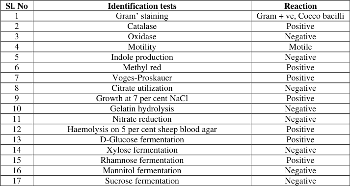 Table 2
 Table 3
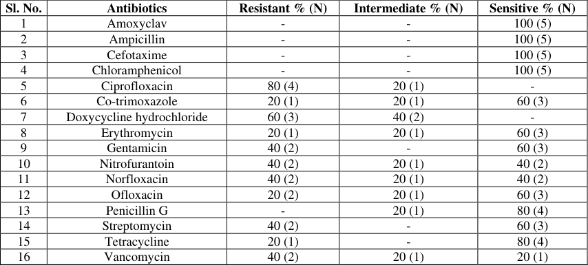 Table 4
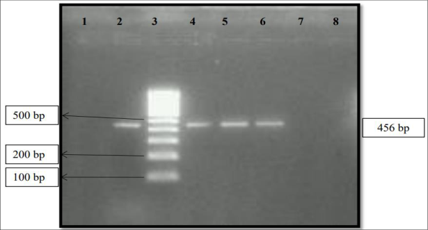 Figure 1
Introduction
Milk is an essential component of the diet of approximately six billion people. In addition to milk, several dairy products such as cream, butter, yogurt, kefir and cheese have been produced and consumed worldwide for millennia. Therefore, the impact of milk and dairy products on human health is quantitatively relevant and has been the subject of several investigations, on both whole products and their isolated components. Milk can carry pathogenic bacteria such as Salmonella, Listeria, Staphylococcus and Escherichia coli which are responsible for numerous foodborne illnesses and outbreaks [2] . Raw milk and other ready-to-eat products can act as a vehicle for listeriosis [5,14] . Being an opportunistic intracellular pathogen L. monocytogenes has become an important cause of human foodborne infections worldwide. The Centre for Disease Control and Prevention (CDC) identifies 31 foodborne pathogens to be the cause of foodborne illnesses and amongst these pathogens; L. monocytogenes is ranked in the top five, contributing to domestically acquired foodborne illness resulting in death [21] . It is mainly associated with a wide range of foods, most often ready-to-eat foods, such as sliced cooked meats, pork pies, crab meat and pate [4,8] . Food borne listeriosis, caused by the pathogen L. monocytogenes, is a relatively rare but serious disease with high fatality rates and hospitalization among susceptible population [17] . L. monocytogenes causes gastroenteritis and mild influenza in immunocompromised individuals [9] . Among the different forms of listeriosis, abortion and meningitis are the most common forms observed. Considering the above facts the present study was undertaken to detect the presence of L. monocytogenes from milk and milk products in Puducherry.
Samples were collected aseptically and transported under refrigerated condition to the laboratory. The collected samples were processed in the Biosafety level-II laboratory in the Department of Veterinary Public Health and Epidemiology, RIVER, Puducherry.
Isolation and identification of L. monocytogenes
Isolation and identification of L. monocytogenes was carried out as described by Parihar et al. (2007) [15] . For primary enrichment, 5 ml of each sample was added to 45 ml of University of Vermont I broth (UVM I) [containing Acriflavin (six mg) and Nalidixic acid (ten mg) as selective agents] and incubated at 37 o C for 24 h. In secondary enrichment, 0.1 ml UVM I broth culture was transferred to 10 ml of UVM II broth [containing higher concentration of Acriflavin (12.5 mg) and Nalidixic acid (10 mg) as selective agents] and incubated at 37 o C for 24 h. After the period of incubation in UVM II broth, a loopful of inoculum was streaked on Polymyxin Acriflavin Lithium-chloride Ceftazidime Esculin Mannitol (PALCAM) agar plates and incubated at 37 o C for 48 h. Samples showing pinpoint greyish green colour colonies surrounded by black zone of aesculin hydrolysis with centre halo were preliminarily designated as L. monocytogenes. The suspected colonies of L. monocytogenes were subjected to various morphological and biochemical tests as described by Barrow and Feltham [3] .
Molecular characterization of L. monocytogenes
Molecular confirmation of L. monocytogenes was done through detection of hlyA gene by Polymerase Chain Reaction (PCR) as described by Paziak-Domanèska et al. (1999) [16] . The PCR assay was performed in 25 µl reaction volume containing 12.5 µl of master mix, 1 µl of forward and reverse primers each (10 pmol), 5 µl of DNA template and 5.5 µl of nuclease free water. The cyclic condition of the PCR included primary denaturation at 95 o C for 2 min followed by 30 cycles of denaturation at 95 o C for 15 sec, annealing at 60 o C for 30 sec and extension at 72 o C for 90 sec. Final extension was carried out at 72 o C for 10 min. The amplified PCR products were run in 1.5% agarose gel and analyzed using gel documentation system. The primers used in this study are given in the Table 1. Biofilm production assay: The qualitative test tube method was employed for determining the adherence and biofilm forming ability of the isolates as described by Christensen et al. (1982) [6] with slight modification. Briefly, a loop full of the bacterial culture from the agar plate was inoculated into a sterile glass test tube containing five ml of BHI broth and incubated at 37 °C for 48 hrs. Each tube was decanted and stained with 0.25% safranin, gently rotated to ensure uniform staining and then the contents were decanted. The tubes were then placed upside-down to ensure draining. The tube's inner surface colour was observed and used for interpretation. An adherent film on the surface of the glass tube was taken as an evidence of slime formation. Based on slime production, the positive results were recorded as strong (+++), moderate (++), weak (+) and the absence of film was represented as negative (-) [12] .
Antibiogram: Antibiotic susceptibility tests were performed for L. monocytogenes by Kirby-Bauer disc diffusion method [7] using commonly used anibiotics. Briefly the isolates were grown in nutrient broth at 37 o C for 16 h. Individual broth cultures were smeared on the Mueller-Hinton (MH) agar plates with the help of sterile cotton swabs. The plates were allowed to dry for few minutes. The antibiotic discs were placed on the agar surface within 15 minutes of inoculation of plates and incubated overnight at 37 o C. The isolates were tested against ampicillin (10 mcg), amoxyclav (30 mcg), chloramphenicol (30 mcg), co-trimaxazole (10 mcg), ciprofloxacin (30 mcg), cefotaxime (30 mcg), gentamicin (30 mcg), penicillin G (10 units), tetracycline (30 mcg), erythromycin (15 mcg), vancomycin (5 mcg), streptomycin (10 mcg), nitrofurantoin (300 mg), norfloxacin (10 mcg), doxycycline hydrochloride (30 mcg) and ofloxacin (5 mcg).
Based on the zone of inhibition, the isolates were graded as sensitive, intermediate and resistant as per the interpretative chart provided by the manufacturer (HiMedia Laboratories, Mumbai). All the isolates obtained in this study were screened by PCR for the presence of virulent hlyA gene. Results revealed the presence of virulent hlyA gene with a desired product size of 456 bp in all the isolates (Fig 1). Biofilm production assay revealed that all the five isolates (100%) had the ability to produce biofilm, of which, three (60%) were weak biofilm producers and two (40%) were moderate biofilm producers. The details about the biofilm producing potential of L monocytogenes isolates are shown in the Table 3. Antibiogram studies showed that 80% of isolates were resistant to ciprofloxacin, 60% of the isolates showed resistant to doxycycline hydrochloride and 40% of the isolates were resistant to norfloxacin, nitrofurantoin, streptomycin, vancomycin, and gentamicin. All isolates showed sensitivity towards ampicillin, chloramphenicol, amoxyclav and cefotaxime, whereas 80% of the isolates were sensitive to tetracycline and penicillin G. Also 60% of the isolates were sensitive to gentamicin, erythromycin, streptomycin, co-trimoxazole and ofloxacin and 40% were sensitive to gentamycin and vancomycin (Table 4).
| Sl. No. | Antibiotics | Resistant % (N) | Intermediate % (N) | Sensitive % (N) |
|---|---|---|---|---|
| 1 | Amoxyclav | - | - | 100 (5) |
| 2 | Ampicillin | - | - | 100 (5) |
| 3 | Cefotaxime | - | - | 100 (5) |
| 4 | Chloramphenicol | - | - | 100 (5) |
| 5 | Ciprofloxacin | 80 (4) | 20 (1) | - |
| 6 | Co-trimoxazole | 20 (1) | 20 (1) | 60 (3) |
| 7 | Doxycycline hydrochloride | 60 (3) | 40 (2) | - |
| 8 | Erythromycin | 20 (1) | 20 (1) | 60 (3) |
| 9 | Gentamicin | 40 (2) | - | 60 (3) |
| 10 | Nitrofurantoin | 40 (2) | 20 (1) | 40 (2) |
| 11 | Norfloxacin | 40 (2) | 20 (1) | 40 (2) |
| 12 | Ofloxacin | 20 (2) | 20 (1) | 60 (3) |
| 13 | Penicillin G | - | 20 (1) | 80 (4) |
| 14 | Streptomycin | 40 (2) | - | 60 (3) |
| 15 | Tetracycline | 20 (1) | - | 80 (4) |
| 16 | Vancomycin | 40 (2) | 20 (1) | 20 (1) |
Table text extracted from the original PDF; superscript letters are shown inline. The original table image is below.
View original table image
| Biofilm forming potential | Raw milk isolates, N=4 (%) | Ice cream, N=1 (%) | Total, N=5 (%) |
|---|---|---|---|
| Strong | 0 (0) | 0 (0) | 0 (0) |
| Moderate | 2 (50) | 0 (0) | 2 (40) |
| Weak | 2 (50) | 1 (100) | 3 (60) |
| Negative | 0 (0) | 0 (0) | 0 (0) |
Table text extracted from the original PDF; superscript letters are shown inline. The original table image is below.
View original table image
Discussion
L. monocytogenes remains a significant cause of foodborne illness. The occurrence of L. monocytogenes in the present study was lower than the findings of Sharma et al. (2024) [19] who reported 9.7% of L. monocytogenes in milk and milk products in Karnataka whereas presence of L. monocytogenes in raw milk in this study (4%) was higher than that reported by Suryawanshi et al. (2023) [20] who demonstrated 3% in raw milk samples in Maharashtra.
In 1998, Paziak-domeska compared Analytical Profile Index (API) Listeria test, production of phosphatidylinositolspecific phospholipase C (PI-PLC) and PCR for a DNA fragment of the hlyA gene encoding listeriolysin-O and reported that API test had 100 per cent agreement with PCR with hlyA gene [16] . PCR revealed that all the isolates in this study harboured hlyA gene. Similar finding was obatined by Nayak et al. (2015) [13] on screening 200 food who found three positive isolates of L. monocytogenes and all showed the presence of hlyA virulence genes. In 2008, Kalorey and his coworkers [10] employed haemolysin gene (hlyA) for identification of L. monocytogenes in 2060 bovine milk samples in Central India and reported the same in 105 samples (5.1%). Although this pathogen is effectively controlled during pasteurization, its presence to a level of 2% in ice cream samples can be attributed to postpasteurization contamination from sources in the plant environment.
The presence of L. monocytogenes in raw milk with biofilm producing ability can contaminate kitchen utensils or surfaces which in turn can act as a source for cross contamination. In concordance with the present study, several studies have reported that most of the L. monocytogenes were either moderate or weak biofilm producers. In USA, biofilm production assay of L. monocytogenes isolates from 203 milk samples collected from buffaloes and cows showed that two isolates from cow milk were able to strongly form biofilm on glass surface while the only isolate from buffalo was moderately adherent [23] . Kayode et al. (2022) [11] on examining 12 L. monocytogenes isolates from milk and milk products in South Africa found that most of the isolates were weak or moderate biofilm formers. All the isolates obtained in the current study had virulent gene hlyA and were moderate to weak biofilm producers which is of concern since the biofilms can be more resistant to disinfectants. Over the last 60 years, due to widespread and irrational use of antimicrobial drugs, bacteria have evolved towards antibiotic resistance posing a great concern to public health especially in developing countries [18] . In the present investigation, L. monocytogenes isolates had shown increased resistance to gentamicin, vancomycin and penicillin G which are commonly used in the treatment of listerial infection which is a matter of public health importance. The levels of resistance vary and are influenced by antimicrobial use and geographical differences [1] . In India, antibiotic resistance strains of L. monocytogenes from food and clinical sources were reported sporadically. A study conducted by Zafar et al. (2020) [22] in milk and milk products in Faisalabad recorded that L. monocytogenes strains were highly resistant to Penicillin (63.15%) while 100% isolates were found susceptible to ampicillin, chloramphenicol and vancomycin. Isolation of increasing number of antibiotic-resistant L. monocytogenes strains could be due to the ability of the pathogen to rapidly acquire a wide variety of antibiotic resistance genes from the commensal bacteria found in foods and food processing environment.
Conclusion
The presence of L. monocytogenes in raw milk and milk products in Puducherry is a matter of public health concern and warrants regulatory mechanism to be put in place to reduce the contamination. The study clearly indicates the need for proper hygienic measures before and after collection of milk, proper processing of milk and dairy products and proper storage to reduce the contamination level.
Acknowledgement
AcknowledgementThe authors are thankful for the support and financial aid provided by Dean, Rajiv Gandhi Institute of Veterinary Education and Research, Puducherry and Head of Departments of VPE and VBC to carry out this study.
Figures and tables from the paper
| Sl. No | Identification tests | Reaction |
|---|---|---|
| 1 | Gram’ staining | Gram + ve, Cocco bacilli |
| 2 | Catalase | Positive |
| 3 | Oxidase | Negative |
| 4 | Motility | Motile |
| 5 | Indole production | Negative |
| 6 | Methyl red | Positive |
| 7 | Voges-Proskauer | Positive |
| 8 | Citrate utilization | Negative |
| 9 | Growth at 7 per cent NaCl | Positive |
| 10 | Gelatin hydrolysis | Negative |
| 11 | Nitrate reduction | Negative |
| 12 | Haemolysis on 5 per cent sheep blood agar | Positive |
| 13 | D-Glucose fermentation | Positive |
| 14 | Xylose fermentation | Negative |
| 15 | Rhamnose fermentation | Positive |
| 16 | Mannitol fermentation | Negative |
| 17 | Sucrose fermentation | Negative |
Table text extracted from the original PDF; superscript letters are shown inline. The original table image is below.
View original table image
References
21 in the original paper- Abdollahzadeh E, Ojagh SM, Hosseini H, Ghaemi EA, Irajian G, Heidarlo NM. Antimicrobial resistance of Listeria monocytogenes isolated from seafood and humans in Iran. Microb Pathog. 2016;100:70-74.
- Asfaw T, Genetu D, Shenkute D, Shenkutie TT, Amare YE, Habteweld HA, et al. Pathogenic bacteria and their antibiotic resistance patterns in milk, yoghurt and milk contact surfaces in Debre Berhan Town, Ethiopia. Infect Drug Resist. 2023;16:4297-4309.
- Rekha BV, Mallik SVS, Chaudhari SP, Barbuddhe SB. Listeriolysin-O based diagnosis of Listeria monocytogenes infection in experimentally and naturally infected goats. Small Rumin Res. 2006;66(13):70-75.
- Castro H, Ruusunen M, Lindström M. Occurrence and growth of L. monocytogenes in packaged raw milk. Int J Food Microbiol. 2017;261:1-10.
- Christensen DG, Simpson WA, Alan LB, Edwin HB. Adherence of slime-producing strains of Staphylococcus epidermidis to Smooth Surfaces. Infect Immun. 1982;37(1):318-326.
- Bauer AW, Kirby WM, Sherris JC, Turck M. Antibiotic susceptibility testing by a standardized single disk method. Am J Clin Pathol. 1966;45:493-496.
- EFSA and ECDC (European Food Safety Authority and European Centre for Disease Prevention and Control). The European Union One Health 2020 Zoonoses Report. EFSA Journal 2021;19(12):83-86.
- Gurgu GL, Bucur FI, Mihalache OA Nicolau AI. Comprehensive Review on the Biocontrol of Listeria monocytogenes in Food Products. Foods. 2024;13(5):734.
- Kalorey DR, Warke SR, Kurkure NV, Rawool DB, Barbuddhe SB. Listeria species in bovine raw milk: A large survey of Central India. Food Control. 2008;19(2):109-112.
- Kayode AJ, Okoh AI. Assessment of the molecular epidemiology and genetic multiplicity of Listeria monocytogenes recovered from ready-to-eat foods following the South African listeriosis outbreak. Sci Rep. 2022;12(1):20129.
- Murugan K, Usha M, Malathi P, Sohaibani SALS, Chandrasekaran M. Biofilm forming multi drug resistant Staphylococcus spp. among patients with Conjunctivitis. Pol J Microbiol. 2010;59(4):233-239. 13. Nayak DN, Savalia CV, Kalyani IH, Kumar R, Kshirsagar DP. Isolation, identification, and characterization of Listeria spp. from various animal origin foods. Vet World. 2015; 8(6):695-701. International Journal of Agriculture and Food Science https://www.agriculturaljournals.com
- Pal M, Shuramo MY, Shiferawu F, Parmar BC. Listeriosis: an emerging foodborne disease of public health concern. J Adv Microbiol Res. 2022;3(2):29-33.
- Parihar VS, Barbuddhe SB, Chakurkar EB, Danielsson T, Tham, W. Isolation of Listeria species from farm bulk milk at the receiving dairy plant and cervico vaginal swabs. Ind J Comp Immunol Microbiol Infect Dis. 2007; 28:53-55.
- Domanèska PB, BogusIawska E, Szakiel WCM, KotIowski R, RoèzÍalska B, Chmiela M, et al. Evaluation of the API test, phosphatidylinositol-speci¢c phospholipase C activity and PCR method in identification of Listeria monocytogenes in meat foods. FEMS Microbiol. Lett. 1999; 171(2):209-214.
- Quereda JJ, García MA, Gorba PC, Dessaux C, Del Portillo GF, Pucciarelli MG, Ortega AD. Pathogenicity and virulence of Listeria monocytogenes: A trip from environmental to medical microbiology. Virulence. 2021;12(1):2509-2545.
- Basu RS, Mondal S, Kundu C, Rai A, Roy, A. Biofilm formation potentiality of some antibiotic resistant Bacillus cereus isolates from fresh raw chhana in Kolkata, India. Ann Biol Res. 2016;7(2):1-6.
- Sharma RK, Jalalpure SS, Pathak S, Ganapathy S, Desvaux M, Roy S, Hegde S. Molecular detection of Listeria monocytogenes from different dairy and street food sources in North Karnataka, India. J Infect Public Health. 2024;17(4):696-703.
- Suryawanshi R, Patil M, Shinde O, Jogdand A, Bhosale P, Deshmukh O, Babar A. Listeria monocytogenes isolated from raw milk: phenotypic and molecular characterization, pathogenicity testing, and multidrug resistance profiling. Indian J. Dairy Sci. 2023;76(6):542-548.
- Tirumalai PS. Listeriosis and Listeria monocytogenes in India. Wudpecker J Food Tech. 2013;1(6): 98-103.
- Zafar N, Zeeshan Nawaz SA, Ali A, Mudassar M, Javid AMT. Prevalence, molecular characterization and antibiogram study of Listeria monocytogenes isolated from raw milk and milk products. Pure Appl Biol. 2020;9(3):1982-1987.
- Osman KM, Zolnikov TR, Samir A, Orabi A. Prevalence, pathogenic capability, virulence genes, biofilm formation and antibiotic resistance of Listeria in goat and sheep milk confirms need of hygienic milking conditions. Pathog Global Health. 2014; 108(1):21-29.