
Research article · 2024 · Full text
International Journal of Advanced Biochemistry ResearchIsolation, molecular characterization and evaluation of probiotic potential of lactic acid bacteria isolated from beet root and sweet corn
Volume 8(3) · Pages 67–74 · DOI: 10.33545/26174693.2024.v8.i3a.678 ↗
Research areasFood composition and propertiesMicrobial Metabolites in Food BiotechnologyGABA and Rice Research
Abstract
Author-writtenThe objective of this study was to isolate, identify and characterize lactic acid bacterial strains from beet root and sweet corn as potential probiotics with antimicrobial activity against some human pathogenic strains. A total of 36 bacterial strains were isolated and morphologically these isolates were rods, cocci, cocco-bacilli in shape with small-medium milky white, creamy colonies; biochemically Gram positive, negative for catalase and endospore production test, positive for exopolysaccharide production and acid production test. None of the isolates including reference strains (Lactobacillus acidophilus MTCC10307 and Lactobacillus plantarum NCIM 2656) produced gas in Durham’s tube and hence characterized as homofermentative lactic acid bacteria. Further these isolates were then screened for their probiotic activities. Results showed that some of these isolates were viable at varied pH (2-3), 0.1-0.4% bile salt concentrations and displayed marked phenotypic resistance against the antibiotics like Kanamycin, Streptomycin, Ciprofloxacin and Gentamycin and low-level resistance to Tetracycline, Chloramphenicol, Ampicillin and Azithromycin. In addition, these isolates also showed good antimicrobial activities against the tested pathogenic strains of humans. Out of 3 selected lactic acid bacterial isolates, BRLB22 and SCLB12 isolates showed maximum zone of inhibition against Staphylococcus aureus and Escherichia coli followed by other isolates. Finally, the best two selected LAB isolates (SCLB12 and BRLB22) were identified as Lactobacillus brevis and Lactobacillus plantarum respectively by molecular and phylogenetic analysis using 16S rRNA gene based molecular method.
Summary
Source-checkedPlain-language reading of the full paper, prepared with AI and checked against the text. Each point links to the authors' own words.
Why it matters
- Probiotics are microorganisms that offer health benefits to a host, with Lactic acid bacteria (LAB) being the most common and generally safe type used in food. View evidence ↗
- Probiotics are documented to help treat various health conditions, such as inflammatory bowel disease, hypertension, diabetes, and certain types of diarrhea. View evidence ↗
- Lactic acid bacteria are suitable probiotics because they can survive harsh conditions in the human body, such as low pH, and can colonize the gut. View evidence ↗
The question
- The study aimed to isolate and characterize lactic acid bacteria from sweet corn and beet root to evaluate their potential as probiotics with antimicrobial properties. View evidence ↗
What they did
- Beet root and sweet corn samples were collected from various locations in Bengaluru and enriched using MRS broth. View evidence ↗
- The researchers isolated 36 bacterial strains, which were Gram-positive, produced acid and exopolysaccharides, and tested negative for catalase and endospore production. View evidence ↗
- To test pH tolerance, bacterial cultures were incubated at pH levels 1, 2, and 3 for 24 hours at 37 °C. View evidence ↗
- Bile salt tolerance was measured by incubating cultures in MRS broth with bile salt concentrations ranging from 0.1% to 0.4% for 4 hours. View evidence ↗
- The antimicrobial activity of the isolates was tested against Escherichia coli and Staphylococcus aureus using an agar well diffusion assay. View evidence ↗
What they found
- Ten of the 22 pH-tolerant isolates remained viable across bile salt concentrations of 0.1% to 0.4%. View evidence ↗
- Isolate BRLB22 had the highest inhibition zone against S. aureus (25.3 mm), while SCLB12 had the highest against E. coli (19.66 mm). View evidence ↗
- Molecular analysis identified the two most promising isolates, SCLB12 and BRLB22, as Lactobacillus brevis and Lactobacillus plantarum, respectively. View evidence ↗
Visual summary
3 visualsIllustrations explain the paper; charts are replotted from the paper's own table values.
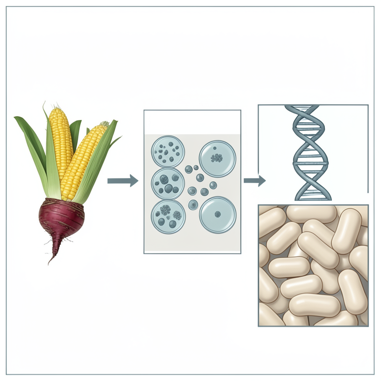
Probiotic Isolation Workflow
AI-generated conceptual illustration (beet root, sweet corn, and lactic acid bacteria). It explains the idea and does not show study data.
View source evidence ↗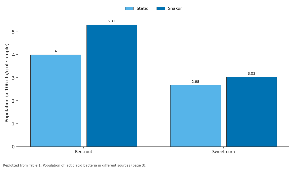
Static and Shaker: Beetroot vs Sweet corn
Replotted from Table 1 (page 3) of the original paper. Every bar is the paper's own table value.
View source evidence ↗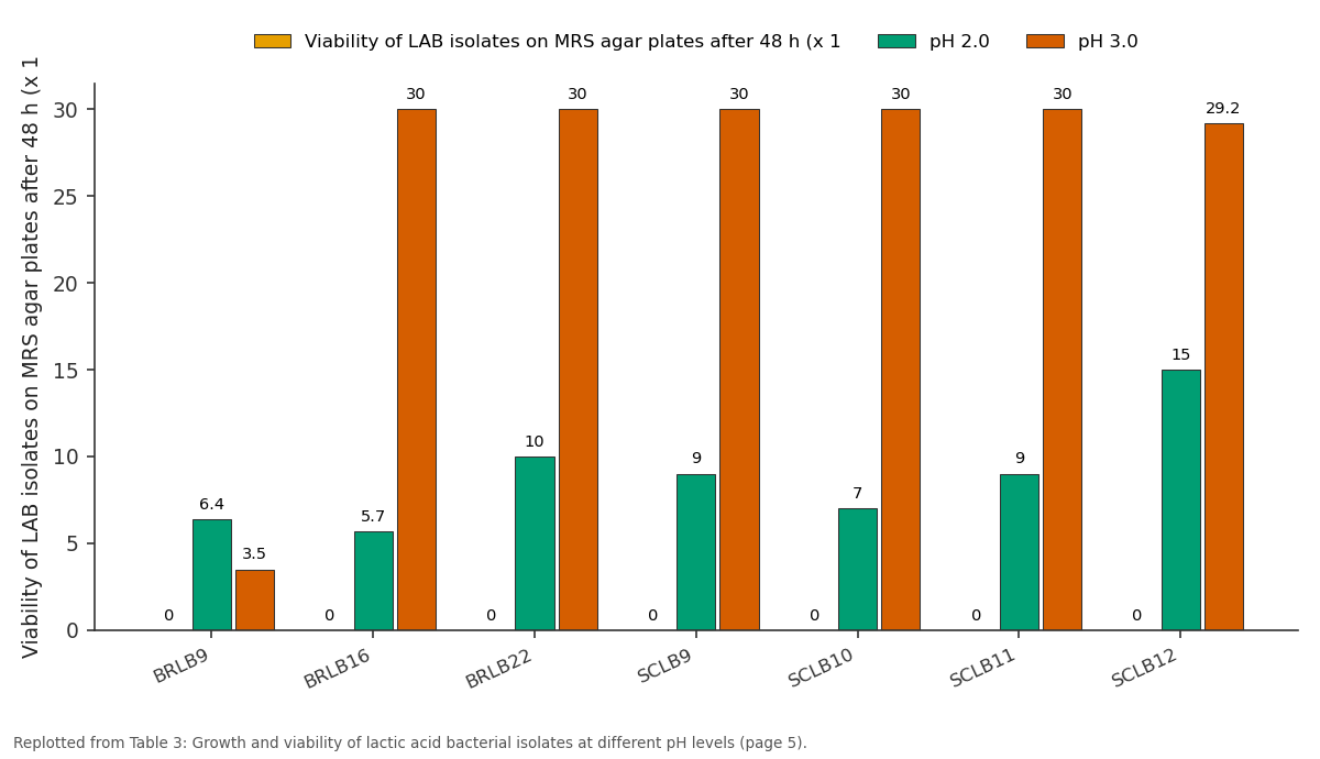
Viability of LAB isolates on MRS agar plates... and pH 2.0 and more: BRLB9 vs BRLB16 vs BRLB22 vs SCLB9 vs SCLB10
Replotted from Table 3 (page 5) of the original paper. Every bar is the paper's own table value.
View source evidence ↗Figures & tables
From the original paper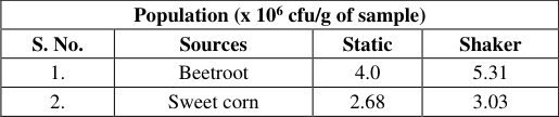 Table 1
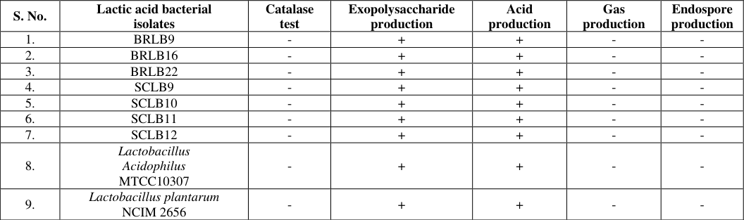 Table 2
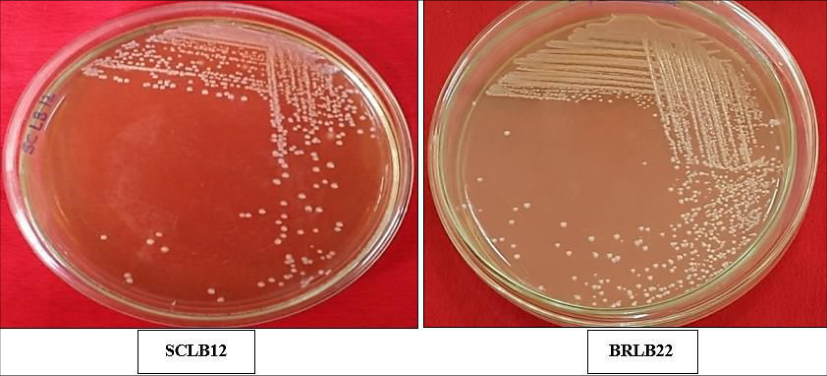 Figure 1
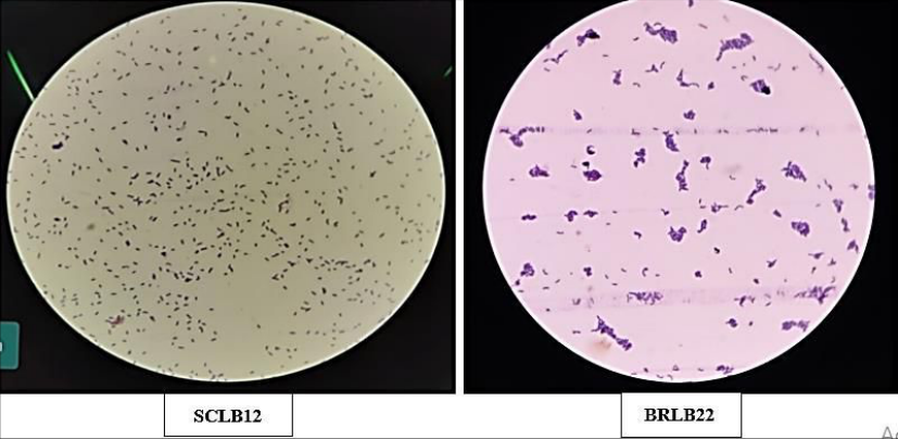 Figure 2
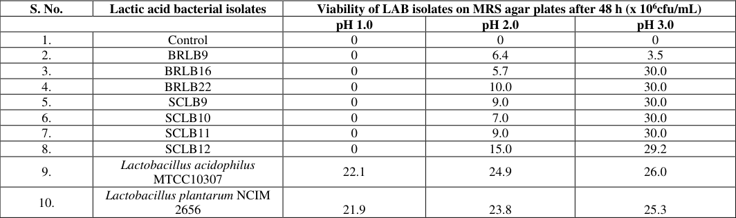 Table 3
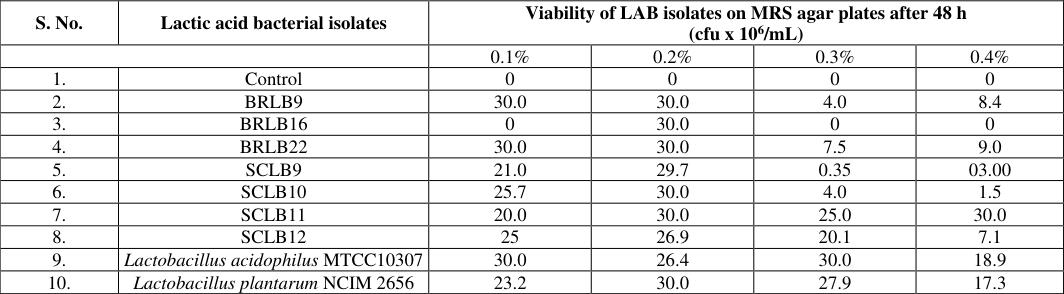 Table 4
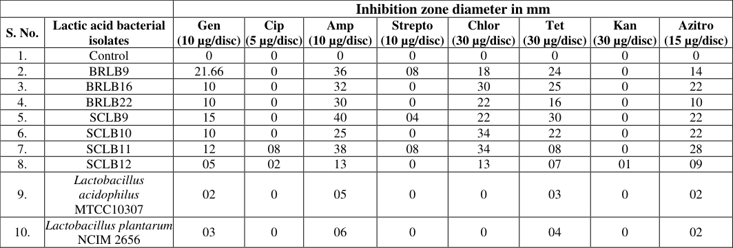 Table 5
 Table 6
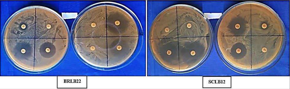 Fig. 3
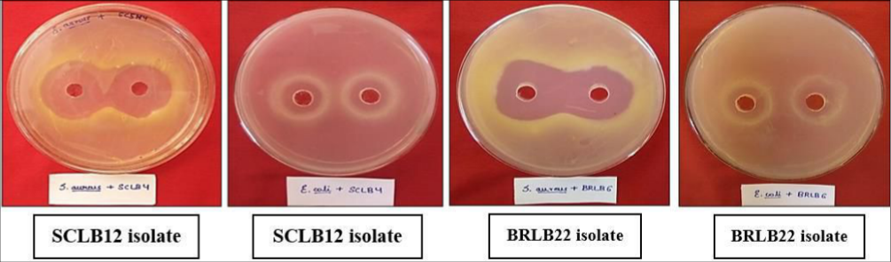 Figure 4
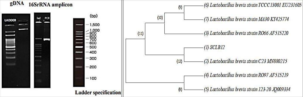 Figure 5
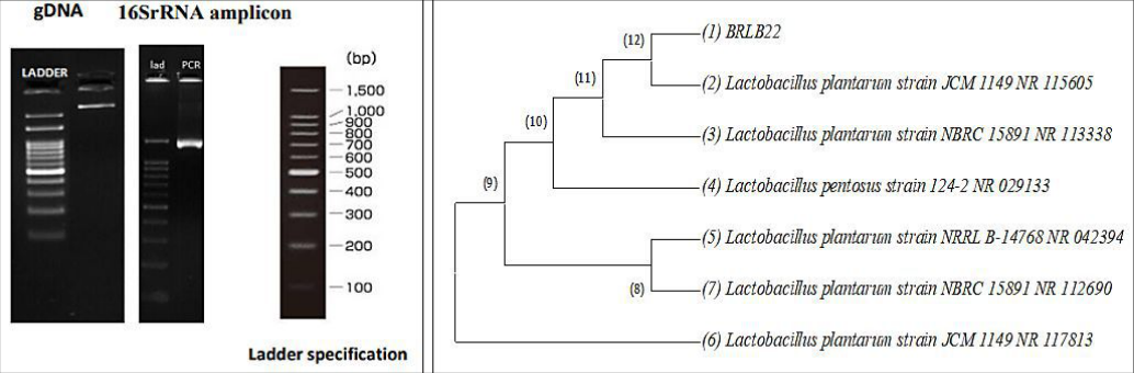 Fig. 6
Introduction
The term Probiotic means 'for life' is derived from the Greek language. Probiotics are defined as microorganisms that provide health benefits to the host when administered in appropriate amounts (FAO/WHO, 2001) [8] . The most common probiotic microorganisms used and marketed in food worldwide belong to Lactic acid bacteria (LAB), Generally Recognized as Safe (GRAS). LAB are a group of Gram-positive bacteria, cocci or rods, produce lactic acid as the major end product of fermentation of carbohydrates. This genera includes Lactobacillus, Lactococcus, Leuconostoc, Streptococcus and Pediococcus as the main species, exert a biopreservative or exhibit inhibitory effect against other microorganisms as a result of competition for nutrients and produce of bacteriocins and other antagonistic compounds that serve as probiotic organisms conferring the health benefit when ingested (Johan and Jesper, 2005) [15] . These microorganisms are found in many nutrient rich environments and occur naturally in production of fermented foods and are also part of intestinal microflora. The health benefits of probiotics in treating disorders, including inflammatory bowel disease, irritable bowel syndrome, constipation, antibiotic-associated and acute diarrhea, allergyrelated conditions, hypertension, and diabetes, have been well-documented by numerous esteemed scientific reports and systematic reviews (Hill et al., 2014) [9] . LAB possesses many typical probiotic characteristics, including the ability to withstand extreme conditions in the human body (e.g. low pH and pancreatic enzymes), colonize gut epithelial cells, and contribute to the health of the host environment. Attempts to screen for new LAB bacteria that possess excellent probiotic characteristics from various food sources are ongoing (Maleki et al., 2019) [20] . This study aimed to evaluate the in vitro probiotic properties of lactic acid bacteria isolated from sweet corn and beet root and identify them by molecular characterization.
Materials and Methods
Materials and Methods
Sample collection
Different samples of beet root and sweet corn were collected from the different locations of Bengaluru for the isolation of lactic acid bacteria (LAB) and were inoculated to de Man, Rogosa and Sharpe (MRS) broth for enriching of lactic acid bacteria.
Isolation and purification of lactic acid bacteria from beet root and sweet corn Lactic acid bacteria were isolated from beet root and sweet corn by the method described by Pundir et al. (2013) [23] . Fresh samples were collected in sterile beaker and conical flask and then 10 g of each sample were suspended in 100 mL sterile MRS broth and was kept in static and shaking condition. After 24 h the samples were serially diluted up to 10 -6 . One ml from each 10 -4 , 10 -5 and 10 -6 dilutions were plated out using De Man, Rogosa and Sharpe (MRS) agar medium (De Man et al., 1960) [6] by standard plate count method and plates were incubated at 37 °C for 24-48 h. Enumeration of LAB isolates were done based on their colony morphology. Colonies embedded in the agar medium and at the beneath of the Petri plate were transferred to sterilized MRS broth and incubated for 48 h. Further, the colonies were subjected to Gram staining and colonies with Gram-positive characteristics were selected, purified by streak plate method on MRS agar and maintained in refrigerator for further studies.
Bacterial reference strains
Reference cultures of LAB strains used in the study were
Biochemical characterization of lactic acid bacterial isolates
Catalase test: (Balazevic and Ederes, 1975) [1] . A drop of bacterial broth culture was added to a clean grease free slide followed by 3% hydrogen peroxide solution and observed immediately for effervescence formation. If bubbles are produced then the results were indicated as catalase positive.
Exopolysaccharide production: (Paulo et al., 2012) [12] A drop of 48 h old broth culture was added to clean glass slide followed by absolute alcohol and observed immediately for precipitate formation.
Acid production: (Seeley and Vandemark, 1970) [25] . The 48 h old LAB cultures were inoculated to the lactose broth with the reagent Bromocresol purple (BCP), incubated for 48 h and observed for the colour change from pink to yellow.
Gas production: (Seeley and Vandemark, 1970) [25] .
The 48 h old LAB culture was inoculated to lactose broth added with BCP containing Durham's tubes, incubated for 48 h, formation of air bubble in the Durham's tubes were observed.
Endospore staining: (Murray et al., 1994) [21] . The lactic acid bacterial isolates of 30 days old were used for endospore staining. A thin smear of bacterial culture was made on a clean grease free glass slide, air-dried followed by heat fixation. The smear was covered with a blotting paper and was flooded with malachite green and kept on hot water bath for 5-7 minutes. Stain was added continuously to prevent drying of slide and later the slide was cooled and washed with water followed by counter staining with safranin for 30 seconds. Finally, safranin was washed with water, the slide was air-dried and observed under oil immersion objective and observation was recorded.
Classification of isolates into homo and hetero fermentative groups
Based on the tests for acid and gas production (colour change of lactose broth and bubble formation in the Durham's tubes), the isolates were classified into homo and hetero-fermentative groups (Zuniga et al., 1993) [27] .
In vitro screening of lactic acid bacterial isolates for Probiotic activity pH tolerance test
The isolated LAB isolates were tested for pH tolerance according to Ishaq et al. (2019) [12] . Overnight bacterial cultures (0.1 mL) were inoculated into 10 mL sterile MRS broth tubes of varying pH 1, 2 and 3 respectively by adjusting the pH using 1N HCl and 1N NaOH and incubated at 37 °C for 24 h. Growth was measured using spectrophotometer at 600 nm and viability of LAB isolates was determined by pouring 1 mL of culture to MRS agar medium by pour plate method and incubated at 37 °C for 48 h. The growth of LAB on MRS agar was used to designate the isolates as pH tolerant.
Bile salt tolerance test
Overnight bacterial cultures (0.1 mL) were inoculated into 10 mL sterile MRS broth containing 0.1%, 0.2%, 0.3% and 0.4% of bile salt respectively and incubated at 37°C for 4 h. Spectrophotometer reading of the inoculated broths at 600 nm was taken after 4hrs of inoculation and viability of LAB isolates was determined by pouring 1 mL of culture to MRS agar medium by pour plate method and incubated at 37 °C for 48 h. MRS broth without bile salt was used as control (Berebon et al., 2019) [3] .
Antibiotic sensitivity of lactic acid bacteria
Each of acid-bile tolerant lactic acid bacterial isolates was assessed for its antibiotic resistance by the disc diffusion method as described by Zhang et al. (2016) [26] . Against some antibiotics that included gentamycin (10 µg/disc), ciprofloxacin (5 µg/disc), ampicillin (10 μg/disc), streptomycin (10 μg/disc), chloromphenicol (30 µg/disc), tetracycline (30 μg/disc), kanamycin (25 μg/disc) and azithromycin (15 µg/disc). Thus, a volume of 100 μL of actively growing cultures of each acid-bile-tolerant lactic acid bacteria was spread evenly over the surface of MRS agar plates. After drying, the antibiotic discs were placed on the solidified agar surface and then incubated at 37 °C for 48 h. Resistance was defined according to the disc diffusion method by using the above antibiotic discs and the diameters of inhibition zones were measured. The zone of inhibition (diameter in mm) for each antibiotic was measured and expressed as susceptible (≥21 mm); intermediate (16-20 mm) and resistance (≤15 mm).
Antimicrobial activity
The inhibitory effect of LAB isolates against human bacterial pathogen (Escherichia coli NCIM 2065 and Staphylococcus aureus NCIM 2079) was carried out by agar well diffusion assay (Bali et al., 2011) [2] . Petri dishes with nutrient agar that was previously inoculated with 0.
Agarose gel electrophoresis
Agarose gel electrophoresis was performed to resolve the amplified product using 0.8% agarose in 1X TAE buffer, 0.5 μg/mL of ethidium bromide and loading buffer (0.25% bromophenol blue prepared in 40% sucrose). Five μL of loading dye was added to 40 μL of PCR product and loaded to the well. Electrophoresis was carried out at 100 V for 2 h.
The gel was visualized under UV trans illuminator and documented using gel documentation unit.
Gel elution
The Gene JET™ Gel Extraction Kit (Thermo Scientific)
was used for rapid and efficient separation of DNA fragments from agarose gel. The gel slice containing DNA was excised using a clean razor blade and was placed into a pre-weighed 1.5 mL tube. Into it, equal volume of binding buffer was added (100 mg gel slice: 100 μL of binding buffer). The mixture was incubated at 60 °C till the gel slice was completely dissolved. The solubilized gel solution was added to the Gene JET™ purification column and centrifuged at 10,000 rpm for 60 seconds. The flow-through was discarded and the column was placed back into the same collection tube, washed by adding 700 μL of wash buffer by centrifugation at 10,000 rpm for 60 seconds. The tube was again centrifuged for 60 seconds to remove the wash buffer remained as residue. Then purification column was placed into a clean 1.5 mL micro centrifuge tube and added with 20 μL of elution buffer and centrifuged at 10,000
rpm for 60 seconds. The DNA eluted was checked for its concentration using nano drop and got sequenced by Barcode Bio Sciences, Bengaluru, Karnataka. The sequence data received from the company was analyzed for homology.
Sequence analysis and homology search
Sequence analysis and homology search sequence results were analyzed by online software from National Centre for Biotechnology Information (NCBI). The sequences were aligned by CLUSTAL W Multiple Sequence Alignment Programme and a phylogenetic tree was constructed with bootstrap support based on a neighbour-joining analysis in MEGA X.
Results and Discussion
Results and Discussion
Isolation and purification of lactic acid bacteria
In beet root, lactic acid bacterial population varied from 4.0 x 10 6 cfu/g in static condition and 5.31 x 10 6 cfu/g in shaking condition. In sweet corn, lactic acid bacterial population varied from 2.68 x 10 6 cfu/g in static condition and 3.03 x 10 6 cfu/g in shaking condition. Among the shaking and static conditions, the population of LAB was found to be higher in shaking conditions, similar results were reported by Ibrahim et al. (2010) [11] . A total of 36 lactic acid bacterial isolates were isolated from beet root and sweet corn by various morphological and biochemical traits. Similar finding was reported by Jalali et al. (2012) [14] , who isolated two lactic acid bacteria from radish and tomato and were identified as Lactobacillus plantarum and Lactobacillus brevis, respectively.
Biochemical characterization of lactic acid bacterial isolates
All the isolates were negative for catalase and endospore production. Similar results were reported by Ismail et al.
(2018) [13] . All the isolates were positive for exopolysaccharide (EPS) production. The result was in accordance with Emnace and Dizon (2018) [7] . Who identified that LAB produced the highest yield of exopolysaccharides. All the isolates were positive for acid production and none of the isolates produced gas in Durham's tube. Based on this assay, the isolates were grouped into homofermentative and heterofermentative. All the isolates including reference strain (Lactobacillus acidophilus MTCC10307 and Lactobacillus plantarum NCIM 2656) were homofermentative. Similar results i.e LAB is a diverse group of Gram-positive, aero tolerant homofermentative bacteria and lactic acid producer were reported by Liu et al. (2010) [19] . isolates were viable at pH 3 only. Reference strains were viable at all the pH levels (pH 1, 2 and 3). The acid tolerance of Lactobacilli is attributed to the presence of a constant gradient between extracellular and cytoplasmic pH. When the internal pH reaches a threshold value, cellular functions are inhibited and the cells die (Kashket, 1987) [16] . The F 0 F 1 -ATPase is a known mechanism that gram-positive organisms use for protection against acidic conditions (Cotter and Hill, 2003) [5] .
Bile salt tolerance test of LAB isolates
The growth and viability of LAB isolates at different bile salt concentrations (0.1%, 0.2%, 0.3% and 0.4%) are represented in Table 5. Out of 22 pH tolerant LAB isolates, 10 isolates were viable at 0.1-0.4% bile salt concentrations.
| — | — | Inhibition zone diameter in mm | — | — | — | — | — | — | — |
|---|---|---|---|---|---|---|---|---|---|
| S. No. | Lactic acid bacterial isolates | Gen (10 µg/disc) | Cip (5 µg/disc) | Amp (10 µg/disc) | Strepto (10 µg/disc) | Chlor (30 µg/disc) | Tet (30 µg/disc) | Kan (30 µg/disc) | Azitro (15 µg/disc) |
| 1. | Control | 0 | 0 | 0 | 0 | 0 | 0 | 0 | 0 |
| 2. | BRLB9 | 21.66 | 0 | 36 | 08 | 18 | 24 | 0 | 14 |
| 3. | BRLB16 | 10 | 0 | 32 | 0 | 30 | 25 | 0 | 22 |
| 4. | BRLB22 | 10 | 0 | 30 | 0 | 22 | 16 | 0 | 10 |
| 5. | SCLB9 | 15 | 0 | 40 | 04 | 22 | 30 | 0 | 22 |
| 6. | SCLB10 | 10 | 0 | 25 | 0 | 34 | 22 | 0 | 22 |
| 7. | SCLB11 | 12 | 08 | 38 | 08 | 34 | 08 | 0 | 28 |
| 8. | SCLB12 | 05 | 02 | 13 | 0 | 13 | 07 | 01 | 09 |
| 9. | Lactobacillus acidophilus MTCC10307 | 02 | 0 | 05 | 0 | 0 | 03 | 0 | 02 |
| 10. | Lactobacillus plantarum NCIM 2656 | 03 | 0 | 06 | 0 | 0 | 04 | 0 | 02 |
Table text extracted from the original PDF; superscript letters are shown inline. The original table image is below.
View original table image
The viability of the LAB isolates decreased in higher concentrations of bile salt. The reason for the reduced growth with increasing level of bile salts could be due to the binding of probiotic organism with bile salts (Patel et al., 2004) [22] .
Antibiotic sensitivity of LAB isolates
The isolates which were viable at varying pH and bile salt concentrations have shown a varying range of resistance to various different antibiotics at different rates (Fig. 3). Most of the LAB isolates have shown resistant to some of the antibiotics like Kanamycin (30 µg/disc), Streptomycin (10 µg/disc), Ciprofloxacin (5 µg/disc) and Gentamycin (10 µg/disc) and low-level resistance to Tetracycline (30 µg/disc), Chloramphenicol (30 µg/disc), Ampicillin (10 µg/disc), as well as to Azithromycin (15 µg/disc), similar results were reported by Hummel et al. (2007) [10] . The results are represented in the Table 5.Some non-specific mechanisms, such as multidrug transporters (Putman et al., 2000) [24] and defective cell wall autolytic systems (Kim et al., 1982) [18] may contribute to the antibiotic resistance.
[4] . The antimicrobial activity of LAB could have been due to the effect of organic acids (Khunajakr et al., 2008) [17] or the production of bacteriocins, which possesses high antimicrobial activity. Finally considering all the above probiotic tests, the best two promising LAB isolates SCLB12 and BRLB22 were identified by molecular characterization.
Molecular identification of efficient lactic acid bacterial isolates
The LAB isolates (SCLB12 and BRLB22) were identified using 16S rRNA gene based molecular method. NCBI BLAST and phylogeny analysis, resulted in identification of SCLB12 and BRLB22 isolates as Lactobacillus brevis and Lactobacillus plantarum respectively which has shown high similarity based on nucleotide homology and phylogenetic analysis (Fig. 5 and6).
Description
Percent identity Accession Lactobacillus brevis strain C23 16S ribosomal RNA gene, partial sequence 100% MN880215 strains showed strong antagonistic activities against a wide range of pathogens to humans, they could be considered as good potential probiotic candidates for treatment and prevention of infections.
Acknowledgement
AcknowledgementThe authors express their gratitude to the Department of Agricultural Microbiology, UAS, GKVK Bengaluru.
Figures and tables from the paper
| Population (x 106 cfu/g of sample) | — | — | — |
|---|---|---|---|
| S. No. | Sources | Static | Shaker |
| 1. | Beetroot | 4.0 | 5.31 |
| 2. | Sweet corn | 2.68 | 3.03 |
Table text extracted from the original PDF; superscript letters are shown inline. The original table image is below.
View original table image
| S. No. | Lactic acid bacterial isolates | Catalase test | Exopolysaccharide production | Acid production | Gas production | Endospore production |
|---|---|---|---|---|---|---|
| 1. | BRLB9 | - | + | + | - | - |
| 2. | BRLB16 | - | + | + | - | - |
| 3. | BRLB22 | - | + | + | - | - |
| 4. | SCLB9 | - | + | + | - | - |
| 5. | SCLB10 | - | + | + | - | - |
| 6. | SCLB11 | - | + | + | - | - |
| 7. | SCLB12 | - | + | + | - | - |
| 8. | Lactobacillus Acidophilus MTCC10307 | - | + | + | - | - |
| 9. | Lactobacillus plantarum NCIM 2656 | - | + | + | - | - |
Table text extracted from the original PDF; superscript letters are shown inline. The original table image is below.
View original table image
| S. No. | Lactic acid bacterial isolates | Viability of LAB isolates on MRS agar plates after 48 h (x 106cfu/mL) | — | — |
|---|---|---|---|---|
| pH 1.0 | pH 2.0 | pH 3.0 | ||
| 1. | Control | 0 | 0 | 0 |
| 2. | BRLB9 | 0 | 6.4 | 3.5 |
| 3. | BRLB16 | 0 | 5.7 | 30.0 |
| 4. | BRLB22 | 0 | 10.0 | 30.0 |
| 5. | SCLB9 | 0 | 9.0 | 30.0 |
| 6. | SCLB10 | 0 | 7.0 | 30.0 |
| 7. | SCLB11 | 0 | 9.0 | 30.0 |
| 8. | SCLB12 | 0 | 15.0 | 29.2 |
| 9. | Lactobacillus acidophilus MTCC10307 | 22.1 | 24.9 | 26.0 |
| 10. | Lactobacillus plantarum NCIM 2656 | 21.9 | 23.8 | 25.3 |
Table text extracted from the original PDF; superscript letters are shown inline. The original table image is below.
View original table image
| S. No. | Lactic acid bacterial isolates | Viability of LAB isolates on MRS agar plates after 48 h (cfu x 106/mL) | — | — | — |
|---|---|---|---|---|---|
| 0.1% | 0.2% | 0.3% | 0.4% | ||
| 1. | Control | 0 | 0 | 0 | 0 |
| 2. | BRLB9 | 30.0 | 30.0 | 4.0 | 8.4 |
| 3. | BRLB16 | 0 | 30.0 | 0 | 0 |
| 4. | BRLB22 | 30.0 | 30.0 | 7.5 | 9.0 |
| 5. | SCLB9 | 21.0 | 29.7 | 0.35 | 03.00 |
| 6. | SCLB10 | 25.7 | 30.0 | 4.0 | 1.5 |
| 7. | SCLB11 | 20.0 | 30.0 | 25.0 | 30.0 |
| 8. | SCLB12 | 25 | 26.9 | 20.1 | 7.1 |
| 9. | Lactobacillus acidophilus MTCC10307 | 30.0 | 26.4 | 30.0 | 18.9 |
| 10. | Lactobacillus plantarum NCIM 2656 | 23.2 | 30.0 | 27.9 | 17.3 |
Table text extracted from the original PDF; superscript letters are shown inline. The original table image is below.
View original table image
| — | — | Inhibition zone diameter in mm | — |
|---|---|---|---|
| S. No. | Lactic acid bacterial isolates | Escherichia coli NCIM 2065 | Staphylococcus aureus NCIM 2079 |
| 1. | BRLB9 | 17.66 | 12.00 |
| 2. | BRLB22 | 19.33 | 25.33 |
| 3. | SCLB12 | 19.66 | 25.00 |
| 4. | Lactobacillus acidophilus MTCC10307 | 21.00 | 26.66 |
| 5. | Lactobacillus plantarum NCIM 2656 | 20.00 | 26.33 |
Table text extracted from the original PDF; superscript letters are shown inline. The original table image is below.
View original table image
References
26 in the original paper- Balazevic DJ, Ederes GM. Principles of Biochemical tests in Diagnostic Microbiology. Willey and Co., New York. 1975;405-415.
- Bali V, Panesar PS, Bera MB. Isolation, Screening and Evaluation of Antimicrobial Activity of Potential Bacteriocin Producing Lactic Acid Bacteria Isolate. Microbiol J. 2011;1(3):113-119.
- Berebon DP, Ofokansi KC, Attama AA, Eze CO, Onwusoba RC, Ugwoke IC. Preliminary Studies on Isolation, Bile Tolerance and Antibiogram of Potential Probiotics (Probionts) from Locally Fermented Food Products at Beach Market, Nsukka Metropolis, Enugu State, Nigeria. Biotechnol J Int. 2019;22(3):1-10.
- Cortes CG, Suarez H, Buitrago G, Moreno CD. Isolation and evaluation of the antagonist activity of lactic acid bacteria in raw cow milk. Agron Colomb. 2017;35(3):357-364.
- Cotter PD, Hill C. Surviving the acid test: responses of Gram-positive bacteria to low pH. Microbiol Mol Biol Rev. 2003;67:429-453.
- De Man JC, Rogosa M, Sharpe M. A Medium for the Cultivation of Lactobacilli. J Appl Bacteriol. 1960;23:130-135.
- Emnace IC, Dizon EI. Selection of lactic acid bacteria for exopolysaccharide production. Ann Trop Res. 2018;40(2):61-68.
- Hill C, Guarner F, Reid G, Gibson GR, Merenstein DJ, Pot B, et al. Expert consensus document: The International Scientific Association for Probiotics and Prebiotics consensus statement on the scope and appropriate use of the term probiotic. Nat Rev Gastroenterol Hepatol. 2014;11:506.
- Hummel AS, Hertel C, Holzapfel WH, Franz CP. Antibiotic Resistances of Starter and Probiotic Strains of Lactic Acid Bacteria. Appl Environ Microbiol. 2007;73(3):730-739.
- Ibrahim SB, Rahman NA, Mohamad R, Rahim RA. Effects of agitation speed, temperature, carbon and nitrogen sources on the growth of recombinant Lactococcus lactis NZ9000 carrying domain 1 of aerolysin gene. Afr J Biotechnol. 2010;9(33):53925398.
- Ishaq M, Wu T, Ahmad N, Liu C, Fang L, Wang J, et al. Phylogenomic and biochemical assessment of four presumptive probiotic lactic acid bacteria. Int J Biosci. 2019;15(5):315-325.
- Ismail YS, Yulvizar C, Mazhitov B. Characterization of lactic acid bacteria from local cow's milk kefir. IOP Conf Ser Earth Envt Sci. 2018;130(1):012019.
- Jalali MD, Issazadeh K, Mohammad, Faezi G, Sasan SK. Antagonism of Lactobacillus Species against Xanthomonas campestris isolated from different plants. J Appl Environ Biol Sci. 2012;2(9):480-484.
- Johan S, Jesper M. Antifungal lactic acid bacteria as biopreservatives. Trends Food Sci Technol. 2005;16(13):70-78.
- KASHKET ER. Bioenergetics of lactic acid bacteria: cytoplasmic pH and osmotolerance. FEMS Microbiol Rev. 1987;46:233-244.
- Khunajakr N, Wongwicharn A, Moonmangmee D, Tantipaiboonvut S. Screening and identification of lactic acid bacteria producing antimicrobial compounds from pig gastrointestinal tracts. Sci Technol J. 2008;8(1):8-17.
- Kim KS, Morrison J, Bayer A. Deficient autolytic enzyme activity in antibiotic-tolerant lactobacilli. Infect Immun. 1982;36(2):582-585.
- Liu B, Yang M, Qi B, Chen X, Su Z, Wan Y. Optimizing l-(+)-lactic acid production by thermophile Lactobacillus plantarum using alternative nitrogen sources with response surface method. Biochem Eng J. 2010;52(2):212-219.
- Maleki KH, Barzegari A, Hanifan S, Barar J, Omidi Y. Isolation and molecular identification of Lactobacillus with probiotic potential from abomasums driven rennet. Food Chem. 2019;272:709-14.
- Patel HM, Pandiella SS, Wang RH, Webb C. Influence of malt, wheat and barley extracts on the bile tolerance of selected strains of Lactobacilli. Food Microbiol. 2004;21:83-89.
- Pundir RK, Rana S, Kashyap N, Kaur A. Probiotic potential of lactic acid bacteria isolated from food samples: an in vitro study. J Appl Pharma Sci. 2013;3(03):085-093.
- Putman M, Veen HW, Konings WN. Molecular properties of bacterial multidrug transporters. Microbiol Mol Biol Rev. 2000;64(4):672-693.
- Zhang B, Wang Y, Tan Z, Li Z, Jiao Z, Huang Q. Screening of probiotic activities of lactobacilli strains isolated from traditional Tibetan Qula, a raw yak milk cheese. Asian-Australas J Anim Sci. 2016;29(10):14901499.
- Zuniga M, Pardo I, Ferrer S. An improved medium for distinguishing between homofermentative and heterofermentative lactic acid bacteria. Int J Food Microbiol. 1993;18:37-42.
- Paulo AA, Rosa RD, Pereira LS. Climate trends and behaviour of drought indices based on precipitation and evapotranspiration in Portugal. Natural Hazards and Earth System Sciences. 2012 May 16;12(5):1481-1491.
- Paulo AA, Rosa RD, Pereira LS. Climate trends and behaviour of drought indices based on precipitation and evapotranspiration in Portugal. Natural Hazards and Earth System Sciences. 2012 May 16;12(5):1481-1491.
Cited by 2
From OpenAlex, OpenCitations and reference lists read by Scinovex
- Isolation and Characterization of Lactic Acid Bacteria from Raw Cow Milk and Evaluation of Their Probiotic Potential ↗
Goiteom Senay Niguse, Desta Berhe Sbhatu, Gerbreselema Gebreyohannes · 2026
❝ 1 - Comparative Study of Antibiotic Resistance in Lactobacillus Species Isolated from Fermented Cassava and Corn ↗
Olodu Blessing Adoh, Stephen Amadin Enabulele · DAHUDER Medical Journal · 2025
Background: This study aimed to conduct a comparative study of antibiotic resistance in Lactobacillus species isolated from fermented cassava and corn samples. Key objectives included identifying antibiotic resistance profiles and evaluating interspecies correlations to support the development of functional probiotics. Methods: A total of nine (9) bacterial strains, including Lactobacillus fermentum, Lactobacillus ghanensis, Lactobacillus delbrueckii, Lactobacillus plantarum, Lactococcus lactis, Lactobacillus…
❝ 0PDF