
Research article · 2021 · Full text
International Journal of Advanced Chemistry ResearchSynthesis of non-enzymatic biosensor based on selenium dioxide nanoparticles in order to detection of glucose under electrochemical method
Volume 3(2) · Pages 12–16 · DOI: 10.33545/26646781.2021.v3.i2a.36 ↗
Research areasElectrochemical sensors and biosensorsElectrochemical Analysis and ApplicationsAdvanced biosensing and bioanalysis techniques
Abstract
Author-writtenIn this project, a novel electrochemical biosensor was produced for the glucose recognition. Selenium dioxide Nanoparticle (SeNPs) synthesized a two-step process with a hydrothermal method. The SeNPs were characterized by X-ray diffraction (XRD). Also, the size distributions of nanoparticles were determined by dynamic light scattering (DLS), that more than 90 percent in the size range of 58–100 nm. The electrochemical performance of the SeNPs electrode for detection of glucose was investigated by cyclic voltammetry (CV). SeNPs show high electrocatalytic activity toward glucose oxidation in alkaline medium and are used for non-enzymatic electrochemical detection of glucose. The biosensor also had a linear response to glucose in the range of 0.5-2.5 mM and a low detection limit of 1.3 μM glucose (S/N=3). Therefore, the sensor shows great promise for simple, sensitive, and quantitative detection and screening of glucose and real sample (glucose in the human serum). This novel structured electrode holds great promise for the development of biosensors and other electrochemical devices.
Summary
Source-checkedPlain-language reading of the full paper, prepared with AI and checked against the text. Each point links to the authors' own words.
Why it matters
- Developing reliable and sensitive biosensors for biomolecule detection is critical for various fields, including clinical diagnostics, medical applications, and the food industry. View evidence ↗
- Enzymatic glucose sensors are often limited by their sensitivity to environmental factors like temperature, pH, and humidity. View evidence ↗
- Selenium dioxide is a promising material for non-enzymatic sensing due to its low cost, chemical stability, and electro-catalytic properties. View evidence ↗
The question
- The study aimed to produce a novel electrochemical biosensor for the recognition of glucose. View evidence ↗
What they did
- Selenium dioxide nanoparticles (SeNPs) were synthesized using a hydrothermal method involving the heating of SeCl2·6H2O and urea at 100°C for 12 hours. View evidence ↗
- The final SeNPs were obtained by calcining a pink selenium precursor at 400°C for 2 hours. View evidence ↗
- The biosensor was constructed by dropping a 10 μL suspension of SeNPs, water, ethanol, and Nafion onto a polished glassy carbon electrode (GCE). View evidence ↗
- Electrochemical measurements were conducted using a three-electrode system consisting of a SeNPs modified working electrode, a Pt wire counter electrode, and an Ag/AgCl reference electrode. View evidence ↗
What they found
- The synthesized SeNPs had an average diameter of 99 nm, with over 90% of particles falling between 58 and 100 nm. View evidence ↗
- The SeNPs/Nafion/GCE electrode exhibited redox peaks with an anodic peak at approximately 0.542 V and a cathodic peak at 0.502 V. View evidence ↗
- Redox peak currents increased linearly with scan rates from 0.1 to 0.35 V/s, indicating that the electrochemical process is surface-controlled. View evidence ↗
- The addition of 0.5 mM glucose caused an increase in anodic peak current and a decrease in cathodic peak current, demonstrating the electrode's electrocatalytic activity for glucose oxidation. View evidence ↗
- The anodic peak current increased as the glucose concentration was raised from 0 to 2.5 mM. View evidence ↗
- The oxidation peak current showed a linear relationship with glucose concentration in the range of 0.0 to 20 mM. View evidence ↗
- The biosensor achieved a detection limit of 1.3 μM for glucose. View evidence ↗
What it means
- The authors conclude that SeNPs are an effective candidate for non-enzymatic glucose sensors in routine analysis due to their high electrocatalytic activity in alkaline media. View evidence ↗
Visual summary
1 visualsIllustrations explain the paper; they do not show study data.
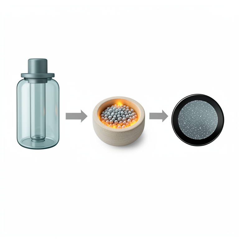
Biosensor Fabrication Workflow
AI-generated conceptual illustration (selenium dioxide nanoparticles on a glassy carbon electrode). It explains the idea and does not show study data.
View source evidence ↗Figures & tables
From the original paper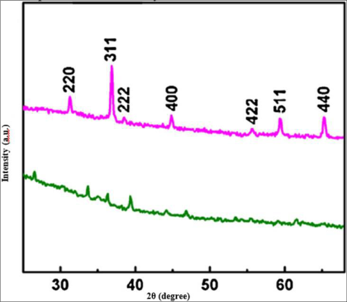 Figure 1
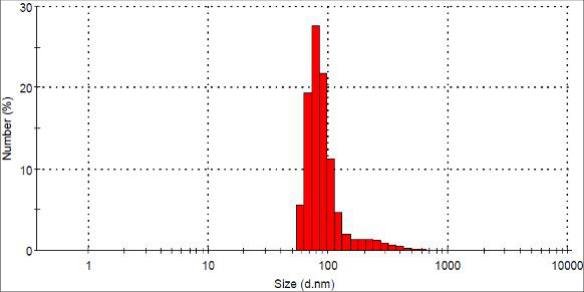 Figure 2
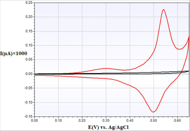 Figure 3
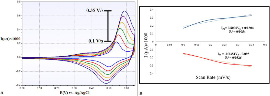 Figure 4
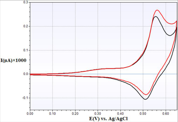 Figure 5
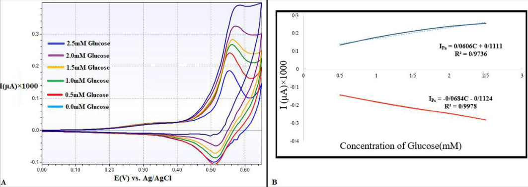 Figure 6
Introduction
Nowadays the development of sensitive, selective, fast and reliable biosensors for detection of biomolecules is of almost importance for use in the biotechnology, bio-processing, clinical diagnostics, medical application, food industry and many other fields [1,2] . Also in the development of renewable and sustainable fuel cells [2] . Comparing with other determination methods such as fluorescent, optical and electronic techniques, electrochemical techniques have been extensively applied due to their advantages as low cost, simple preparation, easy miniaturization and automation, high stability, etc [3] . Enzyme glucose sensors are based on the electrocatalysis of the enzyme glucose oxidase (GOx) to glucose oxidation. On the contrary, non-enzymatic glucose sensors which rely on the current response of glucose oxidation directly on the electrode surface. The performance of enzyme glucose sensors is susceptible to the chemical functional group and environmental factors including humidity, pH value and temperature due to the intrinsic nature of enzymes [4] . Although enzymatic sensors show high selectivity and sensitivity, the pursuit of non-enzymatic sensing with rapid response and precise measurement is a competitive area of research. Enzyme-free sensors can avoid some of the inevitable enzyme electrodes drawbacks such as the chemical and thermal instabilities originated from the intrinsic nature of enzymes without the need of immobilized enzyme on the sensor [5] . In recent years, various metal materials and their derivatives have been investigated for non-enzymatic biosensors [6] such as Co [7] , Ni(OH) 2 [8] , MnO 2 [9] , have been broadly explored as the electrode modified-materials for developing non-enzymatic glucose sensors. However, these electrodes still suffer from inertia behavior and surface contamination due to the absorption of intermediates and chloride ion [10] which result in poor operational stability of these biosensors. In recent years, researchers have shown an increasing interest in development of non-enzymatic glucose sensors based on metallic nanoparticles such as Pt [11] , Pd [12] , Pt/Pb [13] and Au [14] , which showed some unique features including high specific surface area, improved electrocatalytic activity, good biocompatibility [15] . However, studies reveal that glucose oxidation on some noble metals including Pt and Au suffers from slow kinetics with decreased faradaic current [5] . The high price of noble materials and easy poisoning of their surfaces are also concerned. Therefore, there are increasing interests to use transition metals such as Ni, Cu, Ti, Co and Fe [16][17][18] for glucose sensing. Among various transition metals, nickel-based electrodes have been the most widely used sensors for the non-enzymatic determination [19] . NiO, CuO, CoO based non-enzymatic glucose sensors have been developed successfully in recent years by several groups [17] . Selenium dioxide has been proved as a promising material for nonenzymatic glucose sensing because of its low cost, chemical stability environmentally benign nature, relatively good conductivity and electro-catalytic property [20] . In this work, SeO 2 nanoparticles (SeNPs) were prepared by a twostep strategy, with a hydrothermal method. X-ray diffraction (XRD) and dynamic light scattering (DLS) analysis were applied to characterize the as-prepared sample. SeNPs modified electrode showed high catalytic activity for glucose electro-oxidation in alkaline medium. The effects of NaOH concentration and the applied potential were investigated and optimized. Cyclic voltammetry (CV) detection of glucose was performed. Results show the proposed SeNPs exhibit high sensitivity and selectivity toward glucose detection and can be applied successfully in human serum.
Material and Methods
Material and Methods
Chemicals
Selenium chloride hexahydrate (SeCl 2 •6H 2 O), urea (CO (NH 2 ) 2 ), Nafion (5%) and D-(+)-glucose were purchased from Sigma-Aldrich. Glucose solutions with different concentrations were diluted from a stock solution (1 M). All the chemical reagents were used as received without further purification and all aqueous solutions were prepared with deionized water.
Preparation of SeNPs
SeNPs were prepared as follows. In brief, 0.59 g SeCl 2 •6H 2 O and 0.15 g CO(NH 2 ) 2 were dissolved in 20 ml distilled water and transferred into a 40 ml Teflon-lined autoclave, which was heated at 100•C for 12 h and then cooled to room temperature. The resulted pink precipitate was collected and washed with distilled water and absolute ethanol to remove the impurities. After this dried under vacuum at 60•C for 8 h. SeNPs were obtained by calcination of the resulting pink Selenium precursor nano-porous at 400•C for 2 h in air.
Preparation of SeNPs modified electrode
Before surface modification, a glassy carbon electrode (GCE) was polished with 1 μm alumina slurries, and then rinsed thoroughly with deionized water and dried at room temperature. To prepare 5 mg/mL SeNPs suspension, 5 mg SeNPs was first dispersed in a mixture solution containing H 2 O (500 μL), ethanol (490 μL) and Nafion (5%, 10 μL), and then sonicated for 1 h. After that, a 10 μL suspension was dropped onto the surface of GCE and dried naturally. The as-prepared electrode was denoted as SeNPs/Nafion/GCE. The Nafion coated GCE (Nafion/GCE) was also prepared with the same process as a control electrode.
Apparatus and electrochemical measurements
Powder XRD data were acquired on a Philips PW1730 diffracto meter with Cu Kα radiation (λ= 1.54060 ˚A). DLS measurements were performed on a HORIBA Jobin Yvon SZ-100 (Japan). All electrochemical measurements were performed on an Autolab EmStat3 electrochemical workstation (Netherlands). A typical three-electrode system was employed with a saturated Ag/AgCl as a reference electrode, a Pt wire as a counter electrode, and the SeNPs modified electrode as a working electrode. All electrochemical experiments were performed at room temperature and all the potentials are reported against featured Ag/AgCl reference electrode. The solution was stirred to provide convective transport.
Result and discussion
Result and discussion
Characterization of the SeNPs
SeNPs were obtained by calcination of corresponding Selenium precursor. The structure of the sample was determined by XRD analysis (Fig. 1). The Selenium precursor (green curve) show diffraction peaks indexed to Se (CO 3 ) 0.35 Cl 0.2 (OH) 110 (JCPDS 38-0547) [21] . After calcination, only the diffraction peaks (red curve) of the SeO 2 phase are observed at 31•, 37•, 39•, 45•, 56•, 59•, and 65•, which are well indexed to (220), ( 311), ( 222), ( 400), ( 422 Dynamic light scattering studies show a uniform size distribution of the SeNPs. The DLS pattern reveals that Selenium nanoparticles synthesized by a two-step process with a hydrothermal method have an average the diameter of 99 nm (that more than 90 percent in the size range of 58-100 nm) with the polydispersity index (PDI) of 0.525.
Electrochemical behavior of SeNPs/Nafion/GCE
The electrochemical behavior of SeNPs/Nafion/GCE was studied in 0.3 M NaOH solution, and Nafion/GCE was also studied for comparison. Fig. 3 shows the CVs of the Nafion/GCE (black curve) and SeNPs/Nafion/GCE (red curve) in 0.3 M NaOH solution. No obvious oxidation or reduction peak was observed at Nafion/GCE. In sharp contrast, a pair of redox peaks can be observed with the anodic peak at around 0.542 V and the cathodic peak at around 0.502 V at SeNPs/Nafion/GCE, which can be attributed to the reversible conversion between Co (III) and Co (IV) [22] . With an increase of the scan rate, both anodic and cathodic peak currents increased. Fig. 4B shows both of the redox peak currents increase linearly with the scan rate in the range from 0.1 to 0.35 V/s, suggesting a surface controlled electrochemical process of SeNPs/Nafion/GCE in alkaline solution. Linear regression equations: (I Pa = 0.6006V S + 0.1304, R 2 = 0.9054; I Pc = -0.6354V S -0.095, R 2 = 0.9526). This indicates that the modified-electrode reaction of glucose is a surface-confined process. In the scan rates ranging from 0.1 to 3.5 V/s, the linear regression equations of the E pa and E pc vs. the logarithm of the scan rates are expressed as E Pa = 0.0332lnV S + 0.6165 and E Pc = -0.0156lnV S + 0.4669 with R = 0.9889 and 0.9632, respectively. Based on the slopes of the lines RT/ (1-α)nF and -(RT/αnF), the value of the electron-transfer coefficient (α) and the electrontransfer number (n) were calculated as 0.56 and 1. Fig. 3 shows the CVs of the SeNPs/Nafion/GCE in 0.3 M NaOH solution in absence of (black curve) and presence of (red curve) 0.5 mM glucose. Both an increased anodic peak current and a decreased cathodic peak current were also observed upon addition of 0.5 mM glucose, suggesting the electrocatalytic activity of SeNPs/Nafion/GCE for glucose oxidation [23] .
Electro-oxidation of glucose at the SeNPs/Nafion/GCE
Electro-oxidation of glucose at the SeNPs/Nafion/GCE
Conclusion
Following a facile two-step procedure (synthesized a two-step process with a hydrothermal method), SeO 2 nanoparticles were fabricated. SeNPs show high electrocatalytic activity toward glucose oxidation in alkaline medium and are used as an effective sensor for glucose detection. These results make SeNPs a good candidate for the construction of a non-enzymatic glucose sensor in routine analysis.
Figures and tables from the paper
References
22 in the original paper- Luqi K, Daiping H, Lili B. Nanoporous Selenium Dioxide nanowires for non-enzymatic electrochemical glucose detection, Sensors and Actuators B: Chemical, 2015, 888894.
- Mozhdeh M, Hossein A, Abdolreza S. Three-dimensional hybrid graphene/nickel electrodes on zinc dioxide nanorod arrays as nonenzymatic glucose, biosensors, Sensors and Actuators B: Chemical, 2017. http://10.1016/j.snb.2017.05.062
- Zongxu S, Wenyu G, Pei L, Xiaofang W, Qing Z, Hao W, et al. Highly sensitive non-enzymatic glucose sensor based on Nickel nanoparticle-attapulgite-reduced graphene dioxidemodified glassy carbon electrode, Talanta, 2016. http://dx.doi.org/10.1016/j.talanta.2016.06.016
- Hrapovic S, Luong J. Picoamperometric detection of glucose at ultrasmall platinum-based biosensors: preparation and characterization, Analytical Chemistry, 2003, 3308-3315.
- Lu P, Liu Q, Xiong Y, Wang Q, Lei Y, Lu S. Nanosheetsassembled hierarchical microstructured Ni(OH) 2 hollow spheres for highly sensitive enzyme-free glucose sensors. Electrochimica Acta, 2015, 148-156.
- Zhang B, He Y, Liu B, Tang D. Nickel-functionalized reduced graphene dioxide with polyaniline for nonenzymatic glucose sensing, Microchimica Acta,2015:182:625-631.
- Luo J, Jiang S, Zhang H, Jiang J, Liu X. A novel nonenzymatic glucose sensor based on Cu nanoparticle modified graphene sheets electrode, Analytica Chimica Acta,2012:709:47-53.
- Chen J, Zhang W, Ye J. Nonenzymatic electrochemical glucose sensor based on MnO 2 /MWNTs nanocomposite, Electrochemistry Communications,2008:10:1268-1271.
- Ye S, Wen Y, Zhang W, Gan L, Xu G, Sheu F. Nonenzymatic glucose detection using multi-walled carbon nanotube electrodes, Electrochemistry Communications,2004:6:66-70.
- Chang G, Shu H, Huang Q, Oyama M, Ji K, Liu X. Synthesis of highly dispersed Pt nanoclusters anchored graphene composites and their application for non-enzymatic glucose sensing, Electrochim Acta,2015:157:149-157.
- Lin S, Chang S, Hsueh T. ZnO nanowires modified with Au nanoparticles for nonenzymatic amperometric sensing of glucose, Appl Phys Lett,2014:104:193704.
- Cui H, Ye J, Zhang D, Li C, Luong C, Sheu F. Selective and sensitive electrochemical detection of glucose in neutral solution using platinum-lead alloy nanoparticle/carbon nanotube nano composites, Analytica Chimica Acta,2007:594:175-183.
- Li Y, Niu X, Tang J, Lan M, Zhao H. A Comparative Study of Nonenzymatic Electrochemical Glucose Sensors Based on Pt-Pd Nanotube and Nanowire Arrays, Electrochim Acta,2014:130:1-8.
- Gao Z, Han Y, Wang Y, Xu J, Song Y. One-step to prepare self-organized nanoporous NiO/TiO 2 layers and its use in non-enzymatic glucose sensing, Sci Rep, 2013, 3.
- Ding J, Sun W, Wei G, Su Z. Cuprous dioxide microspheres on graphene nanosheets: an enhanced material for nonenzymatic electrochemical detection of H 2 O 2 and glucose, RSC Adv,2015:5:35338-35345.
- Ding Y, Wang Y, Su L, Bellagamba M, Zhang H, Lei Y. Electrospun Co 3 O 4 nanofibers for sensitive and selective glucose detection, Biosens Bioelectron,2010:26:542-548.
- Mei H, Wu W, Yu B, Wu H, Wang B, Xia Q. Nonenzymatic electrochemical sensor based on Fe@Pt core-shell nanoparticles for hydrogen perdioxide, glucose and formaldehyde, Sens Actuators: B,2016:223:68-75.
- Xu Q, Zhao Y, Xu J, Zhu J. Preparation of functionalized Selenium nanoparticles and fabrication of a glucose sensor, Sensors and Actuators B: Chemical,2006:114:379-386.
- Sattarahmady N, Heli H. A non-enzymatic amperometric sensor for glucose based on Selenium dioxide nanoparticles, J. Exp. Nanosci,2012:7:529-546.
- Chen X, Zhang Z, Qiu Z, Shi C, Li X. Hydrothermal fabrication and characterization of polycrystalline linnaeite (Co 3 S 4 ) nanotubes, 2009.
- Zhou M, Zhai Y, Dong S. Electrochemical sensing and biosensing plat-form based on chemically reduced graphene dioxide, Anal. Chem,2009:81:5603-5613.
- Ding Y, Wang Y, Su L, Bellagamba M, Zhang H, Lei Y. Electrospun Co 3 O 4 nanofibers for sensitive and selective glucose detection, Biosens. Bioelectron,2010:26:542-548.
Cited by 4
From OpenAlex, OpenCitations and reference lists read by Scinovex
- Study of mineral profile in patients with hypothyroidism
Bushra, Sangeeta Kapoor, Fozia Jamal · International Journal of Advanced Biochemistry Research · 2022
Background: Thyroid hormones play important role in hemodynamic, thermoregulation and metabolism of the body. As a result, renal hemodynamic, glomerular filtration and electrolyte management may be affected in thyroid related problems. Thyroid disease particularly hypothyroidism has recently received more attention because it may be the primary cause of mineral metabolism disturbance due to its direct effect on bone turnover. Objective: To estimate the serum level of minerals (Calcium, magnesium, phosphorus, iron, zinc and selenium) in hypothyroid patients and compare them with normal healthy
- Paper-based microfluidic devices for electrochemical glucose detection in whole blood: A comprehensive review
Janet L. Walker, R. Lawrence Edwards · International Journal of Biology Sciences · 2023
This review article provides a comprehensive overview of the rapidly evolving field of paper-based microfluidic devices for electrochemical glucose detection in whole blood. We delve into the principles, design strategies, fabrication techniques, and recent advancements in these devices. The advantages of paper-based microfluidics, including cost-effectiveness and portability, are highlighted. Furthermore, we discuss key challenges, such as sample volume requirements and sensitivity improvements, and explore potential applications in point-of-care diagnostics. This review serves as a valuable
- Contamination in meat: A review
Muhammad Umair Sajid · International Journal of Agriculture and Food Science · 2022
Meat is an flesh of animal that’s eaten as a food. Meat has an unprecedented source of energy. Human eat meat of different animals like camels, goats, cow, chicken, fish etc. Meat has some major parts including water, carbohydrates, proteins fats and minor components like vitamins. (Vitamin A and B) and minerals (Iron, Zinc, selenium). Selenium is a mineral that’s play a vital role in metabolism and thyroid function of the body. All of these things are required for human body growth. Due to increases in population of this world, requirement of meat increases day by day. Many factors involve in
- Comparative study of minerals and proximate compositions of selected nuts eaten in south eastern Nigeria ↗
Elsie Chineze Nnanyelugo, Jonathan C. Ifemeje, Kizito Ifeanyi Amaefule, Ekwemba Christian O, Maryjane Oluoma Ifemeje · International Journal of Bioscience and Biochemistry · 2021
Nuts are outstanding source of nutrients and bio active compounds. They are consumed by greater part of the populations of the world in some form or other depending on the accessibility in the geographical area. The study was aimed at investigating and comparing the proximate composition and antioxidant related minerals in four selected local nuts of tiger nut, walnut, cashew