
Research article · 2022 · Full text
International Journal of Advanced Biochemistry ResearchStudy of mineral profile in patients with hypothyroidism
Volume 6(1) · Pages 55–60 · DOI: 10.33545/26174693.2022.v6.i1a.89 ↗
Research areasThyroid Disorders and TreatmentsBiomarkers in Disease Mechanisms
Abstract
Author-writtenBackground: Thyroid hormones play important role in hemodynamic, thermoregulation and metabolism of the body. As a result, renal hemodynamic, glomerular filtration and electrolyte management may be affected in thyroid related problems. Thyroid disease particularly hypothyroidism has recently received more attention because it may be the primary cause of mineral metabolism disturbance due to its direct effect on bone turnover. Objective: To estimate the serum level of minerals (Calcium, magnesium, phosphorus, iron, zinc and selenium) in hypothyroid patients and compare them with normal healthy controls. Material and Methods: The study was carried out from January 2021 to December 2021 in 60 hypothyroid patients and 60 healthy controls (age group 20-60 years), attending the medicine OPD of Teerthanker Mahaveer College & Research Center, Moradabad, Uttar Pradesh. Serum minerals and thyroid profile were measured by using the automated method and their levels were compared with healthy individuals as controls. Results: The serum magnesium and phosphorus levels were significantly elevated whereas the levels of calcium and zinc were significantly decreased in hypothyroid patients compared to healthy subjects. However, iron (p=0.09) and selenium (p=0.38) levels in hypothyroid patients were found to be less than that of healthy individuals but the difference between them was not statistically significant. Conclusion: This study concludes that hypothyroid patients should have their serum mineral levels checked regularly, as early diagnosis and intervention can help to avoid further thyroid problems. It will be beneficial in treating hypothyroidism as well.
Summary
Source-checkedPlain-language reading of the full paper, prepared with AI and checked against the text. Each point links to the authors' own words.
Why it matters
- Thyroid hormones are essential for regulating the body's metabolism, thermoregulation, and hemodynamics. View evidence ↗
- Hypothyroidism is of particular interest because its direct effect on bone turnover can lead to disturbances in mineral metabolism. View evidence ↗
- In India, the prevalence of hypothyroidism is estimated to be between 4% and 11%, with a higher occurrence in women than in men. View evidence ↗
The question
- The study aimed to measure and compare serum levels of calcium, magnesium, phosphorus, iron, zinc, and selenium between hypothyroid patients and healthy controls. View evidence ↗
What they did
- The study took place from January to December 2021 at Teerthanker Mahaveer College & Research Center, involving 60 hypothyroid patients and 60 healthy controls aged 20-60 years. View evidence ↗
- Participants were excluded if they were pregnant or had hepatic disease, renal disease, diabetes mellitus, cardiac disease, or were taking mineral supplements. View evidence ↗
What they found
- Serum calcium levels were significantly lower in hypothyroid patients (8.06±0.99 mg/dl) compared to healthy controls (9.1±0.67 mg/dl), with p < 0.001. View evidence ↗
- Serum magnesium levels were significantly elevated in hypothyroid patients (2.63±0.57 mEq/L) compared to controls (2.36±0.65 mEq/L), with p = 0.02. View evidence ↗
- Serum phosphorus levels were significantly higher in hypothyroid patients (6.79±1.04 mg/dl) than in healthy controls (4.22±1.23 mg/dl), with p < 0.001. View evidence ↗
- Serum zinc levels were significantly decreased in hypothyroid patients (52.52±16.55 µg/dl) compared to controls (95.79±25.34 µg/dl), with p < 0.001. View evidence ↗
- Serum iron levels were lower in hypothyroid patients (59.50±25.29 µg/dl) than in controls (66.61±20.53 µg/dl), but this difference was not statistically significant (p = 0.09). View evidence ↗
- Serum selenium levels were slightly lower in hypothyroid patients (82.11±21.04 ng/ml) compared to controls (84.85±12.54 ng/ml), but the difference was not statistically significant (p = 0.38). View evidence ↗
What it means
- The authors recommend that hypothyroid patients undergo regular serum mineral checks to facilitate early diagnosis and intervention, which may prevent further complications. View evidence ↗
- The authors note that the effect of hypothyroidism on mineral levels is inconsistent, likely due to variables such as dietary intake, absorption rates, subclinical pathologies, and other hormonal influences. View evidence ↗
Visual summary
2 visualsIllustrations explain the paper; charts are replotted from the paper's own table values.
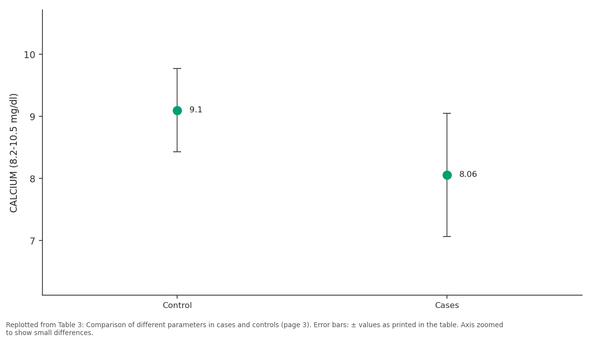
CALCIUM (8.2-10.5 mg/dl): Control vs Cases
Replotted from Table 3 (page 3) of the original paper. Every bar is the paper's own table value.
View source evidence ↗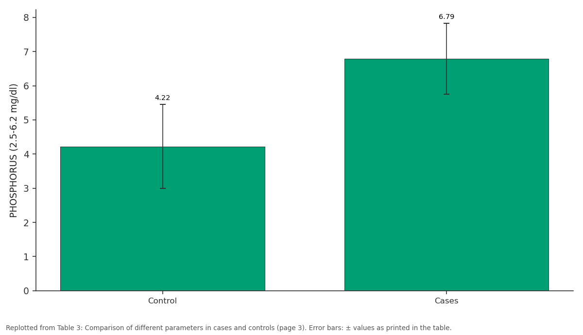
PHOSPHORUS (2.5-6.2 mg/dl): Control vs Cases
Replotted from Table 3 (page 3) of the original paper. Every bar is the paper's own table value.
View source evidence ↗Figures & tables
From the original paper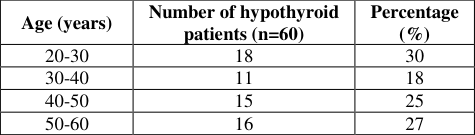 Table 1
 Table 2
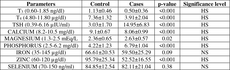 Table 3
Introduction
Thyroid hormones, T3 (triiodothyronine) and T4 (tetraiodothyronine) are two main hormones secreted by the thyroid gland. Biologically, T3 is more active form than T4 which is synthesized by the process of deiodination of T4 by 1, 5 deiodinase within the peripheral tissues [1] . Thyroid Stimulating Hormone (TSH) and Thyrotropin Releasing Hormone (TRH) are released by the pituitary gland and hypothalamus and help to regulate T3 and T4 [2] . Thyroid hormones have a crucial function in the human body regulating body temperature and regulating the metabolism of carbohydrates, proteins, lipids and minerals [3] . Thyroid hormones play important role in hemodynamic, thermoregulation and metabolism of the body. As a result renal hemodynamic, glomerular filtration and electrolyte management may be affected in thyroid related problems [4] . Thyroid disorders can disrupt bone and mineral homeostasis and they are the main causes of secondary osteoporosis [5] . Among all the endocrine disorders, thyroid disorders are the most common disorders worldwide as well as in India out of which hypothyroidism is the most common [6] . Serum T3 and T4 levels are reduced in primary hypothyroidism resulting in pituitary TSH hypersecretion and elevated serum TSH levels. Abnormal activity of thyroid gland slows metabolic processes and causes a variety of clinical psychological and physiological abnormalities [7,8] . According to the latest researches, India has a high prevalence of hypothyroidism which ranges from 4 to 11% with women suffering from the disorders more than males. Hypothyroidism becomes more common as people get older [1] . Thyroid disease particularly hypothyroidism have recently received more attention because they may be the primary cause of mineral metabolism disturbance due to their direct effect on bone turnover [9] . Thyroid hormones cause bone reabsorption and inhibits parathyroid hormone secretion which leads to raise serum calcium levels but hypothyroidism has the reverse effect. Low calcium level in hypothyroidism is caused by poor calcium mobilization into the bone as evidenced by numerous recent studies. Calcitonin synthesis is increased resulting in the reabsorption of phosphate and excretion of calcium from the renal tubules [10,11] . Serum calcium level is regulated by thyroid hormones by releasing it from the parafollicular cells of thyroid gland. In primary hypothyroidism thyroxine level is less so calcium outflux from the cells is decreased [12] . Cyclic 3, 5 nucleotide phosphodiesterase enzyme is stimulated by magnesium. In thyroid gland thyrotropin activity is regulated by cyclic AMP and level of magnesium may be influence by thyroid gland in response to pituitary hormone [13] . Thyroid hormones regulate calcium, phosphorus and magnesium which are divalent metal ions required as a cofactor for metalloenzymes. Thyroid hormones also stimulate numerous metabolic pathways [14] . Iron is needed as a cofactor for thyroid peroxidase (TPO) enzymes. This enzyme catalyzes the two reactions in the production of thyroid hormone in thyroid gland. It operates on the apical plasma membrane and is responsible for iodine oxidation and binding to thyroglobulin tyrosine [15,16,17] . Zinc too has important roles in thyroid metabolism. It participates in the synthesis and mechanism of action of TRH by assisting T3 binding to its nuclear receptor. It is also present in thyroid hormone binding transcription factors in which zinc is bound to cysteine residues [18] . Zinc is crucial for the biological activity of thyroid hormones and associated receptors as well as the peripheral conversion of T3 to T4 hormones. Enzymes in the synthesis of TRH need zinc as a cofactor [19] . Another micromineral selenium acts as a cofactor of iodothyronine deiodinase enzyme which helps to convert T4 to T3 and also important in the regulation of thyroid hormone production. It also serves as a cofactor for glutathione reductase and glutathione peroxidase enzymes which are known to protect the thyroid gland and various other tissues from oxidative stress [20] . Previous research works on the changes in serum mineral levels in hypothyroid patients have yielded mixed outcomes. Some scientists have noticed that there is no change in the circulating levels of these minerals in hypothyroid patients while others have found that the levels of these minerals increase in hypothyroid patients. As a result, this study was done to investigate variations in mineral profile in hypothyroid patients by estimating serum calcium, phosphorus, magnesium, iron, zinc, selenium and analyzing their association with thyroid profile (T3, T4 and TSH) levels.
Materials and Methods
The present study has been done on patients of age group 20-60 years including both males and females [8] , attending the Medicine OPD of Teerthanker Mahaveer College & Research Center, Moradabad, Uttar Pradesh. The participants having age more than 20 years and less than 60 years diagnosed cases of hypothyroidism (TSH more than 4.5 µIU/ml) [10] were selected from the Medicine OPD of Teerthanker Mahaveer Hospital, Moradabad and who have given written consent form will be included in the study group.
Exclusion criteria
Patients on mineral supplementation, patients with Hepatic disease, Renal disease, Diabetic mellitus, Cardiac disease, pregnant women [21] 8 ml of venous blood sample after overnight fasting for 8-12 hours was collected under all aseptic precautions from all the subjects and dispensed into plain vials. After centrifugation at 3000 RPM for 5 minutes, serum sample was used for analysis of serum minerals and serum thyroid profile by different methods. Thyroid hormones (serum T3, T4 and TSH) were determined by Enzyme Linked Fluorescent Assay (ELFA) technique [22] . Serum Calcium was determined by Arsenazo III method [36] . Serum Magnesium was determined by Calmagite method [24] . Serum Phosphorus was determined by Ammonium molybdate method [25] . Serum Iron was determined by Ferrozine method [26] . Serum Zinc was determined by Colorimetric method [27] . Serum Selenium was estimated by Spectrophotometric method [28] . Serum minerals magnesium (p=0.01) and serum phosphorus (p<0.001) levels in the serum were significantly elevated whereas the levels of calcium and zinc were significantly (p<0.001) decreased in hypothyroid patients compared to healthy subjects. However, iron (p=0.09) and selenium (p=0.38) levels in hypothyroid patients were found to be less than that of healthy individuals but the difference between them was not statistically significant.
Statistical analysis
Statistical analysis
Discussion
Thyroid hormones have vital function in human body which includes regulating body hemodynamic, thermodynamics and metabolism. They also have a significant impact on renal hemodynamic and glomerular filtration rate. Hypothyroidism is the most commonly occurring endocrine disorder which can lead to different clinical manifestation including minerals disturbance, osteoporosis, congestive heart failure and coma [4] . As a result, this research was carried out to determine the impact of minerals (calcium, magnesium, phosphorus, iron, zinc and selenium) on hypothyroid patients and also found a possible correlation of these minerals with thyroid profile. In this study maximum number of patients was women and higher prevalence of hypothyroidism was notice in the age group of 20-30 years. Female hypothyroid patients made up almost 78 percent of the total. In table 1 Hence, this shows female patients have a higher prevalence of hypothyroidism than male ones. In table 3 we compared thyroid profile (T3, T4 and TSH) and minerals in hypothyroid cases and controls. We found that the mean value of serum T3 (0.50±0.36 ng/dl) in hypothyroid patients were significantly (p<0.001) reduced when compared with healthy individuals (1.13±0.46 ng/dl) likewise, serum T4 level was also significantly (p<0.001) reduced (3.91±2.04 µg/dl) when compared with control subjects (7.36±1.32 µg/dl). While, serum TSH (14.95±6.83 µIU/ml) was found to be significantly (p<0.001) raised when compared with controls (3.03±1.70 µIU/ml).
| Parameters | Control | Cases | p-value | Significance level |
|---|---|---|---|---|
| T3 (0.60-1.85 ng/dl) | 1.13±0.46 | 0.50±0.36 | <0.001 | HS |
| T4 (4.80-11.80 µg/dl) | 7.36±1.32 | 3.91±2.04 | <0.001 | HS |
| TSH (0.39-6.16 µIU/ml) | 3.03±1.70 | 14.95±6.83 | <0.001 | HS |
| CALCIUM (8.2-10.5 mg/dl) | 9.1±0.67 | 8.06±0.99 | <0.001 | HS |
| MAGNESIUM (1.3-2.5 mEq/L | 2.36±0.65 | 2.63±0.57 | 0.02 | HS |
| PHOSPHORUS (2.5-6.2 mg/dl) | 4.22±1.23 | 6.79±1.04 | <0.001 | HS |
| IRON (35-145 µg/dl) | 66.61±20.53 | 59.50±25.29 | 0.09 | NS |
| ZINC (60-120 µg/dl) | 95.79±25.34 | 52.52±16.55 | <0.001 | HS |
| SELENIUM (70-150 ng/ml) | 84.85±12.54 | 82.11±21.04 | 0.38 | NS |
Table text extracted from the original PDF; superscript letters are shown inline. The original table image is below.
View original table image
| Age (years) | Number of hypothyroid patients (n=60) | Percentage (%) |
|---|---|---|
| 20-30 | 18 | 30 |
| 30-40 | 11 | 18 |
| 40-50 | 15 | 25 |
| 50-60 | 16 | 27 |
Table text extracted from the original PDF; superscript letters are shown inline. The original table image is below.
View original table image
In the present study we found significant low level of serum calcium with mean concentration of 8.06±0.99 mg/dl in cases when compared with healthy controls 9.1±0.67 mg/dl. Decrease in serum calcium level in hypothyroidism could be related to a low level of thyroxine as thyroid hormones play an essential role in calcium homeostasis through their direct influence on bone turnover [29] . This is similar with a study done by Saxena S, et al. 2020 [8] they assessed that in hypothyroid patients, serum calcium level was lowered with a mean value of 8.0±8.4 mg/dl compared with 8.6±9.6 mg/dl healthy individuals. They came to the conclusion that in hypothyroid patients there is a substantial impact of minerals levels.
Our findings are also in similar with Sridevi D, et al. 2016 [30] as they found that serum level of calcium (8.58±0.46 mg/dl) was reduced in cases of hypothyroid patients when compared with healthy subjects (10.04±0.56 mg/dl). Their findings showed that thyroid hormones have a substantial effect on minerals and that regular follow up in thyroid hormone deficit might aid in its management. While the study done by Susanna TY, et al. 2016 [14] observed that there is no change in the levels of serum calcium in hypothyroidism (8.98±0.61 mg/dl) compared with controls (8.98±0.49 mg/dl).
Present study shows that significantly (p=0.02) elevated levels of serum magnesium (2.63±0.57 mEq/L) in hypothyroid patients than in controls (2.36±0.65 mEq/L). Because thyroid hormones have great influence on renal tubules, which causes increased reabsorption of magnesium it could be attributable to increased renal retention of magnesium of up to 15 to 30% from the kidneys. Our findings are consistent with those of Kavita MM, et al. 2014 [10] who reported raised level of serum magnesium (2.37±0.46 mEq/L) in hypothyroid patients compared with healthy subjects (2.1±0.28 mEq/L). Their findings showed that serum magnesium levels were elevated in both subclinical and overt hypothyroidism. Early detection and treatment can help to prevent additional bone complications. Kaur J, et al. 2014 [6] also found similar result in which level of serum magnesium was significantly (p<0.001) raised in hypothyroid cases 2.23±0.31 mEq/L when compared with healthy subjects 1.27±0.28 mEq/L. Their research found that monitoring serum magnesium levels at routine hypothyroid patient checkups can help individuals with hypothyroidism without any clinical manifestation. While Nisa F, et al. 2013 [18] found that the level of serum magnesium was significantly reduced among hypothyroid patients (0.68±0.37 mEq/L) than in controls (1.01±0.26 mEq/L) which are contradictory to our findings. Their research revealed that serum magnesium levels in hypothyroid patients should be monitored reducing the adverse consequences of magnesium insufficiency in hypothyroid individuals.
In this study, we found a statistically significant increase in serum phosphorus (p<0.001) levels in hypothyroid patients (6.79±1.04 mg/dl) when compared with controls (4.22±1.23 mg/dl) which is similar to the findings done by Kavitha MM. 2014 [10] . They found that serum phosphorus levels were significantly elevated in subclinical and overt hypothyroidism than in controls with mean values of 4.1±0.46 mg/dl, 4.42±0.81 mg/dl and for controls 3.6±0.19 mg/dl, respectively. Susanna TY, et al. 2016 [14] found similar findings showing that serum phosphorus levels in hypothyroid patients (5.56±2.03 mg/dl) were significantly higher (p<0.001) than in healthy people (3.10±0.50 mg/dl). Increased serum phosphorus levels could be attributed to increased calcitonin synthesis which increases tubular phosphate reabsorption and tubular calcium excretion resulting in hypocalcemia and Hyperphosphatemia as seen in hypothyroidism [10] .
In this present study, we found statistically no significant (p=0.09) difference in iron concentration among hypothyroid patients when compared with controls with the mean values of 59.54±25.29 μg/dl and 66.61±20.54 μg/dl, respectively. These findings could be related to hypothyroid patients with impaired iron metabolism and interrupted gastrointestinal absorption of iron resulting in a lower level of serum iron in hypothyroid patients. These findings in our study of hypothyroidism cases are in similar with the findings of Talebi S, et al. [31] they reported that there were no significant changes in the levels of serum iron among hypothyroid patients when compared with healthy individuals.
Our findings are also comparable to those of Banday TH, et al. 2017 [32] who found that 34.2% of individuals with primary hypothyroidism had iron insufficiency. Likewise, we also observed significantly decreased levels of zinc in hypothyroid patients (52.67±16.55 μg/dl) as compared to control subjects (95.79±25.34 μg/dl), which is accordance to the study of Arora M, et al. 2018 [34] They observed that the level of serum zinc was decreased in hypothyroid cases than in healthy subjects and their study concluded that patients with hypothyroidism should be given a zinc rich diet to help regulate their thyroid hormone levels and functioning. However, Nisha F, et al. 2013 [18] discovered no statistically significant variation in serum zinc levels between hypothyroid patients and healthy controls in a separate investigation. Zinc deficiency plays a little role in hypothyroid patients, according to their findings. The reason behind the low level of serum zinc is due to reduced gastrointestinal absorption and tubular excretion of zinc in hypothyroid subjects.
In the present study, we observed a statistically insignificant (p=0.38) difference between serum levels of selenium among hypothyroid patients (82.11±21.04 ng/ml) and in healthy subjects (84.85±12.54 ng/ml). The reduction in serum selenium levels in hypothyroidism patients was consistent with previous research by Jinger SK, et al. 2012 [35] . They reported that levels of serum selenium were decreased in cases (53.66±10.31 ng/ml) than in controls (114.35±22.5 ng/ml). They also concluded that correlation of serum selenium level would have a helpful effect on treatment and could prevent complications in hypothyroid patients. This study investigates that serum calcium, iron, zinc and selenium levels were decreased whereas serum phosphorus and magnesium level were increased in cases than in controls. These findings show that altered metabolism of mineral (calcium, magnesium, phosphorus, iron, zinc and selenium) has intimate association with thyroid hormones.
Conclusion
This study concludes that levels of serum magnesium and phosphorus were slightly elevated whereas levels serum, calcium, iron, zinc and selenium were reduced among hypothyroid patients as compared to healthy individuals. In hypothyroidism, it is clear that the lower level of serum T3, T4 and elevation in the TSH level is a consistent finding but their effect on the serum levels of calcium, magnesium, phosphorus, iron, zinc and selenium is inconsistent which is due to the variations in the dietary intake of minerals, renal and intestinal absorption some subclinical pathologies and various other complex hormones which may affect minerals metabolism and cellular uptake. Therefore, hypothyroid patients should have their serum mineral levels checked on a regular basis as early diagnosis and intervention can help to avoid further thyroid problems. It will be beneficial in treating hypothyroidism as well.
Acknowledgement
AcknowledgementI am grateful to all the faculty members and technical staff of the Biochemistry department and laboratory for their kind support.
Figures and tables from the paper
| Gender | Number of hypothyroid patients (n=60) | Percentage (%) |
|---|---|---|
| Female | 47 | 78.3 |
| Male | 13 | 21.6 |
Table text extracted from the original PDF; superscript letters are shown inline. The original table image is below.
View original table image
References
36 in the original paper- Unnikrishnan AG, Kalra S, Sahay RK, Bantral G, John M, Tewari N, et al. Prevalence of hypothyroidism in adults: an epidemiology study in eight cities of India. Indian Journal of Endocrinology and Metabolism. 2013;17(4):647-52.
- Unnikrishnan AG, Menon UV. Thyroid disorders in India: An epidemiology perspective. Indian Journal of Endocrinology and Metabolism. 2011;15(2):78-81.
- Mariani LH, Berns JS. The renal manifestations of thyroid disease. J Am Soc Nephrol. 2012;2(1):22-26.
- Pearce EN. Hypothyroidism and dyslipidemia: Modern concepts and approaches. Current Cardiology Reports. 2004;6(6):451-456.
- Orluwene CG, Mommoh MO. Serum phosphorus, estimated ionized and total serum calcium in industrial females with thyroid dysfunction in Port Harcourt. International Journal of Medical Research & Health Sciences. 2013;3(4):20-24.
- Kaur J, Ahmed N, Gupta A. Changes in the Electrolytes Profile in Patients having Hypothyroidism. Journal of Medical Science and Clinical Research. 2014;2(4):633637.
- Galesanu C, Linsic N, Teslaru R, Apostu L, Zbrancr E. Lipid profile in a group of hypothyroid patients vs. treated hypothyroid patients. Revista MedicoChirurgicala A Societatii De Medici Si Naturalisti Din Iasi. 2004;108(3):554-560.
- Saxena S, Srivastava PC, Katyal R, Das B, Pannu SJ. Evaluation of mineral status in hypothyroidism in a tertiary care center. International Journal of Research in Medical Sciences. 2020;8(2):667-671.
- Shivaleela MB, Poornima RT, Murthy DS. Serum calcium and phosphorus levels in thyroid dysfunction. Indian Journal of Fundamental and Applied Life Sciences. 2012;2(2):179-183.
- Kavitha MM, Pujar S, Hiremath CS, Prasad S, Mahanthesh. Evaluation of serum electrolytes in hypothyroid patients. Medpulse International Medical Journal. 2014;1(8):393-395.
- Suneel B, Nagendra DR, Aparna RR, Balakrishna D, Naidu JN. Mineral status in Thyroid Disorder (Hypo & Hyper). International Journal of Applied Biology and Pharmaceutical Technology. 2011;2(4):423-429.
- Murgod R, Soans G. Changes in Electrolytes and Lipid profile in Hypothyroidism. International Journal of Life science and Pharma Research. 2012;2(3):185-194.
- Erdal M, Sahin M, Hasimi A, Vckaya G, Kuthi M, Saglam K. Trace element levels in Hashimoto thyroiditis patients with subclinical hypothyroidism. Biological Trace Element Research. 2008;123(3):1-7.
- Susanna TY, Sagayaraj A, Shashidhar KN, Gomathi M, Mahesh V. A correlative study of thyroid profile and mineral status in patients with hypothyroidism-a hospital-based case control study. Asian Journal of Pharmaceutical and Clinical Research. 2016;3(9):292294.
- Kammal M, Abdrabo AA. Assessment of thyroid hormone levels in Sudanese females with iron deficiency. Sudan Journal of Medical Sciences. 2014;50(2):98-102.
- Hess SY, Zimmermann MB, Arnold M, Langhans W, Hurrell RF. Iron deficiency anemia reduces thyroid peroxidase activity in rates. Journal of Nutrition. 2002;132(7):1951-1955.
- Akhter S, Nahar ZU, Parvin S, Alam A, Sharmin S, Arslan MI. Thyroid status in patients with low serum ferritin level. Bangladesh Journal of Medical Biochemistry. 2012;5(1):5-11.
- Nisha FU, Mumtaz A, Ulla MI, Atif M, Sami W. Determination of serum zinc and magnesium levels in patients with hypothyroidism. Trace Elements and Electrolytes. 2013;31(4):1-5.
- Maxwell C, Volpe SL. Effect of zinc supplementation on thyroid hormone function-A case study of two college females. Annals of Nutrition and Metabolism. 2007;51(2):188-194.
- Toulis KA Anastasilakis AD, Tzellos TG, Goulis DG, Kouvelas D. Selenium supplementation in the treatment of Hashimoto's thyroiditis: A systematic review and a meta-analysis. Thyroid. 2010;20(10):1163-1173.
- Gaitonde DY, Rowley KD, Sweeney LB. Hypothyroidism: An update. South African Family Practice. 2021;54(5):384-390.
- Beck JR. Laboratory Decision Science Applied to chemometries: Strategic Testing of Thyroid Function Clinical Chemistry. Clinical Chemistry. 1986;32(9):1707-1713.
- Langeroudi NVP, Mollataghi A. Synthesis of nonenzymatic biosensor based on selenium dioxide nanoparticles in order to detection of glucose under electrochemical method. Int. J Adv. Chem. Res. 2021;3(2):12-16. DOI: 10.33545/26646781.2021.v3.i2a.36
- Gindler EM, Heth DA. Colorimetric determination with bound 'Calmagnite' of magnesium in human blood serum. Clinical Chemistry. 1971;17(7):662.
- Fiske CH, Subbarow YJ. The colorimetric determination of phosphorus. Biological Chemistry. 1925;66(4):375-400.
- Siedel J, Wahlefeld AW, Ziegenhorn J. A new iron Ferrozine reagent without deproteinization. Clinical Chemistry. 1984;30:975.
- Akita Abe, Sumiko Yiamashita. Colorimetric method for the estimation of zinc. Clinical Chemistry. 1989;35(4):552-554.
- Cuparigona F, Jtafilov T. Determination of selenium in human blood serum by electrothermal atomic absorption spectrometry. Journal of Chemical Sciences. 2011;46:1-8.
- Rasheed KM, Thivyah Prabha AG, Kumar SK. Study of calcium, magnesium and phosphorus levels among hypothyroid patients in Tricky, Tamil Nadu. Journal of Evidence-Based Medicine and Healthcare. 2021;8(30):2797-2803.
- Sridevi D, Dambal AA, Sidrah, Challa AS, Padaki SK. A study of serum magnesium, calcium and phosphorus in hypothyroidism. International Journal of Clinical Biochemistry and Research. 2016;3(2):236-239.
- Mamatha BV, Chandrakanth KH, Kashinath RT, Rakshitha MN. Assessment of mineral status in subclinical hypothyroidism patients: A case control study. International Journal of Clinical Biochemistry and Research. 2019;6(1):32-35.
- Shukla A, Agarwal S, Gupta A, Sarkar G. Relationship between Body Iron Status and Thyroid Profile in an Adult Population: A Hospital Based Study. National Journal of Laboratory Medicine. 2017;6(2):1-3.
- Modi A, Sahi N. Effect of thyroid hormones on serum calcium and Phosphorus. International Journal of Clinical Biochemistry and Research. 2018;5(4):570573.
- Arora M, Mahat RK, Kumar S, Mustafa I, Sah SP. Study of Trace Elements in Patients of Hypothyroidism with Special Reference to Zinc and Copper. Biomedical Journal of Scientific & Technical Research. 2018;6(2):5190-5194.
- Murgod R, Soans G. Changes in Electrolytes and Lipid Profile in Hypothyroidism. International Journal of Life science and Pharma Research. 2012;2(3):185-194.
- Smith HG, Bauer PJ. Light-induced permeability changes in sonicated bovine disks: Arsenazo III and flow system measurements. Biochem. 1979;18(3):50675073.
Cited by 8
From OpenAlex, OpenCitations and reference lists read by Scinovex
- Estimation of thyroid hormone and adipocytokine levels in men with obesity and type 2 diabetes in Thi-Qar Governorate
Hadeel S. Abd-Alwahab, Zainab Mohammed Farhan, Nesreen Ahmed Nasser, Osama A. Mohsein · International Journal of Advanced Chemistry Research · 2024
Background: Obesity is a common disease around the world and is considered a risk factor for developing type 2 diabetes, which is characterized by the body’s lack of response to the insulin produced, which leads to high blood sugar levels. Thyroid hormones also affect metabolic processes in the body and may play a role in causing obesity. This study is concerned with knowing the levels of thyroid hormones and adipocytokine levels and the relationship between them, to find out the causes of obesity and diabetes, or to identify risk factors for the purpose of developing solutions for them and el
- Serum vitamin B12 levels and some biochemical markers in Iraqi females with subclinical hypothyroidism ↗
A. A. O. Nasir, A. Q. M. A. Qader, N. S. Humadi · Regulatory Mechanisms in Biosystems · 2026
Subclinical hypothyroidism (SCH) is characterized by increased conce n t r ation of thyroid-stimulating hormone (TSH) levels, while thyroid hormones (free thyroxine (T 4 ) and free triiodothyronine (T 3 )) conce n t r ation remain s within the reference ranges. D eficiency in vitamin B 12 is common in patients with autoimmune diseases, including the thyroid disorder autoimmune…
❝ 0PDF - Role of Melissa officalis leaves extract in treatment of induced hypothyroidism in male albino rats ↗
Zainab Naeif Majeed, Abbas Abed Sharhan · International Journal of Clinical and Diagnostic Pathology · 2025
Study was conducted to estimation efficiency of alcoholic Leaves extract from Melissa officinalis in treatment of induced hypothyroidism in male albino rats. The aim of this study was to investigate the effects of oral administration of Melissa officinalis leaves extract on biomarkers of Thyroid hormones and lipid profile in induced hypothyroidism rats. Fifty male rats were divided into five groups,
- Age-Related Variations in Magnesium and Thyroid Hormones among Women with Hypothyroidism in Basrah ↗
Maha Abood Hasani, R Al-Kamil, Falah Hassan Shiri · European Journal of Medical and Health Research · 2025
Background: Recent studies indicate that thyroid function disorders, particularly hypothyroidism, are increasingly recognized as significant contributors to disturbances in mineral metabolism. This is due to their direct influence on various processes in which magnesium plays a critical role, affecting crucial metabolic pathways that are regulated by thyroid hormones, either directly or indirectly. Aim of Study: aim of this study was…
❝ 0PDF - Evaluating the biochemical and clinical features at the time of diagnosis in order to identify eutopic thyroid gland congenital hypothyroidism ↗
Ammar Abdulhasan Aldhalemi, Qais R Lahhob · International Journal of Chemical and Biological Sciences · 2024
Introduction: When left untreated, congenital hypothyroidism (CH), a serious endocrine disease in children, can result in irreversible mental impairment. Recent adjustments to screening systems and demographic considerations have contributed to an increase in CH incidence. More frequently occurring mild variants of CH with a eutopic thyroid gland need for a deeper comprehension of its clinical and molecular characteristics. Methods: In
- Untreated congenital hypothyroidism in limping child: A rare case with epiphyseal dysgenesis and vertebral anomaly ↗
Abhisht Verma, Isha Garg, Anamendra Sharma · International Journal of Case Reports in Orthopaedics · 2024
Hypothyroidism associated with skeletal abnormalities are uncommon these days due to early neonatal screening. In low demanding low socio-economic group in later age of the life, may present with advanced epiphyseal dysgenesis or dysplasia associated with abnormal vertebrae. This many of time confuses with other diagnosis if thyroid profile is not detect. Later in age these children may presented with
- Observational investigation of the clinico-epidemiological features of thyroid dysfunction (TD) in people with metabolic syndrome (MetS) ↗
Harmanpreet Singh, Inderjeet Kaur, Kiratpal Singh, Ravraj Singh Dhaliwal · International Journal of Advanced Research in Medicine · 2023
Aim: The aim of the present study was to assess the prevalence and clinical and epidemiological factors of thyroid dysfunction (TD) in Indian patients diagnosed with metabolic syndrome (MetS). Methods: The study was conducted within the Department of General Medicine for the period of 1 year. In this study, a total of 300 patients diagnosed with Metabolic Syndrome (MetS) were
- A review study of the impacts of vitamins and minerals deficiencies on hypothyroidism
Shaymaa Jaber Hameed · International Journal of Biology Sciences · 2023
Hypothyroidism is a common endocrine disease, characterized by decreased synthesis of thyroid hormones and leading to several metabolic problems. This review aims to find the relationship between hypothyroidism and deficiency of some vitamins and minerals. We comprehensively analysed the existing literature to find and evaluate studies that evaluated this relationship. The study included an analysis of vitamins B12 and D and some minerals, including iron, selenium, iodine, and zinc. The findings of this review indicate that specific inadequacies may play a role in the onset or progression of h