
Research article · 2021 · Full text
International Journal of Biology SciencesSearch for E.coli o157:h7 in stool samples from diahrreagenic patients attending dutsin-ma general hospital, Katsina state, north-western Nigeria
Volume 3(1) · Pages 34–40 · DOI: 10.33545/26649926.2021.v3.i1a.27 ↗
Research areasEscherichia coli research studies
Abstract
Author-writtenEscherichia coli O157 is pathogenic strain of E. coli that is known to cause diarrhea leading to fluid loss, and other severe complications like hemolytic uremic syndrome. This work was therefore aimed at isolating E. coli O157 from human stool with the set objectives of identifying the risk factors associated with diarrhea and determining the biochemical characteristic of E. coli isolates. A total of 30 stool samples were collected from patients with age ranging from zero to thirty (0-30) years, positive E. coli showed that only zero to five years age range of the respondents had the highest positive 10(38.5%).Stool specimens collected from patients were inoculated onto MacConkey and pink and colorless colonies on the media were sub cultured into Eosin methylene blue and Salmonella Shigella agar. The presumptive E. coli isolates that appeared as green metallic sheen on Eosin Methylene Blue agar were picked and confirmed biochemically as E. coli using biochemical test procedure. The confirmed E. coli isolates were then cultured on Rhamnose Sorbitol MacConkey Agar supplemented with Cefixime medium (Oxoid SR 172) and the isolates appeared colorless on CR- SMAC. 26 (72.22%) of the 30 samples yielded E. coli and 8(22.22%) of yielded positive salmonella spp and 2(5.6%) also yield positive Shigellaspp.The prevalence rate of 2 (7.7%) was recorded for Escherichia coli O157:H7. The presence of Enterohaemorhagic E. coli O157:H7 in Dutsin-ma town is no longer in doubt. We advocate that a more intense and well planned public enlightenment be mounted by our sanitary health officials, while cases of gastroenteritis with bloody or non-bloody diarrhea be properly investigated bacteriologically.
Summary
Source-checkedPlain-language reading of the full paper, prepared with AI and checked against the text. Each point links to the authors' own words.
Why it matters
- Escherichia coli O157 is a pathogenic strain that can cause diarrhea and severe complications such as hemolytic uremic syndrome. View evidence ↗
- Acute diarrhea is a leading cause of mortality in developing countries and the second most common cause of infant deaths globally. View evidence ↗
- Approximately 1.3 billion diarrhea episodes occur annually in children under five, resulting in about 760,000 deaths. View evidence ↗
The question
- The study aimed to screen for the presence of Escherichia coli O157:H7 in patients with diarrhea attending the Dutsin-ma general hospital in Katsina state. View evidence ↗
What they did
- The researchers collected 30 stool samples from diarrheal patients at MallamMande General Hospital in Dutsin-ma. View evidence ↗
- Samples were processed using MacConkey agar to separate lactose and non-lactose fermenters, followed by sub-culturing on Eosin Methylene Blue (EMB) agar and Salmonella Shigella agar (SSA). View evidence ↗
- Confirmed E. coli isolates were cultured on Cefixime Rhamnose Sorbitol MacConkey Agar (CR-SMAC), where colorless colonies were identified as presumptive E. coli O157. View evidence ↗
- Biochemical identification was performed using Indole, Methyl red, Voges-Prokauer, and Citrate utilization tests. View evidence ↗
What they found
- E. coli was the most prevalent organism found, appearing in 26 of the 30 samples (72.22%). View evidence ↗
- The prevalence rate of E. coli O157:H7 was 7.7%, with 2 positive cases identified. View evidence ↗
- Salmonella spp was found in 22.22% (8 samples) and Shigella spp in 5.56% (2 samples) of the cases. View evidence ↗
- Children aged 0-5 years had the highest prevalence of E. coli, with 10 positive cases (38.5%). View evidence ↗
- Female patients showed a higher prevalence of E. coli (69.2%, 18 cases) compared to male patients (30.8%, 8 cases). View evidence ↗
What it means
- The authors conclude that Enterohaemorrhagic E. coli O157:H7 is present in Dutsin-ma town. View evidence ↗
- The authors recommend that health inspectors in Dutsin-ma launch public education campaigns to improve sanitary environments. View evidence ↗
- Physicians are advised to investigate E. coli O157:H7 infection in patients presenting with diarrhea to prevent potential outbreaks. View evidence ↗
Visual summary
2 visualsIllustrations explain the paper; charts are replotted from the paper's own table values.
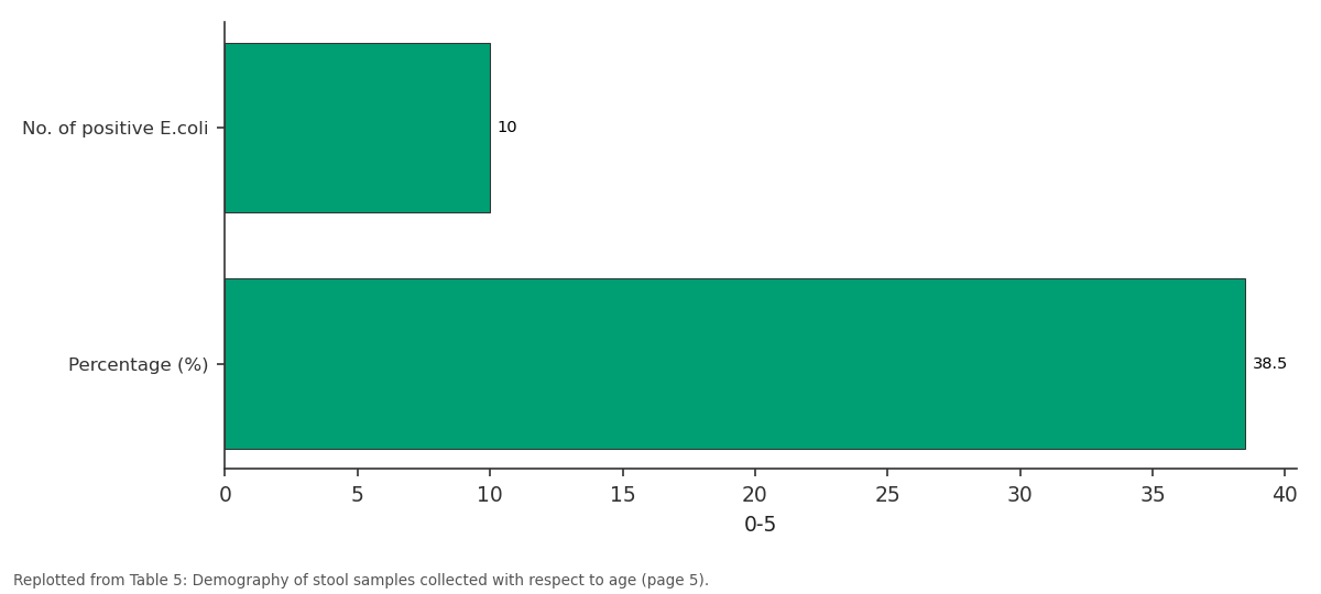
0-5: No. of positive E.coli vs Percentage (%)
Replotted from Table 5 (page 5) of the original paper. Every bar is the paper's own table value.
View source evidence ↗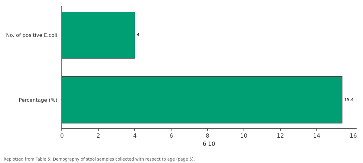
6-10: No. of positive E.coli vs Percentage (%)
Replotted from Table 5 (page 5) of the original paper. Every bar is the paper's own table value.
View source evidence ↗Figures & tables
From the original paper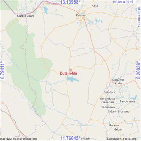 Figure 1
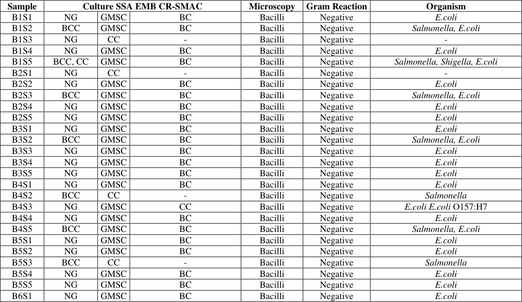 Table 1
 Table 2
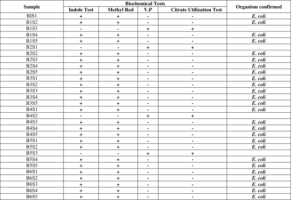 Table 2
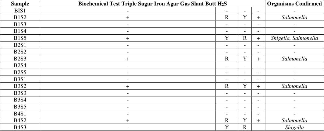 Table 3
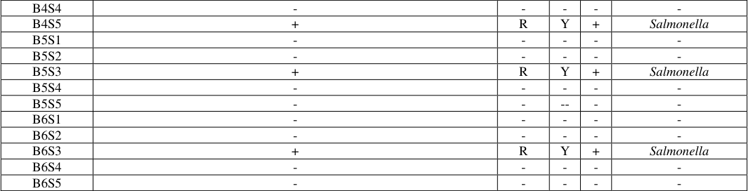 Table 4
 Table 4
 Table 5
 Table 6
 Table 7
 Table 8
Introduction
Enteric pathogens are gastrointestinal organisms known to cause gastrointestinal infection. Gastrointestinal infection also known as gastroenteritis is any infection caused by Viruses, Bacteria or Parasites and is characterized by excessive watery diarrhea and stomach pain. Acute diarrhea is a second most common cause of infant deaths worldwide and is a common cause of mortality in developing countries (Victoria et al., 2008).It is estimated that 1.3 billion episodes of diarrhea occur in children below five years of age with about 760,000 deaths occurring yearly (WHO 2013). Escherichia coli is a common in habitant of the human and animal gut, but canal so be found in the physical environment such as; water, soil and vegetation and are thus referred to as being ubiquitous. Many Escherichia coli strains are usually not harmful and act as commensalism in the intestine of warm blooded animals, but some few strains have been found to cause mild to severe disease in man (WHO 2013). Escherichia coliis the pathogen most commonly associated with endemic forms of childhood diarrhea (Huillanet al., 1991). Diarrhea is a leading cause of morbidity and mortality among children in developing countries (Guerrantet al., 1990;WHO 2005). Among the adult populace, diarrhea diseases can lead to drastic loss in man-hours, thus depleting state and national income (WHO 2011) . E. coli O157:H7isaGram negativebacillus (rod) bacteriuminthe Entero bacteria ceae family. The etiologic agent E.coli O157:H7 have several transmissions that can be spread around to animals and humans. In humans this serotype of E.coli "is transmitted to humans primarily through consumption of contaminated foods, (WHO 2011) mainly undercooked meat like raw beef and milk. With contaminated food products, it can also cause "cross--contamination during food preparation. (WHO 2011). Known contaminated foods that have caused outbreaks of infections besides beef are fruits and vegetables" Where by contamination maybe due to contact with feces from domestic or wild animals at some stage during cultivation or handling. (WHO, 2011). People that visit farms and come into direct contact with farm animals that carry the pathogen can also become infected. Another mode of human transmission is person to person contact through the oral-fecal route is "infected people do not wash their hands after using the toilet. (DHI New York 2006). E. coli have many reservoirs, cattle and small "domesticated ruminants constitute a primary animal reservoir of E.coli O157:H7 (USN 2011) like poultry. The bacterium can also be in vegetables, surface water, fruit and dairy products. Animal manure can also harbor the pathogen where it can contaminate water supply of both DOI: https://doi.org/10.33545/26649926.2021.v3.i1a.27
humans and domesticated animals. E. coli O157:H7 produce a toxin called Shiga toxin that can be deadly in humans and is similar to toxins produced by Shigella dysenteries. E. coli that produce Shiga toxins are also referred as (STEC). E. coli O157:H7 grow with an optimum temperature of 37°C and can grow in temperatures of between 7°C to 50°C. It is a facultative anaerobe, which means it can grow with or without the presence of oxygen (Tarr et al., 1990). The aim is to screen for Escherichia coli O157:H7 in diarrheic patients in L.G.A Dutsin-ma general hospital Katsina state.
Methodology
Methodology
Methods
Methods
Isolation of lactose and non-lactose fermenter
Lactose and non-lactose fermenter were isolated using Mac Conkey agar. Mac Conkey agar was prepared according to the manufacturer instruction. Thereafter, pink and colorless colonies appeared on the media. (Cheesbrough 2006) [11] Isolation of Escherichia coli Those with pink colonies were sub cultured on Eosin methylene blue (EMB) agar. EMB agar was prepared according to the manufacturer instruction. After 24hrs green metallic sheen colonies appeared on EMB which shows positive E. coli. (Cheesbrough 2006) [11] Isolation of salmonella and shigella Those with colorless colonies were sub cultured on salmonella shigella agar (SSA). SSA agar was prepared according to the manufacturer instruction. After 24hrs, creamy colonies with dark centered appeared on SSA which shows positive salmonella and Shigella. (Cheesbrough 2006) [11] Gram staining Smear was prepared on a clean and dry slide from overnight culture, the smear was allowed to dry and then fixation was done. The smear was then covered with crystal violent for 30 to 60 seconds and then washed. Iodine was added too for 30 to 60 seconds and washed. Decolorized with alcohol and washed immediately. Safranin was added for 2 minutes then washed. It was examined using 100xobjective lens with oil immersion.
Biochemical Test
Several biochemical tests were carried out in order to have a presumptive identification of the potential bacteria chosen before. Most of the methods were done according to the microbiology laboratory manual (Cappuccino and Sherman, 2005).
Indole test
Tryptophan broth was inoculated with colony of the test organism in tryptophan broth. Incubated at 37 for 24-48hrs in a incubator. Added 0.5ml of kovac's reagent to the both cultures. Positive results showed pink color in the reagent layer in 1 minute. MacConkey was prepared according to manufacturer instruction and was also diluted with cefrixime. The confirmed E. coli isolates were cultured onRamnose Sorbitol MacConkey Agar plates (CR-SMAC), the colonies that appeared colour less on CR-SMAC were tagged as presumptive E. coli O157.
Methyl red test
Methyl red test
Results
Table 1 shows results for Culture, Microscopy, and Gram reaction for the 30 stool samples collected.
| Sample | Culture SSA EMB CR-SMAC | — | — | Microscopy | Gram Reaction | Organism |
|---|---|---|---|---|---|---|
| B1S1 | NG | GMSC | BC | Bacilli | Negative | E.coli |
| B1S2 | BCC | GMSC | BC | Bacilli | Negative | Salmonella, E.coli |
| B1S3 | NG | CC | - | Bacilli | Negative | - |
| B1S4 | NG | GMSC | BC | Bacilli | Negative | E.coli |
| B1S5 | BCC, CC | GMSC | BC | Bacilli | Negative | Salmonella, Shigella, E.coli |
| B2S1 | NG | CC | - | Bacilli | Negative | - |
| B2S2 | NG | GMSC | BC | Bacilli | Negative | E.coli |
| B2S3 | BCC | GMSC | BC | Bacilli | Negative | Salmonella, E.coli |
| B2S4 | NG | GMSC | BC | Bacilli | Negative | E.coli |
| B2S5 | NG | GMSC | BC | Bacilli | Negative | E.coli |
| B3S1 | NG | GMSC | BC | Bacilli | Negative | E.coli |
| B3S2 | BCC | GMSC | BC | Bacilli | Negative | Salmonella, E.coli |
| B3S3 | NG | GMSC | BC | Bacilli | Negative | E.coli |
| B3S4 | NG | GMSC | BC | Bacilli | Negative | E.coli |
| B3S5 | NG | GMSC | BC | Bacilli | Negative | E.coli |
| B4S1 | NG | GMSC | BC | Bacilli | Negative | E.coli |
| B4S2 | BCC | CC | - | Bacilli | Negative | Salmonella |
| B4S3 | NG | GMSC | CC | Bacilli | Negative | E.coli E.coli O157:H7 |
| B4S4 | NG | GMSC | BC | Bacilli | Negative | E.coli |
| B4S5 | BCC | GMSC | BC | Bacilli | Negative | Salmonella, E.coli |
| B5S1 | NG | GMSC | BC | Bacilli | Negative | E.coli |
| B5S2 | NG | GMSC | BC | Bacilli | Negative | E.coli |
| B5S3 | BCC | CC | - | Bacilli | Negative | Salmonella |
| B5S4 | NG | GMSC | BC | Bacilli | Negative | E.coli |
| B5S5 | NG | GMSC | BC | Bacilli | Negative | E.coli |
| B6S1 | NG | GMSC | BC | Bacilli | Negative | E.coli |
Table text extracted from the original PDF; superscript letters are shown inline. The original table image is below.
View original table image
The media used for the isolation are Eosine Methylene Blue Agar, Salmonella Shigella Agar Cefixime Rhamnose Sorbitol MacConkey Agar for the isolation of Escherichia coli, Salmonella and Shigella spp and Escherichia coli O157:H7 colonies respectively. Microscopy and Gram staining was performed as all shapes for colonies revealed bacilli and Gram reaction, Gram negative.
Sample Biochemical Test Triple Sugar Iron Agar Gas Slant Butt H2S Organisms Confirmed
Sample Biochemical Test Triple Sugar Iron Agar Gas Slant Butt H2S Organisms Confirmed
Discussion
The finding in this study indicates that age remains a major risk factor in diarrhea disease, children between the Age of 0-5 are highly vulnerable to diarrhea. The prevalence of 38.5% diarrhea in respondent 0-5 in this work is higher than the 9.8% obtained by (H.O Abdulazizet al., 2016) [19] , lower than 43.1% obtained by Ifeanyiet al., in Abuja, and higher than 2.6% obtained by Yilgwan andOkolo in Jos plateau. These differences might be due to breaches in sanitation and hygiene infrastructure of the respondents from these cities. The high occurrence rate of diarrhea among children 0-5 years in this study may be due to the fact that children within this age group on their own cannot differentiate between what to eat and what not to eat; they have not learnt the rudiment of adherence to aseptic or hygienic practices. Another reason for their high vulnerability to diarrhea may be due to weaker immunity as a result of them having lost their inborn immunity after being weaned from breast milk. Young children use more water over the course of a day given their higher metabolic rates, also their kidneys are less able to conserve water compared to older children and adults as such diarrhea is usually prevalent and often life threatening too. In this study, it was observed that the number of diarrheic stool gotten from adults was quite small compared to that obtained from children and this might not be unrelated to the fact that, adults in the locality rarely visit health institutions when they have diarrhea unless they perceive the diarrhea as being
Conclusion
The result on this studies indicate that diarrhea is higher among younger children between the age of 0-5 (38.5%) than adult between the age of 26 -30 (3.9%). This is due to the fact that children within this age group on their own cannot differentiate between what to eat and what not to eat. Also the prevalence ofE.coli (72.22%) in this study is higher than the E.coli O157:H7 (7.7%) and the prevalence of salmonella (22.22%) is higher than the prevalence of Shigella (5.6%). Therefore E. coli O157:H7 is an important Aetiology for diarrhea. It is also important to note that an exceptionally low dose of this organism is able to cause infection and once introduced into a closed group or family, it can spread by person to person transmission especially by children.
Recommendations
Recommendations
Health inspector in the local government area of Dutsin-ma should be vigilant and ensures that campaigns are mounted to educate our citizens on ways of improving on the unsanitary environment.
Potential source of infection as well as food and meat inspection must be followed up.
In addition, our physicians should make inquiry on E.coli O157:H7 infection whenever patients present at their clinic with bloody or non-bloody diarrhea so as to subdue possible outbreaks early enough.
Figures and tables from the paper
| B6S2 | NG | GMSC | BC | Bacilli | Negative | E.coli |
|---|---|---|---|---|---|---|
| B6S3 | BCC | GMSC | CC | Bacilli | Negative | Salmonella, E.coli, E.coli O157:H7 |
| B6S4 | NG | GMSC | BC | Bacilli | Negative | E.coli |
Table text extracted from the original PDF; superscript letters are shown inline. The original table image is below.
View original table image
| Sample | Biochemical Tests | — | — | — | Organism confirmed |
|---|---|---|---|---|---|
| Indole Test | Methyl Red | V.P | Citrate Utilization Test | ||
| BIS1 | + | + | - | - | E. coli |
| B1S2 | + | + | - | - | E. coli |
| B1S3 | - | - | + | + | |
| B1S4 | + | + | - | - | E. coli |
| B1S5 | + | + | - | - | E. coli |
| B2S1 | - | - | + | + | |
| B2S2 | + | + | - | - | E. coli |
| B2S3 | + | + | - | - | E. coli |
| B2S4 | + | + | - | - | E. coli |
| B2S5 | + | + | - | - | E. coli |
| B3S1 | + | + | - | - | E. coli |
| B3S2 | + | + | - | - | E. coli |
| B3S3 | + | + | - | - | E. coli |
| B3S4 | + | + | - | - | E. coli |
| B3S5 | + | + | - | - | E. coli |
| B4S1 | + | + | - | - | E. coli |
| B4S2 | - | - | + | + | |
| B4S3 | + | + | - | - | E. coli |
| B4S4 | + | + | - | - | E. coli |
| B4S5 | + | + | - | - | E. coli |
| B5S1 | + | + | - | - | E. coli |
| B5S2 | + | + | - | - | E. coli |
| B5S3 | - | - | + | + | |
| B5S4 | + | + | - | - | E. coli |
| B5S5 | + | + | - | - | E. coli |
| B6S1 | + | + | - | - | E. coli |
| B6S2 | + | + | - | - | E. coli |
| B6S3 | + | + | - | - | E. coli |
| B6S4 | + | + | - | - | E. coli |
| B6S5 | + | + | - | - | E. coli |
Table text extracted from the original PDF; superscript letters are shown inline. The original table image is below.
View original table image
| Sample | Biochemical Test Triple Sugar Iron Agar Gas Slant Butt H2S | — | — | — | Organisms Confirmed |
|---|---|---|---|---|---|
| BIS1 | - | - | - | - | - |
| B1S2 | + | R | Y | + | Salmonella |
| B1S3 | - | - | - | - | - |
| B1S4 | - | - | - | - | - |
| B1S5 | + | Y | R | + | Shigella, Salmonella |
| B2S1 | - | - | - | - | - |
| B2S2 | - | - | - | - | - |
| B2S3 | + | R | Y | + | Salmonella |
| B2S4 | - | - | - | - | - |
| B2S5 | - | - | - | - | - |
| B3S1 | - | - | - | - | - |
| B3S2 | + | R | Y | + | Salmonella |
| B3S3 | - | - | - | - | - |
| B3S4 | - | - | - | - | - |
| B3S5 | - | - | - | - | - |
| B4S1 | - | - | - | - | - |
| B4S2 | + | R | Y | + | Salmonella |
| B4S3 | - | Y | R | Shigella |
Table text extracted from the original PDF; superscript letters are shown inline. The original table image is below.
View original table image
| B4S4 | - | - | - | - | - |
|---|---|---|---|---|---|
| B4S5 | + | R | Y | + | Salmonella |
| B5S1 | - | - | - | - | - |
| B5S2 | - | - | - | - | - |
| B5S3 | + | R | Y | + | Salmonella |
| B5S4 | - | - | - | - | - |
| B5S5 | - | - | -- | - | - |
| B6S1 | - | - | - | - | - |
| B6S2 | - | - | - | - | - |
| B6S3 | + | R | Y | + | Salmonella |
| B6S4 | - | - | - | - | - |
| B6S5 | - | - | - | - | - |
Table text extracted from the original PDF; superscript letters are shown inline. The original table image is below.
View original table image
| Batches | No. of samples analyzed | No. of positive E. coli | Prevalence of E.coli (%) |
|---|---|---|---|
| B1 | 5 | 4 | 15.4 |
| B2 | 5 | 4 | 15.4 |
| B3 | 5 | 5 | 19.2 |
| B4 | 5 | 4 | 15.4 |
| B5 | 5 | 4 | 15.4 |
| B6 | 5 | 5 | 19.2 |
| Total | 30 | 26 | 100 |
Table text extracted from the original PDF; superscript letters are shown inline. The original table image is below.
View original table image
| Age | No. of samples analyzed | No. of positive E.coli | Percentage (%) |
|---|---|---|---|
| 0-5 | 12 | 10 | 38.5 |
| 6-10 | 5 | 4 | 15.4 |
| 11-15 | 4 | 4 | 15.4 |
| 16-20 | 4 | 4 | 15.4 |
| 21-25 | 3 | 3 | 11.5 |
| 26-30 | 2 | 1 | 3.9 |
Table text extracted from the original PDF; superscript letters are shown inline. The original table image is below.
View original table image
| Gender | No. of samples analyzed | No. of positive E.coli | Percentage (%) |
|---|---|---|---|
| Male | 12 | 8 | 30.8 |
| Female | 18 | 18 | 69.2 |
Table text extracted from the original PDF; superscript letters are shown inline. The original table image is below.
View original table image
| Isolate | No. of E.coli | No. of E.coli O157:H7 | Prevalence of E.coli O157:H7 (%) |
|---|---|---|---|
| B1 | 4 | 0 | 0 |
| B2 | 4 | 0 | 0 |
| B3 | 5 | 0 | 0 |
| B4 | 4 | 1 | 3.33 |
| B5 | 4 | 0 | 0 |
| B6 | 5 | 1 | 3.33 |
Table text extracted from the original PDF; superscript letters are shown inline. The original table image is below.
View original table image
| Organisms | Occurrence | Prevalence (%) |
|---|---|---|
| Escherichia coli | 26 | 72.22 |
| Salmonella | 8 | 22.22 |
| Shigella | 2 | 5.56 |
| E.coli O157:H7 | 2 | 7.7 |
| Total | 36 | 100 |
Table text extracted from the original PDF; superscript letters are shown inline. The original table image is below.
View original table image
References
31 in the original paper- Alisky J, Iczkowski K, Rappaport A, Troitsky N. 1998. Bacteriophages show promise as antimicrobial agents. J. Infect,1998:36:5-15.
- Anderson RC, Buckley SA, Kubena LF, Stanker LH, Harvey RB, Nisbet DJ. Bactericidal effect of sodium chlorate on Escherichia coli O157:H7 and Salmonella typhimurium DT104 in rumen contents in vitro. J. Food Prot,2000:63:10381042.
- Armstrong GL, Hollingsworth J, Morris JGJ. Emerging foodborne pathogens: Escherichia coli O157:H7 as a model of entry of a new pathogen into the food supply of the developed world. Epidemiol. Rev,1996:18: 29-51.
- Barker J, Humphrey TJ, Brown MWR. Survival of Escherichia coli O157 in a soil protozoan: implicationsfor disease. FEMS Microbiol. Lett,1999:173:291-295.
- Bell BP, Goldoft M, Griffin PM, Davis MA, Gordon DC, Tarr PI et al. A multistate outbreak of Escherichia coli O157:H7associated bloody diarrhea and hemolytic uremic syndrome from hamburgers. The Washington experience. J. Am. Med. Assoc,1994:272:1349-1353.
- Besser TE, Hancock DD, Pritchett LC, McRae EM, Rice DH, Tarr PII et al. Duration of detection of fecalexcretion of Escherichia coli O157:H7 in cattle. J. Infect. Dis,1997:175:726-729.
- Buchko SJ, Holley RA, Olson WO, Gannon VPJ, Veira DM. The effect of different grain diets on fecal shedding of Escherichia coli O157:H7 by steers. J. Food Prot,2000:63:1467-1474.
- Buchanan RL, Doyle MP. Foodborne disease significance of Escherichia coli O157:H7 and other enterohemorrhagic E. coli. Food Technol,1997:51:69-76.
- Chapman PA, Wright DJ, Siddons CA. A comparison of immune magnetic separation and direct culture for theisolation of verocy to toxin-producing Escherichia Coli O157 frombovinefaeces. J. Med. Microbiol,1994:40:4424-427.
- Chapman PA, Siddons CA, Cerdan-Malo AT, Harkin MA. A 1year study of Escherichia coli O157 in cattle, sheep, pigs and poultry. Epidemiol. Infect,1997:119:245-250.
- Cheesbrough M. District Laboratory Practice in Tropical Countries Part 2, 2006.
- Cray WCJ, Moon HW. Experimental infection of calves and adult cattle with Escherichia coli O157:H7. Appl. Environ. Microbiol,1995:61:1586-1590.
- Doyle MP, Schoeni JL. Survival and Growth Characteristics of Escherichia coli O157:H7 Associated with Haemorrhagic Colitis. Applied and Environmental Microbiology,1984:48(4):855-856.
- Elder RO, Keen JE, Siragusa GR, Barkocy-Gallagher GA, Koohmarie M, Laegreid WW. Correlation of enterohemorrhagic Escherichia coli O157 prevalence in feces, hides, and carcasses of beef cattle during processing Proc Natl Acad Sci,2000:97:29993003.
- Esumeh FI, Isibor JO, Egbagbe IDS. Screening For Escherichia Coli O157:H7 In Diarrheic Patients InBenin City, Nigeria Journal of Microbiology and Biotechnology Research,2011:1(4):1-4.
- Fenlon DR, Wilson J. Growth of Escherichia coli O157 in poorly fermented laboratory silage: a possible environmental dimension in the epidemiology of E. coli O157. Lett. Appl. Microbiol,2000:30:118-121.
- Griffin PM, Tauxe RV. The epidemiology of infections caused by Escherichia coli O157:H7, other enterohemorrhagic E. coli, and the associated hemolytic uremic syndrome Epidemiol Rev,1991:13:6096.
- Hancock DD, Besser TE, Kinsel ML, Tarr PI, Rice DH Paros MG. The prevalence of Escherichia coli O157:H7 in dairy and beef cattle in Washington State Epidemiol Infect,1994:113:199207.
- AbdulAziz HO, Maryam Aminu, Machido DA, "Isolation and Characterisation Esherichia coli O157 in Human Stool Samples from Parts of Kaduna Metropolis Nigeria. American Journal of Food Science and Technology,2016:4:5.
- Isobol SB, Ratnam S. Sorbitol-MacConkey medium for detection of Escherichia coli O157:H7 associated with hemorrhagic colitis J ClinMicrobiol,1896:23:869-87.
- Janisiewicz WJ, Conway WS, Brown MW, Sapers GM, Fratamico P, Buchanan RL al. Fate of Escherichia coli O157:H7 on fresh-cut apple tissue and its potential for transmission by fruit flies Appl Environ. Microbiol,1999:65:1-5.
- Johnson RP, Wilson JB, Michel P, Rahn K, Renwick SA, Gyles CL et al. Human infection with verocytotoxigenic Escherichia coli associated with exposure to farms and rural environments in CS Stewart and HJ Flint, eds Escherichia coli O157 in farm animals. CABI Publications, New York, NY,1999:147-168.
- Kudva IT, Hatfield PG, Hovde CJ. Escherichia coli O157:H7 in microbial flora of sheep J Clin Microbiol,1996:34:431-433.
- MacDonald IA, Gould IM, Curnow J. Epidemiology of infection due toEscherichia coli O157: a 3-year prospective study Epidemiol Infect.1996:116:279-284.
- Nataro JP, Kaper JB Diarrheagenic Escherichia coli. Clin. Microbiol Rev,1998:11:142-201.
- Schmidt H, Beutin L, Karch H. Molecular analysis of plasmidencoded hemolytic of Escherichia coli O157:H7 strain EDL 933 Infect Immun,1995:63:1055-1061.
- Shere JA, Bartlett KJ, Kaspar CW. Longitudinal study of Escherichia coli O157:H7 dissemination on four dairy farms in Wisconsin Appl Environ Microbiol,1998:64:1390-1399.
- Swerdlow DL, Woodruff BA, Brady RC, Griffin PM, Tippen S, Donnell HDJ et al. A waterborne outbreak in Missouri of Escherichia coli O157:H7 associated with bloody diarrhea and death Ann Intern Med,1992:117:812-819.
- Rice DH, Hancock DD, Besser TE.Verotoxigenic E coli O157 colonization of wild deer and range cattle Vet Rec,1995:137:524.
- Riley LW, Remis RS, Helgerson SD, McGee HB, Wells JG, Davis BR et al. Hemorrhagic colitis associated with a rare Escherichia coli serotype N Engl J Med,1983:308:681685.
- Van Donkersgoed, Berg J, Potter A, Hancock,D, Besser T, Rice D et al. Environmental sources and transmission of Esherichia coli O157 in feedlot cattle Can Vet J,2001:42:714-720. 32. World Health Organization. DA,2011.5/1/2014
Cited by 7
From OpenAlex, OpenCitations and reference lists read by Scinovex
- Antibacterial plant extracts inhibit the beta-lactamase of Escherichia coli clinical isolates ↗
a . · Journal of Medicinal Plants Studies · 2023
Beta-lactamase production is the main resistance mechanism to beta-lactam antibiotics by Enterobacteriaceae including Escherichia coli. In this study, we selected Terminalia superba, Annona senegalensis, Ipomoea batatas and Psidium guajava, which antibacterial properties have been proved with the aim to evaluate their beta-lactamase inhibitory activity. Production of β-lactamase was determined by modified tube acidimetric method using benzyl penicillin as substrate and…
❝ 4PDF - Physicochemical characteristics and antimicrobial of composition from linum usitatissimum L seeds oil ↗
Fath Elrahman, Ahmed, Nosaiba Khader, Munged Ibrahim, Basheer Seed, Abass Babiker · International Journal of Chemical Research and Development · 2023
Linum usitatissimum L a member of Linaceae family, which is used for various medical purposes. The aims of this study is the isolation of oil, testing with some microbes and measure some physicochemical properties. The seeds of Linum usitatissimum L was collected from the Omdurman and air-dried at room temperature, the oil extract of seed was prepared using soxhlet extraction
❝ 3Found by Scinovex - Influence of electrical process parameters to inactivate microorganisms in orange juice by PEF technology
S Krishnaveni · International Journal of Agriculture and Food Science · 2023
Pulsed electric field (PEF) is a non-thermal/chemical food processing method for microbial inactivation in liquid food. This method finds its application to prolong the shelf-life of the seasonal fruit juices which helps to maintain the supply chain throughout the year. So, in the present study, the most commonly found microorganisms of Escherichia coli (E. coli), Staphylococcus aureus (S.aureus) and Saccharomyces cerevisiae (S.cerevisiae) inoculated in orange juice are considered. PEF impact on microbial inactivation based on the experimental results are analyzed with respect to pulse frequen
- a ↗
Debeli Tadesse Amente, Shimelis Mangistu Hailu, Dinaol Belina Kitila, Adem Hiko, Sufiyan Abdi Musa · International Journal of Bioscience and Biochemistry · 2022
Escherichia Coli O157:H7 is among the most newly recognized foodborne pathogens. One of the cause for Escherichia Coli O157:H7 pathogens was eating contaminated raw meat commonly in Ethiopia. However, poor hygienic processing and the practices of distribution are danger for contamination of beef meat leading to human infection. The current study was investigating the occurrence of E. coli O157:H7 in
❝ 3Found by Scinovex - Antimicrobial properties and medicinal effects of Andrographis paniculata
Anumihe OC, Etim GR, Ihejieto NT, Chukwuemeka P. Azubuike, Chibu-Ikwuagwu GO, Ahamefula CO · International Journal of Agriculture Extension and Social Development · 2023
Antimicrobial properties and medicinal effects of Andrographis paniculata was carried out in this study. Andrographis paniculata leaves were collected from Federal Polytechnic Nekede, Owerri and identified by a botanist. The clinical isolates of Klebsiella specie, Vibrio cholera and Escherichia coli were obtained from the Microbiology Department of Federal Medical Centre, Owerri, identified and isolates were then sub-cultured on sterile Nutrient agar and the microbial culture were diluted with peptone water until the final suspension that contained about 1.5 x 10⁵cfu/ml of Vibrio cholera and E
- In vivo subacute toxicity and antimicrobial properties of Annickia affinis (Exell) Versteegh and Sosef stem bark ↗
Jean Emmanuel Mbosso Teinkela, Iselin Fipa Pieumeu, Jean Baptiste Hzounda Fokou, Thierry Fokou Nzodjou, Jules Clément Nguedia Assob, Edwige Laure Nguemfo · International Journal of Pharmacognosy and Pharmaceutical Sciences · 2023
Annickia affinis is a medicinal plant of the Annonaceaes family widely used in traditional medicine for the management of typhoid, diarrhoea and urinary tract infections, hence our interest in this plant. Therefore, our work focused on the study of the subacute toxicity and the antimicrobial activities of the methanolic extract of the Annickia affinis (Exell) Versteegh & Sosef stem bark.
- Investigation of β-lactamase-producing Escherichia coli from cancer patients ↗
Aseel Ali Albadery, Amani Abd-Al-Ridha Al-Abdullah, Saad Shakir Mahdi Al-Amara · International Journal of Clinical Biology and Biochemistry · 2023
Background: E. coli is nosocomial bacteria which leading infections of the urinary tract in the patients with cancer. The emergence of β-lactam-resistant strains is a major source of concern in UTI treatment. resistant to β-lactam E. coli is becoming more of an issue everywhere, including in Iraq. The study estimate β -lactam-resistant E. coli prevalence in Basrah province/ Iraq. Methods: