
Research article · 2017 · Full text
International Journal of Veterinary Sciences and Animal HusbandryPathology of an experimental intramammary infection with two isolates of Streptococcus uberis in mice
Volume 2(3) · Pages 10–14
Research areasMilk Quality and Mastitis in Dairy CowsInfant Nutrition and HealthProbiotics and Fermented Foods
Abstract
Author-writtenIn the present study, Streptococcus uberis isolated from bovine milk was inoculated into the mice mammary gland and studied biochemical and pathological changes in time course manner. Thirty lactating Swiss albino mice were inoculated with S. uberis (5 x 10³ cfu in 50µl) by intramammary route on day 7 of lactation. Experimental inoculation was done on abdominal mammary glands L4, L5, R4, R5 and control group with PBS and mice were euthanized at 2, 4, 8, 12, 24 and 48h time points. The mammary gland weight and percentage of body weight revealed significant (P<0.05) increase at initial time periods after inoculation and reduced at 48h. Serum Aspartate transaminase (AST), Alanine transaminase (ALT) and Lactate dehydrogenase (LDH) revealed initial increase indicating mammary gland tissue damage and later decreased at 48h. On gross examination, mammary gland showed reddish coloured and congestion at 12, 24h and normal pinkish appearance with milk at 48h. On histopathology, both isolates at 12, 24h showed mild to moderate infiltrations of neutrophils and alveolar macrophages in the interstitial space. At 48h showed recovery of the mammary gland to normal alveolar architecture. Thus, successfully established S. uberis mastitis in mice, with moderate pathological changes and biochemical changes especially LDH which may be used as an indicator for S. uberis mastitis in mice.
Summary
Source-checkedPlain-language reading of the full paper, prepared with AI and checked against the text. Each point links to the authors' own words.
Why it matters
- Mastitis is considered one of the most expensive diseases affecting the dairy industry's economy and milk productivity. View evidence ↗
- Streptococcus uberis is a significant environmental pathogen causing mastitis, but its specific pathogenesis in India is not yet fully understood. View evidence ↗
- Mice are used as a model for bovine mastitis because they are cost-effective and easier to handle and maintain in controlled environments than other animals. View evidence ↗
The question
- The study aimed to determine the clinical and pathological changes resulting from the experimental intramammary infection of two Streptococcus uberis isolates in mice. View evidence ↗
What they did
- Thirty mice were inoculated with either S. uberis isolate 1 (SU1) or isolate 2 (SU2) at a dose of 5 x 10³ cfu in 50 µl PBS per teat (L4, L5, R4, R5) on the 7th or 9th day of lactation, while a control group received sterile PBS. View evidence ↗
- Mice from both the infected and control groups were euthanized for post-mortem examination at 2, 4, 8, 12, 24, and 48 hours. View evidence ↗
- Serum biochemistry was measured using Modified UV IFCC methods for AST and ALT, and optimized Deutsche Gesellschaft fur Klinische Chemie methods for LDH. View evidence ↗
What they found
- Mammary gland weight significantly increased at 2 hours for both SU1 and SU2 infected mice, then significantly decreased at 24 hours for SU1 and 48 hours for SU2. View evidence ↗
- Serum AST levels increased at 4 and 8 hours, with a significant difference observed at 4 hours compared to 24 hours. View evidence ↗
- A non-significant increase in LDH was observed at 8 and 12 hours, which the authors suggest indicates mammary gland tissue damage. View evidence ↗
- Gross examination showed no changes at 2, 4, and 8 hours, but revealed reddish, hemorrhagic, and congested glands at 12 and 24 hours, returning to a normal pink appearance by 48 hours. View evidence ↗
- Histopathology showed mild neutrophil infiltration at 12 hours, progressing to alveolar epithelial thinning, connective tissue loss, and moderate hyperemia and edema at 24 hours. View evidence ↗
- By 48 hours, the mammary gland showed proliferation of alveolar epithelial cells and increased alveolar structure, though blood vessels remained congested. View evidence ↗
- No histological changes were observed in other organs, including the liver, kidney, spleen, heart, lung, and inguinal lymph nodes. View evidence ↗
What it means
- The authors concluded that S. uberis mastitis was successfully established in mice and that LDH may serve as a biochemical indicator for this infection. View evidence ↗
- The authors state that further research on host-pathogen interactions at the subcellular level is needed to understand the mechanisms of bacterial damage in mice. View evidence ↗
Visual summary
3 visualsIllustrations explain the paper; charts are replotted from the paper's own table values.
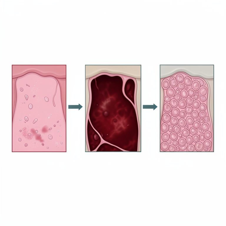
Timeline of Mammary Gland Pathology
AI-generated conceptual illustration (mouse mammary gland tissue). It explains the idea and does not show study data.
View source evidence ↗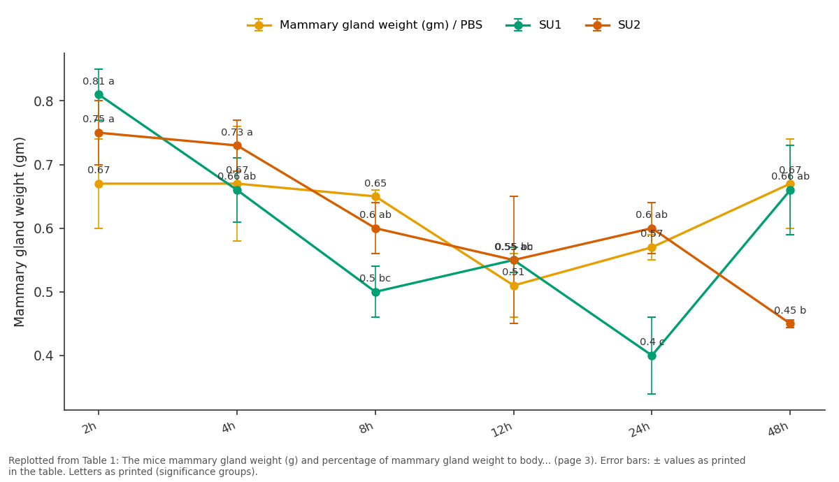
Mammary gland weight (gm) / PBS and SU1 and more: 2h vs 4h vs 8h vs 12h vs 24h
Replotted from Table 1 (page 3) of the original paper. Every bar is the paper's own table value.
View source evidence ↗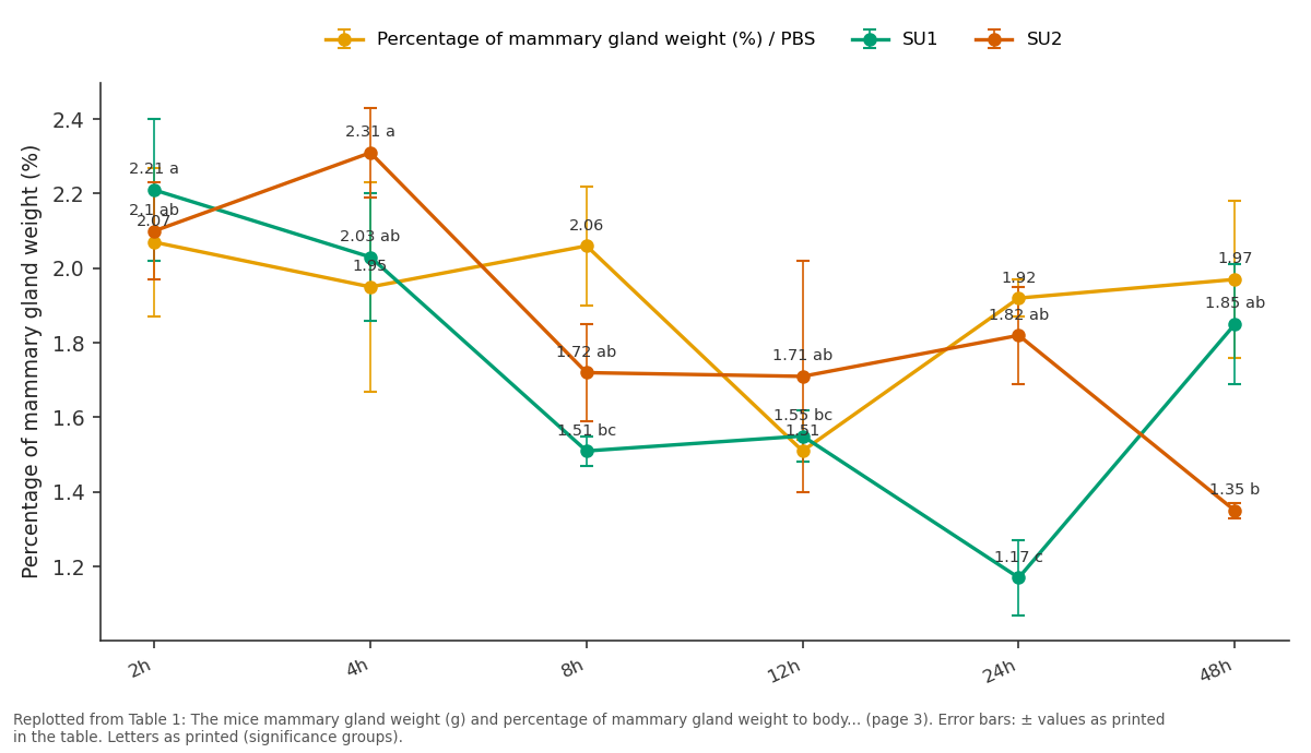
Percentage of mammary gland weight (%) / PBS and SU1 and more: 2h vs 4h vs 8h vs 12h vs 24h
Replotted from Table 1 (page 3) of the original paper. Every bar is the paper's own table value.
View source evidence ↗Figures & tables
From the original paper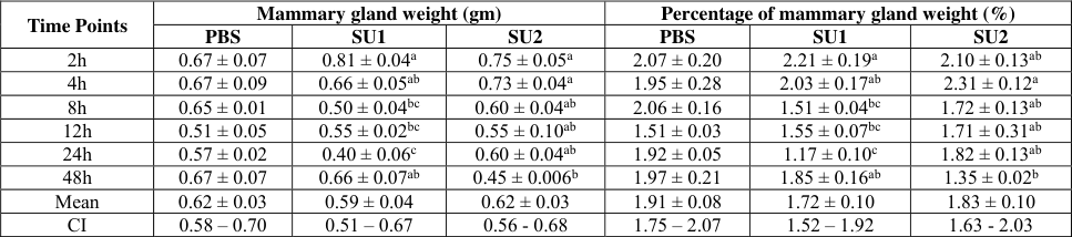 Table 1
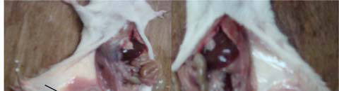 Figure 1
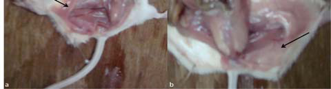 Figure 1
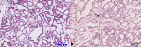 Figure 2
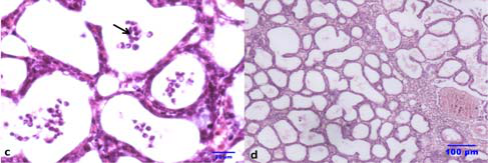 Figure 2
Introduction
India ranks first in cattle and buffalo population in the world and also ranks first in the world milk production, with total milk production of 146.3 million tonnes during 2014 and contributes to 18.6% of world milk production as per Economic Survey of India (2015-16) report. Although several diseases affect the milk productivity and economy, mastitis is regarded as one of the costliest disease confronting the dairy industry. It is estimated that mastitis accounts to about 70 per cent of all avoidable losses incurred during milk production (Sadana, 2006) [19] . The direct effects of mastitis include either temporary or permanent loss in milk production, poor milk quality, reduction in milk price, increase in treatment costs and premature culling (Krishnamoorthy et al., 2017a) [8] . In India, the total annual economic loss due to mastitis was calculated to be 7,165.51 crore rupees (Bansal and Gupta, 2009) [1] . Among the bacteria isolated in bovine mastitis, Staphylococcus species occupies an important place and next comes the Streptococcus species in India. The pooled prevalence estimate for the Streptococcus species was 14 per cent based on the meta-analysis of mastitis prevalence studies in India (Krishnamoorthy et al., 2017b) [12] . The S. uberis is a major environmental mastitis causing pathogen, however it is important pathogen still the pathogenesis is not understood yet in detail in India. The pathogenesis of experimental intramammary infection of S. uberis in bovine has been studied earlier (Rambeaud et al., 2003) [18] . The study of mastitis in bovines is costly and involves various ethical and social issues, especially in India and also keeping the bovines in controlled environment is difficult. Hence, the pathogenesis of mastitis pathogens are mostly studied in laboratory animals like mice, rat and rabbit models. The mouse mastitis model is considered to be very good model to study mastitis compared to other laboratory animals for ease of handling, ease of keeping them in controlled environment and less cost (Krishnamoorthy et al., 2017) [8] . The mouse model is regarded as straight forward and suitable model for the study of bovine mastitis which provides valuable information about pathogenic mechanisms of variety of organisms involved in the intramammary infections (Notebaert and Meyer, 2006) [16] . Perusal of reported literatures revealed scanty information is available on clinical pathology of experimentally induced mastitis in mice by using S. uberis especially in India. Keeping this information in background, the present study was undertaken to know the clinical pathological changes of two S. uberis isolates in experimental intramammary infection in mice.
Materials and Methods
Materials and Methods
Bacteria used for inoculation
Two isolates of Streptococcus uberis was isolated from milk samples of apparently healthy bovines from dairy farms in Karnataka and species identification was done by using Genus, species and 16S RNA PCR by our research team as reported earlier (Shome et al., 2011,2012) [21,22] . The Multi-Locus Sequence Typing of the isolates using seven housekeeping genes of S. uberis was carried out as described earlier (Coffey et al., 2006) [5] .
Experimental Animals and Infection
Thirty five timed pregnant (Day 12 to 15), Swiss albino mice were procured from National Centre for Laboratory Animal Science, Hyderabad. The mice were grouped in to seven groups of five mice in each group consisting of 30 for bacterial inoculation and five for PBS inoculation. The mice were housed in individually ventilated cages (IVC) and temperature and humidity were maintained at 23 ± 3 ºC and 50 to 70 per cent respectively. Mice were provided rodent pellet feed procured from M/s. Nutripet India Private Limited, Bengaluru, India and reverse osmosis purified water ad libitum. The animal experiments were approved by Institutional Animal Ethics Committee (IAEC) of ICAR-National Institute of Veterinary Epidemiology and Disease Informatics, Bengaluru (Registration No. 881/05/R/S/CPCSEA). The experiment was carried out as per the guidelines of Committee for the purpose of Supervision and Experiments on Animals (CPCSEA), Ministry of Environment, Forests and Climate Change, New Delhi. The S. uberis isolate 1 (SU1) and S. uberis isolate 2 (SU2) were inoculated to thirty mice each on 7 th or 9 th day of lactation with dose containing 5 x 10 3 cfu of bacterial suspension in PBS (50 µl) per teat of left fourth and fifth (L4, L5) and right fourth and fifth (R4, R5) teats and control mice were inoculated with sterile PBS as reported earlier (Krishnamoorthy et al., 2014a, b) [9,10] . After one hour of intramammary inoculation (IMI), the pups were allowed to suckle the teat to simulate the natural field conditions of bovines in dairy farms. The mice from both bacteria inoculated and PBS control groups were euthanized at different time intervals at 2, 4, 8, 12, 24 and 48h and thorough post mortem examination was carried out.
Animal weight and mammary gland weight
The mice were weighed using weighing balance (Precisa, Switzerland) and recorded the body weight in grams. The mice mammary glands were carefully dissected out using sterile scissors and foreceps. The left (L4, L5) and right (R4, R5) mammary glands were separately collected, weighed and expressed in grams. The mammary gland weight (average of left and right side) and animal body weight was used to calculate the percentage of mammary gland weight to body weight.
Serum Biochemistry
Blood was collected by intracardiac route by using sterile syringe and needle from mice in dipotassium ethylene diamine tetra acetate (K2EDTA) vials for serum biochemistry. The serum aspartate aminotransferase (AST) was estimated by Modified UV International Federation of Clinical Chemistry (IFCC), alanine aminotransferase (ALT) by Modified UV IFCC (Bergmeyer et al., 1978) [2] and lactate dehydrogenase (LDH) by optimized Deutsche Gesellschaft fur Klinische Chemie methods (Buhl and Jackson, 1978) [3] by using commercially available kits (M/s. Span Diagnostics Limited, Surat, India) and Semiauto analyzer, Biosystems (BTS 320) as reported earlier (Krishnamoorthy et al., 2015) [11] .
Pathology
The gross changes in the mammary gland and various organs was recorded and histopathological studies were carried out on representative tissue samples collected during experimental intramammary infection in mice. The mammary gland tissues and various organs like liver, kidney, spleen, heart, lung and inguinal lymph node were collected and immediately fixed in 10 per cent neutral buffered formalin. After proper fixation, the tissue samples were processed by routine paraffin embedding technique. Sections of four to five micron thickness were cut using rotary microtome with disposable blades. The sections were then stained with routine haematoxylin and eosin method (Luna, 1968) [14] and observed under bright field microscope (Nikon, Japan).
Statistical analysis
Data obtained from mammary gland weight, percentage of mammary gland weight and biochemical parameters were analyzed by using Statistical Analysis System (SAS) software SAS Enterprise Guide version 5.1 (SAS, 2012) [20] and by one way analysis of variance (ANOVA) method (Snedecor and Cochran, 1980) [23] , and obtained the significant difference between different time points. The results were expressed as the Mean ± SE (Standard error) with significant difference at P<0.05 and confidence interval at 95 per cent level.
Results
Results
Bacteria
The two isolates used for inoculation was confirmed by genus, species specific and 16sRNA PCR and identified as S. uberis. The Multi-Locus Sequence Typing of the two isolates of S. uberis was done and found to be Sequence type (ST) 475, which is prevalent in India.
Clinical signs and Mice mammary gland weight
The clinical signs of mastitis were not apparent at 2, 4 8, 12, 24 and 48h after intramammary inoculation with SU1 and SU2 in mice. The mean ± SE mammary gland weight and percentage of mammary gland weight at different time points after IMI is presented in Table 1. No significant difference in the mammary gland weight and percentage of mammary gland weight was observed in PBS inoculated mice. The mammary gland weight showed significant (P<0.05) increase in SU1 and SU2 infected mice at 2h and significant (P<0.05) decrease at 24h for SU1 and 48h for SU2 when compared to other time points. The percentage of mammary gland weight revealed significant (P<0.05) increase at 2h in SU1 and 4h in SU2 and significant (P<0.05) decrease at 24h in SU1 and 48h in SU2.
| Time Points | Mammary gland weight (gm) | — | — | Percentage of mammary gland weight (%) | — | — |
|---|---|---|---|---|---|---|
| PBS | SU1 | SU2 | PBS | SU1 | SU2 | |
| 2h | 0.67 ± 0.07 | 0.81 ± 0.04a | 0.75 ± 0.05a | 2.07 ± 0.20 | 2.21 ± 0.19a | 2.10 ± 0.13ab |
| 4h | 0.67 ± 0.09 | 0.66 ± 0.05ab | 0.73 ± 0.04a | 1.95 ± 0.28 | 2.03 ± 0.17ab | 2.31 ± 0.12a |
| 8h | 0.65 ± 0.01 | 0.50 ± 0.04bc | 0.60 ± 0.04ab | 2.06 ± 0.16 | 1.51 ± 0.04bc | 1.72 ± 0.13ab |
| 12h | 0.51 ± 0.05 | 0.55 ± 0.02bc | 0.55 ± 0.10ab | 1.51 ± 0.03 | 1.55 ± 0.07bc | 1.71 ± 0.31ab |
| 24h | 0.57 ± 0.02 | 0.40 ± 0.06c | 0.60 ± 0.04ab | 1.92 ± 0.05 | 1.17 ± 0.10c | 1.82 ± 0.13ab |
| 48h | 0.67 ± 0.07 | 0.66 ± 0.07ab | 0.45 ± 0.006b | 1.97 ± 0.21 | 1.85 ± 0.16ab | 1.35 ± 0.02b |
| Mean | 0.62 ± 0.03 | 0.59 ± 0.04 | 0.62 ± 0.03 | 1.91 ± 0.08 | 1.72 ± 0.10 | 1.83 ± 0.10 |
| CI | 0.58 – 0.70 | 0.51 – 0.67 | 0.56 - 0.68 | 1.75 – 2.07 | 1.52 – 1.92 | 1.63 - 2.03 |
Table text extracted from the original PDF; superscript letters are shown inline. The original table image is below.
View original table image
Serum Biochemistry
The serum Aspartate transaminase (AST), Alanine transaminase (ALT) and Lactate dehydrogenase (LDH) values at different time points after IMI with S. uberis is presented in Graph 1. The AST showed initial increase at 4 and 8h after IMI with S. uberis isolates in mice. The increase was significant (P<0.05) difference at 4h when compared to the 24h in mice. ALT values revealed non-significant increase at 4h after IMI when compared to other time periods. At 8 and 12h after IMI with S. uberis in mice revealed non-significant increase of LDH and indicated that it might be resulted from mammary gland tissue damage caused by the infecting organisms.
Graph 1: Graph showing serum Alanine transaminase (ALT), Aspartate transaminase (AST) and Lactate dehydrogenease (LDH) values at different time points after intramammary inoculation with
Streptococcus uberis in mice.
Gross pathology
The PBS inoculated mice revealed normal pink colour of mammary gland structure and on incision normal milk oozed out. The gross changes were not observed during 2, 4 and 8h after IMI, but reddish colour, haemorrhagic and congested mammary glands was observed at 12 and 24h (Fig. 1a). The mammary gland appeared pink colour with milk on incision and appeared normal at 48h (Fig. 1b) after IMI of SU1 and SU2 isolates in mice. On palpation, at 24h the gland was firm to palpate and upon incision thick, slightly discoloured milk oozed out from the infected mice mammary glands when compared to PBS group. There was no difference between the two isolates SU1 and SU2 in the gross changes of mammary gland in mice. There was no gross changes in liver, kidney, spleen, lung, heart and inguinal lymph nodes.
Histopathology
The mammary glands in mice inoculated with sterile PBS as control did not reveal any histological changes at 2, 4, 8, 12, 24 and 48h. The microscopic appearance of the mammary gland was normal in which several lobules showed secretory acini and were distended with eosinophilic secretory material in the alveolar lumen and separated by interlobular septa. Mammary gland of mice infected with SU1 and SU2 revealed no observable histopathological lesions at 2, 4, 8h after IMI. The SU1 and SU2 showed histopathological lesions at 12h with mild changes with infiltration of few inflammatory cells mainly neutrophils in the interstitial space and between the lobules (Fig. 2a). The lesions at 24h included thinning of alveolar epithelial cells and loss of connective tissue with enlarged lobules, moderate degree of hyperemia with perilobular and interlobular edema. The lumen of acini was filled with varying amount of eosinophilic secretory material along with desquamated epithelial cells and moderate infiltration of inflammatory cells mainly neutrophils and macrophages (Fig. 2b). There was moderate infiltration of neutrophils with multi-lobed nucleus and mononuclear cells mainly macrophages and lymphocytes in the alveolar lumen which was shown in Fig. 2c. The mammary gland revealed proliferation of alveolar epithelial cells with increase in alveolar structure and congestion of blood vessels at 48h after IMI (Fig. 2d). No observable histological changes were observed in the liver, kidney, spleen, heart, lung and inguinal lymph node of mice after IMI with SU1 and SU2.
Discussion
There was no clinical signs reported in mice infected with two isolates of S. uberis SU1 and SU2 in the present study. The Lasagno et al., (2012) [13] reported no signs or systemic symptoms evoked by S. uberis inoculation in experimental goats and concurred with the present study. The absence of clinical signs in this study may be due to low dose of organisms as compared to other reported studies in which the dose used was above 10 5 cfu. In the present study, the mammary gland weight showed significant increase at 2 and 4 h after IMI in SU1 and SU2, respectively. The slight increase in the mammary gland weight was observed when compared to the PBS control mice. The increased mammary gland weight may be due to the inflammatory processes, congestion and infiltration of inflammatory cells at early time points. However, the slight reduction in mammary gland weight at 24h was observed, may be due to loss of milk in mammary gland and compensated by the infiltration of inflammatory cells.
Aspartate aminotransferase (AST) has been used to detect the extent of liver cell damage during disease conditions and recent studies have shown the relationship between the AST values and development of clinical mastitis. The AST showed non-significant increase at 4 and 8h after IMI with S. uberis isolates in mice and concurred with previous reports with other mastitis pathogens (Krishnamoorthy et al., 2015, Zaki et al.,2008, Moyes et al., 2009) [11,24,15] . However, Zaki et al., (2008) [24] reported a highly significant increase in AST values after infection with Streptococcus agalactia in buffaloes. The aspartate aminotransferase may be potential marker for the risk of mastitis in early lactation in dairy cattle (Krishnamoorthy et al., 2015, Moyes et al., 2009) [11,15] . The increase of AST values was attributed to the stressful conditions after IMI in mice (Lasagno et al., 2012) [13] . ALT values revealed non-significant increase at 4h after IMI when compared to other time periods. The results of the present study concurred with the previous report (Zaki et al., 2008) [24] . At 8 and 12h after IMI with S. uberis in mice revealed non-significant increase of LDH activity and concurred with previous studies with various mastitis causing organisms (Krishnamoorthy et al., 2015, Zaki et al., 2008, Chagunda et al., 2006, Kermori et al., 1989) [11,24,4,7] . The increase can be attributed to the participation of leukocytes which have LDH activity at the 1,000 U/mg protein level in mastitic milk. This indicated that it might be resulted from mammary gland tissue damage caused by the infecting SU1 and SU2 in mice.
In the present study, the gross lesions were not observed in liver, kidney, spleen, lung, heart and inguinal lymph nodes at different time points. The reason for the same might be due to low dose of organisms (5 x 10 3 ) inoculated in mice. Thus the dose and species of the organisms plays an important role in establishment of infection and generation of lesions in natural infections even in dairy cattle. However, mammary gland showed the reddish discoloration of mammary gland at 12 and 24h after IMI with SU1 and SU2 which indicated the congestion and infiltration of blood cells in the mammary gland. The severity of histopathological lesions revealed no variation between the two isolates of SU1 and SU2 in mice model of mastitis in the present study. The histological changes observed in this study was supported by the gross lesions in the S. uberis infected mice mammary gland. The histological changes observed were moderate infiltration of inflammatory cells mainly neutrophils and mononuclear cells in the interstitial space and alveolar lumen at 12 and 24h after IMI and concurred with previous study (Krishnamoorthy et al., 2017a, Lasagno et al.,2012) [8,13] . However, the SU1 and SU2 could not resist phagocytosis and eventually got cleared from mammary gland through phagocytosis by neutrophils and macrophages at 48h after IMI. The observations in the present study clearly indicated that there is mammary tissue damage in the mice infected with S. uberis. The mammary tissue damage has been shown to be induced by either apoptosis or necrosis and both host and bacterial factors contribute to epithelial tissue damage. During infection of mammary glands, the tissue damage can be initially caused by bacteria and their products. Mastitis is characterized by an influx of somatic cells, primarily neutrophils into the mammary gland which leads to break down of blood milk barrier; damage to the alveolar epithelium worsens.
Polymorphonuclear neutrophils can harm the mammary tissue by releasing reactive oxygen intermediates and proteolytic enzymes (Zhao et al., 2008) [25] . The oxidative stress can damage all types of biomolecules like DNA, proteins, lipids and carbohydrates which perpetuates tissue injury. The PMN cells have primary, secondary and tertiary granules which contain bactericidal peptides, proteins and enzymes such as elastase, proteinases and myeloperoxidases which are released into the extracellular environment and cause tissue destruction during mastitis (Paape et al., 2003) [17] . The mammary tissue damage could also be caused by the proteinases and collagenolytic enzymes which degrade the extracellular matrix compounds (Haddadi et al., 2005) [6] . The tissue damage was induced either by apoptosis or necrosis in the mammary epithelial cells indirectly through induction of proteases or proinflammatory cytokines (Zhao et al., 2008) [25] . Peptidoglycan fraction of cell wall of bacteria is involved in hypersensitivity reaction of the mammary gland. The cytokines also promote a wide variety of function of PMN cells, including adhesion, surface receptor expression, free radical production and release of lysosomal constituents (Paape et al., 2003) [17] .
Conclusions
In the present study, successfully induced mastitis by using two isolates of Streptococcus uberis from cattle in organized
Figures and tables from the paper
References
23 in the original paper- Bansal BK, Gupta DK. Economic analysis of bovine mastitis in India and Punjab -a review. Indian J Dairy Sci. 2009; 62:337-45.
- Bergmeyer HU, Scheibe P, Wahlefeld AW. Optimization of methods for aspartate aminotransferase and alanine aminotransferase. Clin Chem. 1978; 24:58-73.
- Buhl SN, Jackson KY. Optimal conditions and comparison of lactate dehydrogenase catalysis of the lactate-to-pyruvate and pyruvate-to-lactate reactions in human serum at 25, 30, and 37 ºC. Clin Chem 1978; 24:828-831.
- Chagunda MG, Larsen T, Bjerring M, Ingvartsen KL. Llactate dehydrogenase and N-acetyl-betaglucosaminase activities in bovine milk as indicator of non-specific mastitis. J Dairy Res. 2006; 73(4):431-440.
- Coffey TJ, Pullinger GD, Urwin R, Jolley KA, Wilson SM, Maiden MC et al. First insights into the evolution of Streptococcus uberis: a multilocus sequence typing scheme that enables investigation of its population biology. Appl Environ Microbiol 2006; 72:1420-1428.
- Haddadi K, Moussaoui F, Hebia F, Laurent F, Leroux Y. Escherichia coli proteolytic activity in milk and casein breakdown. Reprod Nutrition Develop. 2005; 45:485496.
- Kermori K, Kanae M, Mario K. Contribution of leucocytes to the origin of lactate dehydrogenase by isoenzymes in milk of bovine mastitis. Japanese J Vet Sci. 1989; 51(3):530-539.
- Krishnamoorthy P, Satyanarayana ML, Shome BR, Rahman H. Pathological changes in experimental intramammary infection with different staphylococcal species in Mice. J Microscopy Ultrastructure, 2017. 10.1016/j.jmau.2017.05.001.
- Krishnamoorthy P, Satyanarayana ML, Shome BR, Rao S, Rahman H. Haemato-Biochemical Changes in Staphylococcal Species induced Mastitis in Mouse Model. Research & Reviews: J Vet Sci Tech. 2015; 4(3):8-14.
- Krishnamoorthy P, Suresh KP, Saha S, Govindaraj G, Shome BR, Roy P. Meta-analysis of prevalence of Subclinical and Clinical Mastitis, Major Mastitis Pathogens in Dairy Cattle in India. Intl J Current Microbiol Appl Sci. 2017; 6(3):1214-1234.
- Lasagno MC, Vissio C, Reinosso EB, Raspanti C, Yaciuk R, Larriestra AJ et al. Development of an experimentally induced Streptococcus uberis subclinical mastitis in Goats. Vet Microbiol 2012; 154:376-383.
- Luna LG. Manual of the histological staining methods of Armed Forces Institute of Pathology. 3 rd Edn, London, United Kingdom: Mcgraw Hill Book Company, 1968.
- Moyes KM, Larsen T, Friggens NC, Drackley JK, Ingvartsen KL. Identification of potential markers in blood for the development of subclinical and clinical mastitis in dairy cattle at parturition and during early lactation. J Dairy Sci. 2009; 92(11):5419-5428.
- Notebaert S, Meyer E. Mouse models to study the pathogenesis and control of bovine mastitis. A review. Vet Quart 2006; 28:2-13.
- Paape MJ, Bannerman DD, Zhao X, Lee JW. The bovine neutrophil: structure and function in blood and milk. Vet Res 2003; 34:597-627.
- Rambeaud M, Almeida R, Pighetti G, Oliver S. Dynamics of leukocytes and cytokines during experimentally induced Streptococcus uberis mastitis. Vet Immun Immunopathol 2003; 96:193-205.
- Sadana V. Dairy scenario vision 2020. New Delhi, India: Dairy year book, 2006, 47-48.
- SAS. Statistical Analysis System. Software Version 9.3. Mumbai, India: SAS India Limited, 2012.
- Shome BR, Mitra SD, Bhuvana M, Krithiga N, Shome R, Velu D et al. Multiplex PCR for the detection of five important Staphylococcus sp. in bovine subclinical mastitis milk. Indian J Anim Sci. 2012; 82:9-14.
- Shome BR, Mitra SD, Bhuvana M, Krithiga N, Velu D, Isloor S et al. Multiplex PCR assay for species identification of bovine mastitis pathogens. J Appl Microbiol. 2011; 111:1349-1356.
- Edn, Ames, Iowa, USA: The Iowa State University Press, 1980, 196-252.
- Zaki MS, Sharaf NE, Mostafa SO, Fawzi OM, Batrawy NE. Effect of subclinical mastitis on some biochemical and clinicopathological parameters in buffalo. Am Eurasian J Agri Environmental Sci. 2008; 3(2): 200-204.
- Zhao X, Lacasse P. Mammary tissue damage during bovine mastitis: causes and control. J Anim Sci. 2008; 86: 57-65.