
Research article · 2026 · Full text
International Journal of Advanced Chemistry ResearchSchiff base metal complexes as potential anticancer agents: Biological evaluation, and in silico studies
Volume 8(8) · Pages 111–120 · DOI: 10.33545/26646781.2026.v8.i8b.738 ↗
Research areasMetal complexes synthesis and propertiesSynthesis and biological activityInorganic and Organometallic Chemistry
Abstract
Author-writtenThe development of novel anticancer agents with improved efficacy and selectivity remains a significant challenge in medicinal chemistry. In this study, a series of Schiff base ligands (IAP, TAP, and FAP) and their corresponding cobalt(II) and nickel(II) complexes were synthesized and evaluated for their anticancer potential. The cytotoxic activity of the synthesized compounds was investigated against human cancer cell lines (MCF-7, MDA-MB-231, HeLa, and PC-3), along with a normal cell line (HEK-293) at 10 µM concentration. The results demonstrated that metal complexation significantly enhances cytotoxic activity compared to the free ligands. Among the tested compounds, Co-TAP and Co-FAP complexes exhibited the most promising anticancer activity, showing higher inhibition of cancer cell proliferation while maintaining relatively lower toxicity toward normal cells.In addition, in silico ADME analysis revealed that the ligands possess favorable physicochemical properties, good gastrointestinal absorption, and compliance with drug-likeness criteria. Toxicity prediction suggested moderate acute toxicity with low risks of carcinogenicity and immunotoxicity, although potential hepatotoxicity and mutagenicity were indicated for some compounds. Overall, the combined experimental and computational findings suggest that these Schiff base metal complexes represent promising candidates for further development as anticancer agents.
Summary
Source-checkedPlain-language reading of the full paper, prepared with AI and checked against the text. Each point links to the authors' own words.
Why it matters
- Cancer is a primary cause of death globally, creating a significant burden for healthcare systems. View evidence ↗
- Standard chemotherapy often fails to distinguish between healthy and cancerous cells, which leads to systemic toxicity and makes it harder for patients to adhere to treatment. View evidence ↗
- Combining organic ligands with transition metals can improve a drug's ability to enter cells and interact with DNA or proteins, potentially increasing its effectiveness against cancer. View evidence ↗
The question
- The study aims to design, synthesize, and evaluate the anticancer potential of new Schiff base ligands and their cobalt(II) and nickel(II) complexes. View evidence ↗
What they did
- The researchers synthesized three Schiff base ligands, IAP, TAP, and FAP, and created corresponding complexes using cobalt(II) and nickel(II). View evidence ↗
- Cytotoxicity was tested at a 10 µM concentration over 48 hours against four human cancer cell lines (MCF-7, MDA-MB-231, HeLa, and PC-3) and one normal cell line (HEK-293). View evidence ↗
- Computational tools SwissADME and ProTox-3.0 were used to predict the pharmacokinetic properties and toxicity of the ligands. View evidence ↗
What they found
- Binding the ligands to metal ions significantly increased their ability to kill cancer cells. View evidence ↗
- The Co-TAP and Co-FAP complexes showed the strongest anticancer activity and remained relatively safe for normal cells. View evidence ↗
- Tamoxifen, the reference drug, showed the highest inhibition rates against MCF-7 (56.62%) and MDA-MB-231 (44.46%) cells. View evidence ↗
- Computational analysis indicated that the IAP, TAP, and FAP ligands have good gastrointestinal absorption and favorable drug-like properties. View evidence ↗
- The ligands were predicted to have moderate acute toxicity but low risks of causing cancer, immune reactions, or damage to the heart and kidneys. View evidence ↗
- Predictions suggested potential risks of liver damage, nerve damage, and mutagenicity, indicating a need for more experimental testing. View evidence ↗
What it means
- The authors conclude that these Schiff base metal complexes are promising starting points for developing new anticancer drugs. View evidence ↗
- The authors state that further research is needed, specifically in vivo testing and detailed studies on how the drugs work (mechanisms), to fully understand their potential. View evidence ↗
Visual summary
2 visualsIllustrations explain the paper; charts are replotted from the paper's own table values.
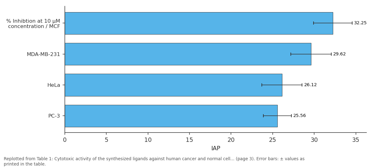
IAP: % Inhibtion at 10 µM... vs MDA-MB-231 vs HeLa vs PC-3
Replotted from Table 1 (page 3) of the original paper. Every bar is the paper's own table value.
View source evidence ↗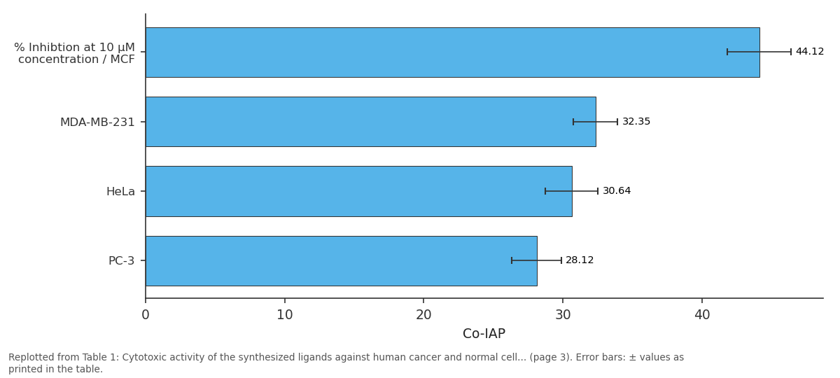
Co-IAP: % Inhibtion at 10 µM... vs MDA-MB-231 vs HeLa vs PC-3
Replotted from Table 1 (page 3) of the original paper. Every bar is the paper's own table value.
View source evidence ↗Figures & tables
From the original paper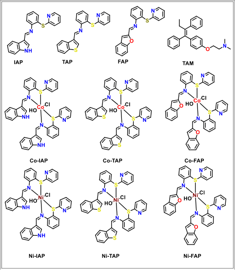 Figure 1
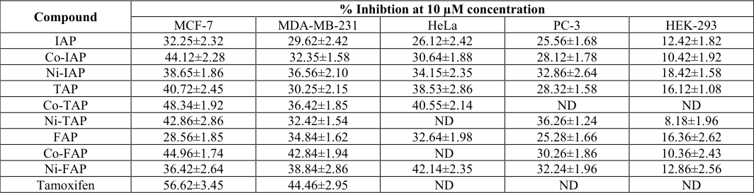 Table 1
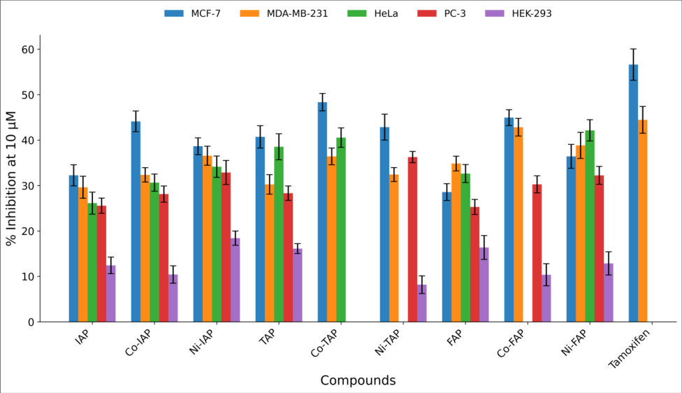 Figure 2
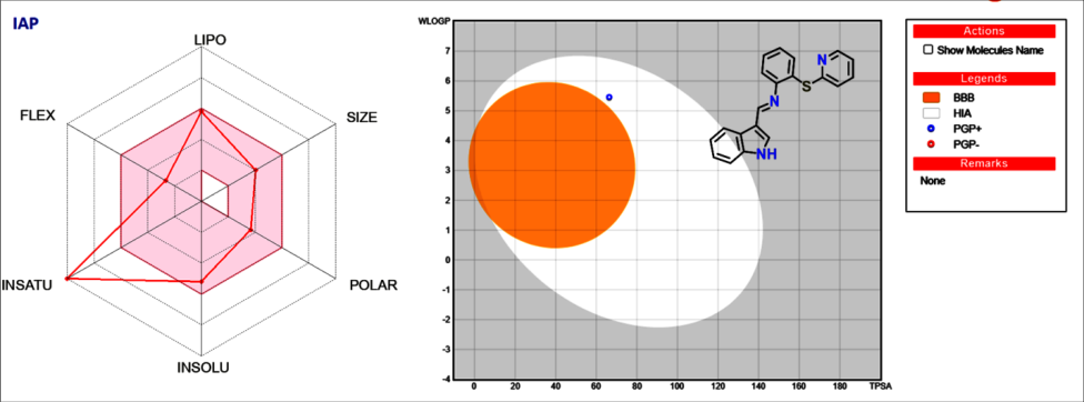 Figure 3
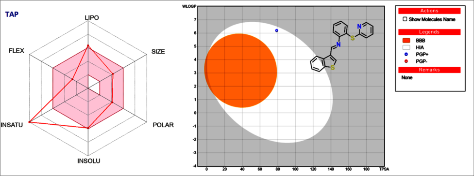 Figure 4
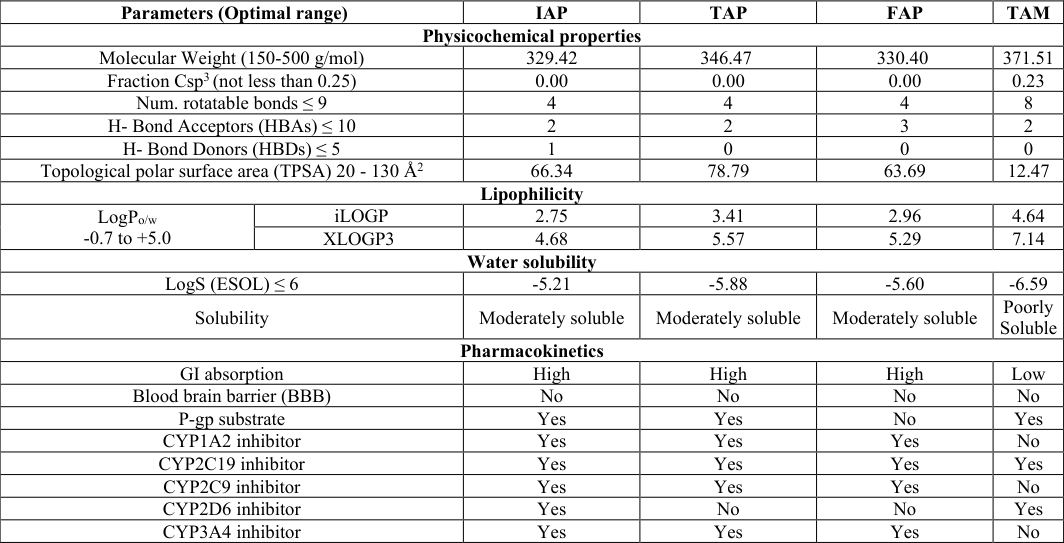 Table 2
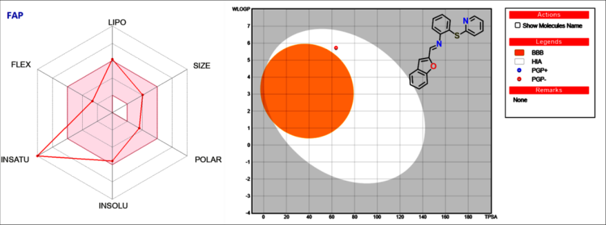 Figure 5
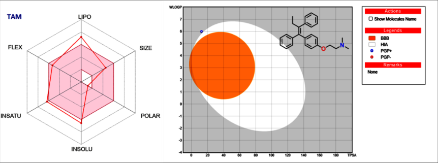 Figure 6
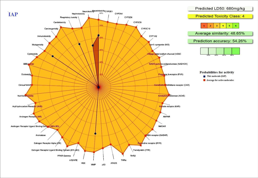 Figure 7
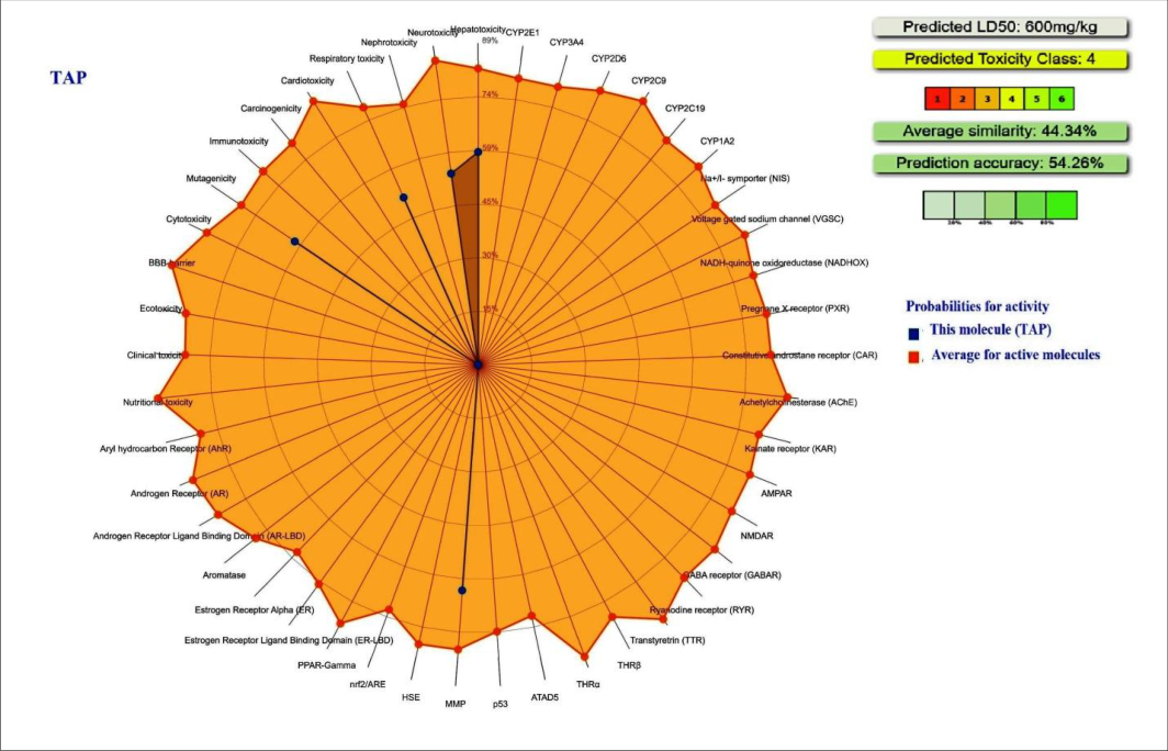 Figure 8
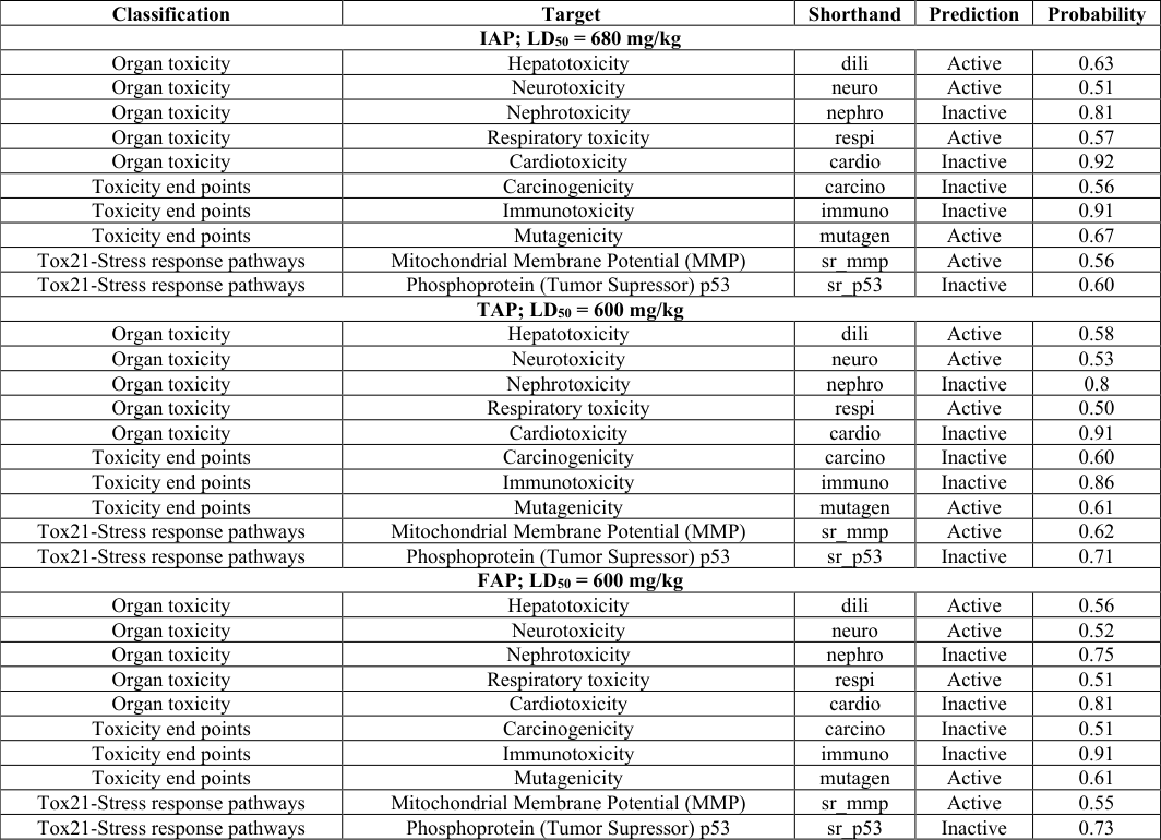 Table 3
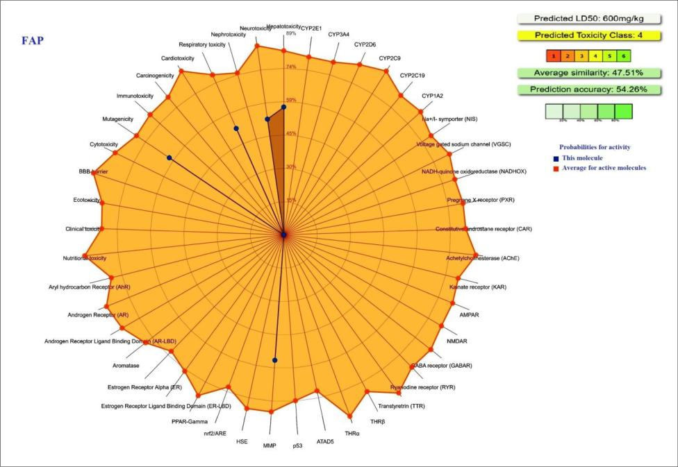 Figure 9
 Table 4
Introduction
Cancer continues to be one of the leading causes of mortality worldwide, posing a major challenge to global healthcare systems. Despite substantial progress in early diagnosis and therapeutic strategies, the effectiveness of current anticancer treatments remains limited by several factors, including drug resistance, poor selectivity, and severe adverse effects on normal tissues. Conventional chemotherapeutic agents often lack the ability to discriminate between cancerous and healthy cells, resulting in systemic toxicity and reduced patient compliance. In addition, tumor heterogeneity and the emergence of multidrug resistance further compromise therapeutic outcomes, highlighting the urgent need for the development of novel anticancer agents with improved efficacy, selectivity, and safety profiles [1,2] Heterocyclic compounds play a pivotal role in medicinal chemistry and constitute the core structure of many clinically approved drugs [3][4][5] . Among them, Schiff base derivatives have gained significant attention due to their structural versatility, ease of synthesis, and diverse biological activities, including anticancer, antimicrobial, antiviral, and anti-inflammatory properties. The presence of the azomethine (-C=N-) functional group enables Schiff bases to interact with various biological targets through hydrogen bonding and coordination interactions, thereby enhancing their pharmacological potential. Furthermore, the incorporation of different substituents within the ligand framework allows fine-tuning of their electronic and steric properties, which is crucial for optimizing biological activity [6][7][8][9][10] In recent years, metal-based drug design has emerged as a promising approach in anticancer research. The coordination of organic ligands with transition metal ions can significantly alter their physicochemical and biological properties. Metal complexes often exhibit enhanced lipophilicity, improved cellular uptake, and the ability to interact with biomolecules such as DNA and proteins, leading to improved cytotoxic activity [11][12][13][14] In particular, cobalt(II) and nickel(II) complexes have attracted considerable interest due to their redox properties and their ability to induce oxidative stress, DNA damage, and apoptosis in cancer cells. Several studies have demonstrated that metal complexes of Schiff base ligands exhibit superior anticancer activity compared to their corresponding free ligands, emphasizing the importance of metal coordination in drug development [15][16][17][18] . Alongside experimental approaches, computational methods have become indispensable tools in modern drug discovery. The evaluation of absorption, distribution, metabolism, and excretion (ADME) properties at an early stage helps in identifying compounds with favorable pharmacokinetic profiles and reduces the likelihood of late-stage failure [19][20][21][22] . SwissADME is widely used for predicting key physicochemical and pharmacokinetic parameters, such as lipophilicity, gastrointestinal absorption, and drug-likeness [23, 24] . Similarly, toxicity prediction tools such as ProTox (III) provide valuable insights into the safety profile of compounds, including acute toxicity, organ toxicity, and potential mutagenicity [25,26] . These in silico approaches offer a cost-effective and efficient strategy to complement experimental studies and guide the rational design of drug candidates.
In this context, the present study focuses on the design, synthesis, and biological evaluation of novel Schiff base ligands and their corresponding cobalt(II) and nickel(II) complexes as potential anticancer agents (Figure 1). The synthesized compounds were evaluated for their cytotoxic activity against multiple human cancer cell lines, along with a normal cell line to assess selectivity. Furthermore, in silico ADME and toxicity analyses were performed to investigate their pharmacokinetic behavior and safety profiles. The integration of synthetic chemistry, biological evaluation, and computational modeling in this study is expected to provide valuable insights into the development of new metal-based anticancer agents with improved therapeutic potential.
Results and Discussion
The cytotoxic activity of the synthesized ligands and their corresponding metal complexes was evaluated against human cancer cell lines including MCF-7, MDA-MB-231, HeLa, and PC-3, along with the normal cell line HEK-293 at a concentration of 10 µM after 48 h incubation. The obtained results are summarized in Table 1 andFigure However, its metal complexes showed improved activity. The Co-FAP complex exhibited significant inhibition against MCF-7 (44.96%), MDA-MB-231 cells (42.84%), while Ni-FAP showed comparatively higher activity against HeLa cells (42.14%). Importantly, most of the synthesized compounds showed lower inhibition toward the normal HEK-293 cell line compared with cancer cell lines, suggesting selective cytotoxic effects.For comparison, the reference drug Tamoxifen exhibited the highest inhibition against MCF-7 (56.62%) and MDA-MB-231 (44.46%) cell lines, which is consistent with its well-known anticancer activity against breast cancer cells (Table 1 and Figure 2). Overall, the results indicate that coordination of the Schiff base ligands with transition metals, particularly cobalt and nickel, generally enhances cytotoxic activity against the tested cancer cell lines. Among the investigated compounds, Co-TAP and Co-FAP complexes exhibited comparatively stronger anticancer activity, while maintaining relatively lower toxicity toward normal HEK-293 cells, highlighting their potential as promising candidates for further anticancer studies.
| Compound | % Inhibtion at 10 µM concentration | — | — | — | — |
|---|---|---|---|---|---|
| MCF-7 | MDA-MB-231 | HeLa | PC-3 | HEK-293 | |
| IAP | 32.25±2.32 | 29.62±2.42 | 26.12±2.42 | 25.56±1.68 | 12.42±1.82 |
| Co-IAP | 44.12±2.28 | 32.35±1.58 | 30.64±1.88 | 28.12±1.78 | 10.42±1.92 |
| Ni-IAP | 38.65±1.86 | 36.56±2.10 | 34.15±2.35 | 32.86±2.64 | 18.42±1.58 |
| TAP | 40.72±2.45 | 30.25±2.15 | 38.53±2.86 | 28.32±1.58 | 16.12±1.08 |
| Co-TAP | 48.34±1.92 | 36.42±1.85 | 40.55±2.14 | ND | ND |
| Ni-TAP | 42.86±2.86 | 32.42±1.54 | ND | 36.26±1.24 | 8.18±1.96 |
| FAP | 28.56±1.85 | 34.84±1.62 | 32.64±1.98 | 25.28±1.66 | 16.36±2.62 |
| Co-FAP | 44.96±1.74 | 42.84±1.94 | ND | 30.26±1.86 | 10.36±2.43 |
| Ni-FAP | 36.42±2.64 | 38.84±2.86 | 42.14±2.35 | 32.24±1.96 | 12.86±2.56 |
| Tamoxifen | 56.62±3.45 | 44.46±2.95 | ND | ND | ND |
Table text extracted from the original PDF; superscript letters are shown inline. The original table image is below.
View original table image
In-silico ADME Analysis
Absorption, distribution, metabolism, and excretion (ADME) properties play a crucial role in determining the pharmacokinetic behavior, bioavailability, and therapeutic effectiveness of drug candidates. Evaluation of these parameters at an early stage of drug discovery helps in identifying compounds with favorable drug-like characteristics and reduces the risk of late-stage failure. Several physicochemical properties, including molecular weight, lipophilicity, polarity, hydrogen bonding capacity, and molecular flexibility, significantly influence membrane permeability, solubility, and oral absorption of drug molecules. In recent years, computational approaches have become valuable tools for predicting ADME properties prior to experimental studies. Among these, SwissADME is widely used to estimate physicochemical descriptors, pharmacokinetic parameters, and drug-likeness of small molecules [23,24] Therefore, in the present study, the ADME properties of the investigated ligands were predicted using SwissADME to evaluate their pharmacokinetic behavior and potential as drug-like molecules.
Discussion
The in-silico ADME analysis was carried out to evaluate the physicochemical and pharmacokinetic properties of the ligands IAP, TAP and FAP in comparison with the reference drug Tamoxifen (TAM). The predicted parameters summarized in Table 2 provide insight into the druglikeness, absorption behavior and pharmacokinetic characteristics of the investigated compounds. For ligand IAP, the molecular weight is 329.42 g/mol, which lies within the acceptable range for drug-like molecules. The compound possesses four rotatable bonds, indicating moderate molecular flexibility. The hydrogen bonding parameters also satisfy Lipinski criteria, with two hydrogen bond acceptors and one hydrogen bond donor. The TPSA value of 66.34 Ų falls within the optimal range for good membrane permeability and oral absorption. 2). For ligand TAP, the molecular weight is 346.47 g/mol, which also lies within the recommended range for drug candidates. TAP contains four rotatable bonds and two hydrogen bond acceptors with no hydrogen bond donors.
| Parameters (Optimal range) | — | IAP | TAP | FAP | TAM |
|---|---|---|---|---|---|
| Physicochemical properties | |||||
| Molecular Weight (150-500 g/mol) | 329.42 | 346.47 | 330.40 | 371.51 | |
| Fraction Csp3 (not less than 0.25) | 0.00 | 0.00 | 0.00 | 0.23 | |
| Num. rotatable bonds ≤ 9 | 4 | 4 | 4 | 8 | |
| H- Bond Acceptors (HBAs) ≤ 10 | 2 | 2 | 3 | 2 | |
| H- Bond Donors (HBDs) ≤ 5 | 1 | 0 | 0 | 0 | |
| Topological polar surface area (TPSA) 20 - 130 Å2 | 66.34 | 78.79 | 63.69 | 12.47 | |
| Lipophilicity | |||||
| LogPo/w -0.7 to +5.0 | iLOGP | 2.75 | 3.41 | 2.96 | 4.64 |
| XLOGP3 | 4.68 | 5.57 | 5.29 | 7.14 | |
| Water solubility | |||||
| LogS (ESOL) ≤ 6 | -5.21 | -5.88 | -5.60 | -6.59 | |
| Solubility | Moderately soluble | Moderately soluble | Moderately soluble | Poorly Soluble | |
| Pharmacokinetics | |||||
| GI absorption | High | High | High | Low | |
| Blood brain barrier (BBB) | No | No | No | No | |
| P-gp substrate | Yes | Yes | No | Yes | |
| CYP1A2 inhibitor | Yes | Yes | Yes | No | |
| CYP2C19 inhibitor | Yes | Yes | Yes | Yes | |
| CYP2C9 inhibitor | Yes | Yes | Yes | No | |
| CYP2D6 inhibitor | Yes | No | No | Yes | |
| CYP3A4 inhibitor | Yes | Yes | Yes | No |
Table text extracted from the original PDF; superscript letters are shown inline. The original table image is below.
View original table image
The TPSA value of 78.79 Ų indicates suitable polarity for efficient intestinal absorption. The predicted iLOGP value of 3.41 suggests moderate lipophilicity. TAP is predicted to be moderately soluble in water and exhibits high gastrointestinal absorption. Similar to IAP, TAP is not predicted to cross the blood-brain barrier. In the BOILED-Egg plot, TAP is also located in the white region, indicating good gastrointestinal absorption with low probability of brain penetration (Fig. 4, Table 2). 2). In comparison, the reference drug tamoxifen (TAM) shows a molecular weight of 371.51 g/mol and eight rotatable bonds, indicating higher molecular flexibility. The TPSA value of 12.47 Ų reflects its highly lipophilic nature, which is consistent with its higher lipophilicity values. Tamoxifen is predicted to be poorly soluble and shows low gastrointestinal absorption in the model. It also exhibits one Lipinski rule violation, mainly due to its high lipophilicity, although the predicted bioavailability score remains comparable to the ligands (Fig 6, Table 2). Overall, the ADME predictions and BOILED-Egg model analysis indicate that the synthesized ligands IAP, TAP and FAP possess favorable physicochemical properties, good gastrointestinal absorption and acceptable pharmacokinetic profiles, supporting their potential as promising candidates for further pharmacological investigation.
In-silico Toxicity Assessment
Evaluation of the toxicity profile of newly designed compounds is a crucial step in drug discovery, as potential toxic effects can significantly influence their therapeutic applicability and safety. Early prediction of toxicological properties helps to identify possible adverse effects and reduce the risk of failure during later stages of drug development. In recent years, computational toxicity prediction has emerged as an efficient and cost-effective approach for evaluating the safety of chemical compounds prior to experimental studies. Various in-silico tools are now widely used to estimate different toxicity parameters such as acute toxicity, organ toxicity, carcinogenicity, mutagenicity, and immunotoxicity based on the molecular structure of compounds. Among these tools, ProTox-3.0 provides reliable predictions of oral LD 50 values, toxicity classes, organ toxicity, and stress response pathways using advanced machine-learning models [25,26] . These predictions offer valuable insights into the potential toxicological behavior of compounds and assist in evaluating their safety profiles. Therefore, in the present study, the toxicity properties of the investigated ligands were predicted using ProTox-3.0 to assess their potential toxicological risks. The predicted parameters including LD₅₀ values, organ toxicity, toxicity end points, and stress response pathways were analyzed to evaluate the safety profile of the studied compounds.
The toxicity profile of the investigated ligands was predicted using the ProTox-3.0 platform. The predicted toxicity parameters, including LD₅₀ values, organ toxicity, and toxicity end points, are summarized in Table X and illustrated in Figures 567.For compound IAP, the predicted oral LD 50 value was 680 mg/kg, indicating moderate acute toxicity. The compound showed predicted hepatotoxicity and neurotoxicity with moderate probability values, suggesting possible effects on liver and nervous system functions. Respiratory toxicity was also predicted to be active. However, nephrotoxicity and cardiotoxicity were predicted to be inactive, indicating a lower risk of kidney and heart toxicity. In terms of toxicity end points, IAP was predicted to be inactive for carcinogenicity and immunotoxicity, while mutagenicity was predicted to be active. Additionally, the compound showed potential interaction with the mitochondrial membrane potential stress response pathway, while the p53 signaling pathway remained inactive (Fig 7, Table 3). For compound TAP, the predicted LD 50 value was 600 mg/kg, suggesting slightly higher toxicity compared with IAP but still within a moderate toxicity range. Similar to IAP, TAP exhibited predicted hepatotoxicity, neurotoxicity, and respiratory toxicity. However, nephrotoxicity and cardiotoxicity were predicted to be inactive. The compound was also predicted to be inactive for carcinogenicity and immunotoxicity, indicating a favorable toxicity profile in these aspects. Mutagenicity was predicted to be active with moderate probability, while mitochondrial membrane potential stress response was also predicted to be active. The p53 pathway remained inactive for this compound as well (Fig 8, Table 3). For compound FAP, the predicted LD₅₀ value was 600 mg/kg, similar to TAP, indicating moderate toxicity. The compound showed predicted hepatotoxicity, neurotoxicity, and respiratory toxicity, while nephrotoxicity and cardiotoxicity were predicted to be inactive. The toxicity end point analysis indicated that FAP is likely noncarcinogenic and non-immunotoxic. However, mutagenicity was predicted to be active. Similar to the other ligands, FAP was predicted to influence the mitochondrial membrane potential stress response pathway, while no significant interaction with the p53 pathway was predicted (Fig 9,
Table 3). Overall, the toxicity prediction results indicate that the investigated ligands exhibit moderate acute toxicity with relatively low risks of carcinogenicity, immunotoxicity, nephrotoxicity, and cardiotoxicity. However, the predicted hepatotoxicity, neurotoxicity, and mutagenicity suggest that further experimental toxicological evaluation is required to confirm the safety profile of these compounds.
Conclusion
In summary, a series of Schiff base ligands and their corresponding cobalt(II) and nickel(II) complexes were successfully designed, synthesized, and evaluated for their anticancer potential. The biological studies revealed that metal complexation significantly enhances cytotoxic activity compared to the free ligands, highlighting the importance of coordination chemistry in drug design. Among the investigated compounds, Co-TAP and Co-FAP complexes demonstrated the most promising anticancer activity against tested cancer cell lines, along with relatively lower toxicity toward normal cells, indicating a favorable selectivity profile. Furthermore, in silico ADME analysis suggested that the ligands possess desirable physicochemical properties and good gastrointestinal absorption, supporting their drug-likeness. Toxicity prediction indicated moderate acute toxicity with low risks of carcinogenicity and immunotoxicity, although potential hepatotoxicity and mutagenicity warrant further experimental validation.
Overall, the combined experimental and computational findings suggest that these Schiff base metal complexes represent promising scaffolds for the development of new anticancer agents. Further studies, including detailed mechanistic investigations and in vivo evaluation, are required to fully explore their therapeutic potential.
Materials and methods
Materials and methods
in-silico study
The in-silico pharmacokinetic properties of the studied ligands were evaluated using the SwissADME web server. Initially, the chemical structures of the ligands were drawn and converted into SMILES (Simplified Molecular Input Line Entry System) notation using chemical drawing software (Table 4). The generated SMILES strings for each ligand were then used as input in the SwissADME online platform [23,24] .The SwissADME tool was employed to predict various physicochemical descriptors and pharmacokinetic parameters including molecular weight, number of rotatable bonds, hydrogen bond donors and acceptors, topological polar surface area (TPSA), lipophilicity (iLOGP and XLOGP3), water solubility (LogS), gastrointestinal absorption, blood-brain barrier permeability, P-glycoprotein interaction, cytochrome P450 enzyme inhibition, and skin permeation coefficient (log Kp). In addition, drug-likeness was evaluated using Lipinski's rule of five and bioavailability score. Furthermore, SwissADME generated graphical representations such as the bioavailability radar and BOILED-Egg model to visualize the drug-likeness profile and the probability of gastrointestinal absorption and blood-brain barrier penetration of the investigated compounds. The predicted results were compiled and summarized in Tables 2, while the radar plots and BOILED-Egg diagrams are presented in Fig 3456.
| S. No | Ligands | SMILES |
|---|---|---|
| 1 | IAP | C1(/N=C/C2=CNC3=C2C=CC=C3)=CC=CC=C1SC4=CC=CC=N4 |
| 2 | TAP | C1(/N=C/C2=CSC3=C2C=CC=C3)=CC=CC=C1SC4=CC=CC=N4 |
| 3 | FAP | C1(/N=C/C2=CC(C=CC=C3)=C3O2)=CC=CC=C1SC4=CC=CC=N4 |
| 4 | TAM | CN(CCOC1=CC=C(/C(C2=CC=CC=C2)=C(CC)\C3=CC=CC=C3)C=C1)C |
Table text extracted from the original PDF; superscript letters are shown inline. The original table image is below.
View original table image
MTT assay
The antiproliferative activity of the compounds was evaluated using the MTT reduction assay [27,28] . Cells (2.5 × 10³ per well) were seeded in 96-well plates containing 100 µL of DMEM supplemented with 10% FBS and incubated for 24 h at 37 °C in a CO₂ incubator. The compounds were then added at the desired concentrations, and cells were further incubated for 48 h. Subsequently, the medium was removed and 100 µL of MTT solution (0.5 mg mL⁻¹) was added to each well, followed by incubation for 3 h. The supernatant was carefully discarded, and the resulting formazan crystals were dissolved in 100 µL of DMSO.
Absorbance was measured at 540 nm using a Labman LMSPUV1900 spectrophotometer.
Acknowledgement
AcknowledgmentsThe authors sincerely acknowledge the necessary support from B. N. Mandal University Madhepura, Bihar.
Figures and tables from the paper
References
28 in the original paper- Siegel RL, Miller KD, Wagle NS, Jemal A. Cancer statistics, 2023. CA Cancer J Clin. 2023;73:17-48. doi:10.3322/caac.21763.
- Feng Y, Spezia M, Huang S, Yuan C, Zeng Z, Zhang L, et al. Breast cancer development and progression: risk factors, cancer stem cells, signaling pathways, genomics, and molecular pathogenesis. Genes Dis. 2018;5:77-106. doi:10.1016/j.gendis.2018.05.001.
- Jampilek J. Heterocycles in medicinal chemistry III. Molecules. 2026;31:661. doi:10.3390/molecules31040661.
- Kabir E, Uzzaman M. A review on biological and medicinal impact of heterocyclic compounds. Results Chem. 2022;4:100606. doi:10.1016/j.rechem.2022.100606.
- Martins P, Jesus J, Santos S, Raposo L, RomaRodrigues C, Baptista P, et al. Heterocyclic anticancer compounds: recent advances and the paradigm shift towards the use of nanomedicine's tool box. Molecules. 2015;20:16852-16891. doi:10.3390/molecules200916852.
- Nidhi, Siddharam, Rao DP, Gautam AK, Verma A, Gautam Y. Schiff bases and their possible therapeutic International Journal of Advanced Chemistry Research https://www.chemistryjournals.net applications: a review. Results Chem. 2025;13:101941. doi:10.1016/j.rechem.2024.101941.
- Sankar R, Sharmila TM. Schiff bases-based metallo complexes and their crucial role in the realm of pharmacology: a review. Results Chem. 2023;6:101179. doi:10.1016/j.rechem.2023.101179.
- Sudha A, Askar Ali SJ. Investigation of new Schiff base transition metal (II) complexes: theoretical, antidiabetic and molecular docking studies. J Mol Struct. 2022;1259:132700. doi:10.1016/j.molstruc.2022.132700.
- Nath BD, Islam MM, Karim MR, Rahman S, Shaikh MAA, Georghiou PE, et al. Recent progress in metalincorporated acyclic Schiff-base derivatives: biological aspects. ChemistrySelect. 2022;7. doi:10.1002/slct.202104290.
- Malav R, Ray S. Recent advances in the synthesis and versatile applications of transition metal complexes featuring Schiff base ligands. RSC Adv. 2025;15:22889-22914. doi:10.1039/D5RA03626G.
- Ndagi U, Mhlongo N, Soliman M. Metal complexes in cancer therapy: an update from drug design perspective. Drug Des Devel Ther. 2017;11:599-616. doi:10.2147/DDDT.S119488.
- Adhikari S, Nath P, Das A, Datta A, Baildya N, Duttaroy AK, et al. A review on metal complexes and its anti-cancer activities: recent updates from in vivo studies. Biomed Pharmacother. 2024;171:116211. doi:10.1016/j.biopha.2024.116211.
- Temesgen A, Ananda Murthy HC, Enyew AZ, Revathi R, Venkatesha Perumal R. Emerging trends in metalbased anticancer agents: drug design to clinical trials and their mechanism of action. ChemistrySelect. 2023;8. doi:10.1002/slct.202302113.
- Bugalia S, Atal K, Dhayal Y, Phageria U. Macrocyclic complexes containing transition metal ions: fascinating scaffolds for the development of potential anticancer agents. 2025. p. 41-80. doi:10.1021/bk-20251492.ch003.
- Yadav A, Yadav S, Kamboj A, Phogat B, Poonia K. Bioactivity profile of Schiff base complexes of copper, cobalt, zinc, and nickel: a five-year review. Inorg Chem Commun. 2025;181:115151. doi:10.1016/j.inoche.2025.115151.
- Prabaharan R, Rengan R, Thangavel SK, Małecki JG. Exploration of antiproliferative activity and apoptosis induction of new nickel(II) complexes encompassing carbazole ligands. ACS Omega. 2023;8:12584-12591. doi:10.1021/acsomega.3c01252.
- Jungwirth U, Kowol CR, Keppler BK, Hartinger CG, Berger W, Heffeter P. Anticancer activity of metal complexes: involvement of redox processes. Antioxid Redox Signal. 2011;15:1085-1127. doi:10.1089/ars.2010.3663.
- Uddin E, Sardar MN, Reza MS, Hasan MS, Talukder MT, Hoque MM, et al. Emerging pharmaceutically active drugs: synthesis and pharmacology of Schiff base ligands with their metal complexes. Discov Chem. 2025;2:153. doi:10.1007/s44371-025-00228-6.
- Wu F, Zhou Y, Li L, Shen X, Chen G, Wang X, et al. Computational approaches in preclinical studies on drug discovery and development. Front Chem. 2020;8. doi:10.3389/fchem.2020.00726.
- Sliwoski G, Kothiwale S, Meiler J, Lowe EW. Computational methods in drug discovery. Pharmacol Rev. 2014;66:334-395. doi:10.1124/pr.112.007336.
- Paul D, Sanap G, Shenoy S, Kalyane D, Kalia K, Tekade RK. Artificial intelligence in drug discovery and development. Drug Discov Today. 2021;26:80-93. doi:10.1016/j.drudis.2020.10.010.
- Ouma RBO, Ngari SM, Kibet JK. A review of the current trends in computational approaches in drug design and metabolism. Discov Public Health. 2024;21:108. doi:10.1186/s12982-024-00229-3.
- Daina A, Michielin O, Zoete V. SwissADME: a free web tool to evaluate pharmacokinetics, drug-likeness and medicinal chemistry friendliness of small molecules. Sci Rep. 2017;7:42717. doi:10.1038/srep42717.
- Afridi MB, Sardar H, Serdaroğlu G, Shah SWA, Alsharif KF, Khan H. SwissADME studies and density functional theory (DFT) approaches of methyl substituted curcumin derivatives. Comput Biol Chem. 2024;112:108153. doi:10.1016/j.compbiolchem.2024.108153.
- Banerjee P, Kemmler E, Dunkel M, Preissner R. ProTox 3.0: a webserver for the prediction of toxicity of chemicals. Nucleic Acids Res. 2024;52-W520. doi:10.1093/nar/gkae303.
- Alomar HA, El Kady WM, Mandour AA, Naim AA, Ghali NI, Ibrahim TA, et al. Computational antidiabetic assessment of Salvia splendens L. polyphenols: SMOTE, ADME, ProTox, docking, and molecular dynamic studies. Results Chem. 2025;14:102081. doi:10.1016/j.rechem.2025.102081.
- Stockert JC, Blázquez-Castro A, Cañete M, Horobin RW, Villanueva Á. MTT assay for cell viability: intracellular localization of the formazan product is in lipid droplets. Acta Histochem. 2012;114:785-796. doi:10.1016/j.acthis.2012.01.006.
- Ganot N, Meker S, Reytman L, Tzubery A, Tshuva EY. Anticancer metal complexes: synthesis and cytotoxicity evaluation by the MTT assay. J Vis Exp. 2013. doi:10.3791/50767.