
Research article · 2026 · Full text
International Journal of Biology SciencesWound Infections in Critically Burned Patients: Microbiology, Cytokine Response, and Clinical Outcomes
Volume 8(6) · Pages 57–64 · DOI: 10.33545/26649926.2026.v8.i6a.861 ↗
Research areasBurn Injury Management and OutcomesWound Healing and TreatmentsSepsis Diagnosis and Treatment
Abstract
Author-writtenBurn injuries continue to be one of the most serious form of trauma with infection being the most common cause of morbidity and mortality in patients with burns. The loss of the skin barrier, the body's main defence against microbial invasion, coupled with deep immunosuppressive therapy, the normal microbiome is disturbed and a wide range of invasive supportive therapies are used, all of which make the body very susceptible to microbial colonization and invasion. This review thoroughly discusses the microbiology of burn wound infections, the host's cytokine response to burn injury and secondary infection, and the effects of burn wound infections on outcomes. We examine the epidemiology and virulence of the most clinically important pathogens in burn units such as Staphylococcus aureus (MRSA), Pseudomonas aeruginosa, Klebsiella pneumoniae, Acinetobacter baumannii, and fungal species, as well as the evolution of pro- and anti-inflammatory cytokines that characterise the post-burn immunological environment. The review also includes coverage of the growing problem of multidrug-resistant organisms (MDROs) in burn units, diagnostic studies, treatment options including both system and topical antimicrobials, and infection prevention and control. The cytokine profile is a particular focus both as a prognosis and as a therapeutic target, while clinical outcomes of specific infectious complications such as sepsis, bacteraemia, ventilator-associated pneumonia and fungal superinfection are considered.
Summary
Source-checkedPlain-language reading of the full paper, prepared with AI and checked against the text. Each point links to the authors' own words.
Why it matters
- Burn injuries are a severe form of trauma where infection is the primary driver of patient illness and death. View evidence ↗
- In hospitalized burn patients, infection is the leading cause of death, contributing to 50-75% of total mortality. View evidence ↗
- Patients are highly susceptible to infection due to the loss of the skin barrier, the use of immunosuppressive therapies, and the disruption of the normal microbiome. View evidence ↗
The question
- The review aims to summarize clinically relevant research from 2020-2025 regarding the microbiology, immunopathology, diagnosis, treatment, and prevention of burn wound infections. View evidence ↗
What they did
- The authors conducted a review of literature published between 2020 and 2025. View evidence ↗
What they found
- Pseudomonas aeruginosa is the most frequent gram-negative pathogen, appearing in 25-40% of ICU burn patient cultures. View evidence ↗
- Staphylococcus aureus, including MRSA, accounts for 30-45% of the gram-positive organisms isolated from burn wound infections. View evidence ↗
- The magnitude of the cytokine response depends on burn size: burns over 20% total body surface area (TBSA) cause systemic elevations, and those over 40% TBSA can trigger a cytokine storm with IL-6 levels 100-1000 times normal. View evidence ↗
- Elevated levels of the anti-inflammatory cytokine IL-10 at day 7 post-burn are an independent predictor of pneumonia and bacteraemia. View evidence ↗
- Bacteraemia occurs in 5-28% of hospitalized burn patients and increases mortality by 2 to 5 times compared to those without bacteraemia. View evidence ↗
- Ventilator-associated pneumonia (VAP) affects 20-50% of mechanically ventilated burn patients. View evidence ↗
- VAP increases ICU stay by 7-14 days and is associated with 15-30% attributable mortality. View evidence ↗
- A quantitative wound biopsy culture threshold of 10⁵ CFU/g is a sensitive and specific predictor of invasive infection and graft failure. View evidence ↗
- Using procalcitonin (PCT)-guided protocols for antibiotic duration and de-escalation reduces antibiotic exposure by 20-30% without negatively affecting outcomes. View evidence ↗
What it means
- The authors recommend investing in new strategies like phage therapy, anti-virulence agents, and bioengineered dressings to improve results for burn patients. View evidence ↗
Visual summary
1 visualsIllustrations explain the paper; they do not show study data.
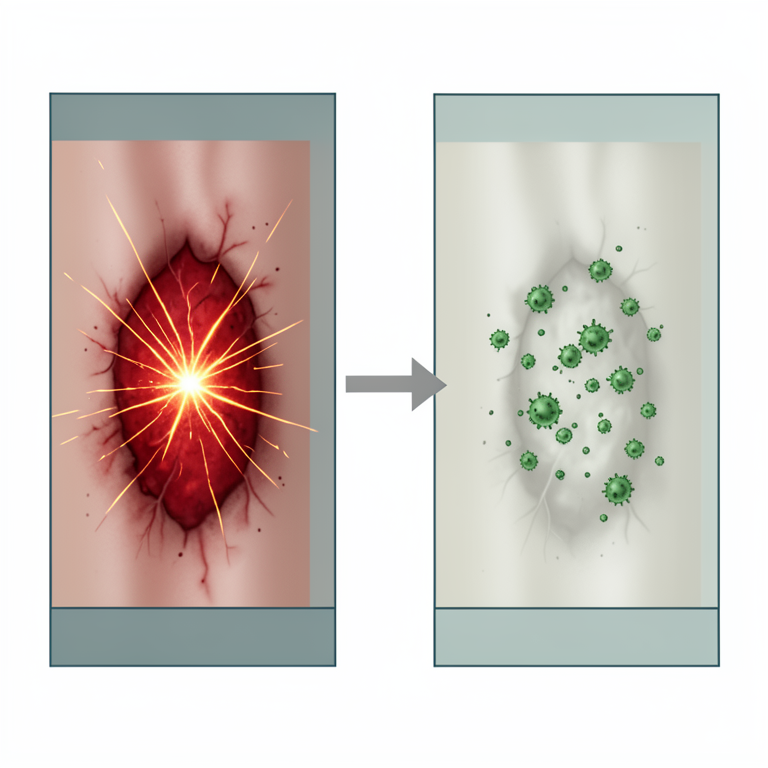
Biphasic Cytokine Response
AI-generated conceptual illustration (human skin with severe burn wounds). It explains the idea and does not show study data.
View source evidence ↗Figures & tables
From the original paper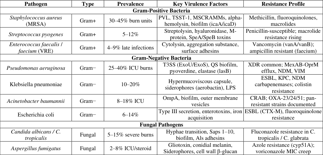 Table 1
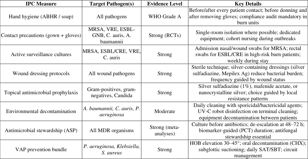 Table 2
Introduction
Burns are one of the most difficult clinical problems in current medical practice, both with regard to the degree of tissue damage and to the extensive systemic physiological upset resulting from such damage. Burns are estimated to account for 180,000 deaths globally a year, and most are in low and middle income countries where specialist advice on burn care is unavailable. Infection is the leading cause of death among burnt patients, and the leading cause of death in burn centres around the world in hospitalized cases, accounting for 50-75% of the mortality [1,2] . In addition to its mechanical barrier role, the skin is an active immune organ and tightly regulates innate and adaptive responses to microbial challenge. Additionally thermal injury destroys the barrier to colonization, washes away the resident microbiome that interferes with pathogen colonization, and reduces the ability of Langerhans cells and dermal dendritic cells to provide immune surveillance, as well as creating a warm, protein-rich, avascular eschar that serves as a growth matrix for bacterial and fungal colonization [3,4] . The post-burn immune response is paradoxical because of its destruction. The early proinflammatory phases (involving the secretion of high levels of TNF-α, IL-1β, IL-6 and IL-8) result in systemic inflammatory response syndrome (SIRS), vascular leak and injury in other organs. This is followed by a prolonged compensatory anti-inflammatory response syndrome (CARS) involving IL-10, IL-4 and TGF-β that affects neutrophils and macrophages, reduces numbers of T lymphocytes and results in acquired immunodeficiency status for weeks to months [5] .
Disruption of barrier function, severe immunodeficiency, and the intensive care environment with its central venous lines, urinary lines, mechanical ventilation and long-term antibiotic use, facilitate the acquisition of infection with multidrug resistant organisms. In the past few decades, the epidemiology of burn wound infections has changed significantly, with gram-negative organisms and MDR pathogens increasing in prevalence in modern times compared to gram-positive organisms, which were more prevalent in the early post-burn era [6,7] . It aims to condense existing research related to the microbiology, immunopathology, diagnosis, treatment, and prevention of burn wound infections by summarizing recent literature published in the last 5 years (2020-2025) that would have clinical relevance to those caring for critically burned patients [8] .
Epidemiology and Microbiology of Burn Wound Infections 2.1 Incidence and Temporal Trajectory of Infection
Burn wounds are sterile at the time of injury, but they progress in predictable sequence of colonization over the following days. Surface colonization by gram-positive organisms (mainly Staphylococcus aureus and coagulasenegative staphylococci) of the wound eschar occurs within 24-48 hours, primarily from the patient's own skin flora and healthcare workers. Gram-negative organisms, such as Pseudomonas aeruginosa, Klebsiella pneumoniae and Escherichia coli, become present on days 3-5, usually via translocation from the gastrointestinal tract of the patient or from an environment source in the burn unit. When patients survive for more than 10-14 days, fungal colonization, mostly by Candida spp, is more common, especially when broad-spectrum antibiotic therapy is used [9,10] . Histological criteria for invasive wound infection vs surface colonization are: ≥ 105 cfus/g and invasion beyond viable tissue-eschar junction. This is the point at which failure of the skin graft occurs, and local signs of wound infection start to appear (Exudates increase, the wound turns red or blue, surrounding skin becomes red and there is oedema), as well as a risk of haematogenous dissemination leading to bacteraemia and sepsis [10] .
The ESKAPE Pathogens in Burn Units
The ESKAPE pathogens (Enterococcus faecium, S. aureus, Klebsiella pneumoniae, Acinetobacter baumannii, Pseudomonas aeruginosa, Enterobacter spp.) are all associated with a high prevalence of nosocomial infections in the burn unit, and are known for their high virulence and multi-drug resistance. P. aeruginosa is the most common gram-negative pathogen in burn units around the world, with 25-40% of cultures from patients with a burn injury in an ICU setting being positive for this organism. It is a highly adaptable organism, a result of its ability to acquire resistance, its ability to form biofilms and its quorum sensing networks. [7,11] Most series report that Staphylococcus aureus/burn infection, including MRSA, accounts for 30-45% of grampositive organisms isolated from burn wound infections. The prevalence of MRSA in burn units varies from 20-60% of all S. aureus isolates, according to geographic location and burn unit control measures. MRSA is a very virulent burn pathogen due to its combination of potent virulence factors (adhesins, toxins, biofilm) and broad beta-lactam resistance. With its capacity to survive for many weeks on environmental surfaces and its high rates of carbapenem resistance, A. baumannii has been responsible for epidemic outbreaks in burn units worldwide, especially in the context of conflicts and in ICUs with poor infection control [11,12] .
Table 1; Common microbial pathogens in burn wound infections: type, prevalence, key virulence factors, and resistance profile [10,11,[13][14][15][16][17] MSCRAMMs are surface-expressed proteins that include fibronectin-binding proteins (FnBPA, FnBPB), clumping factors A and B (ClfA, ClfB), and collagen-binding protein (CNA), which all contribute to the adhesion to exposed extracellular matrix components in the burn wound bed. The ica gene locus that encodes PIA/PNAG is responsible for the formation of biofilms, 100-1000-fold more resistant to antibiotics than planktonic cells, that can be grown on wound surfaces, eschar and wound dressings by S. aureus [13, 14] .
Pathogen
A set of toxins is of special importance in burn patients. Alpha-hemolysin (Hla) is an enzyme that forms heptameric pores in the hosts cell membranes, leading to the lysis of erythrocytes and leukocytes, and to direct tissue necrosis in the wound. PVL is a bicomponent leukocidin that is capable of lysing neutrophils and macrophages that are linked to more severe and rapidly progressive cellulitis and wound infections. In addition, cytokines are set free in response to massive non-specific activation of T-cells by superantigens, such as toxic shock syndrome toxin-1 (TSST-1) and staphylococcal enterotoxin A-E, which further contributes to the cytokine storm and the already dysregulated inflammatory state of the burned patient [13,15] .
Pseudomonas aeruginosa
Three integrated systems including the type III secretion system (T3SS), quorum sensing (QS) regulatory networks, and biofilm formation are responsible for orchestration of P. aeruginosa virulence in burn wound. The T3SS injects effector proteins directly into host cell cytoplasm: ExoU, a phospholipase A2, causes rapid membrane lysis; ExoS and ExoT disrupt actin cytoskeleton and phagocytic function; ExoY raises intracellular cAMP, disrupting endothelial barrier integrity. ExoU-producing strains are associated with particularly high mortality and should prompt consideration of aggressive combined antimicrobial therapy [16,17] . The las/rhl/pqs quorum sensing circuits coordinately regulate production of elastase (LasB), alkaline protease (AprA), rhamnolipids, hydrogen cyanide, and pyocyanin-a redox-active phenazine that directly suppresses neutrophil oxidative killing. Iron acquisition through the pyoverdine siderophore, which also signals exotoxin A (ExoA) expression, provides the growth advantage in the ironlimited wound environment. Alginate overproduction creates a mucoid biofilm phenotype that is particularly resistant to both host immune clearance and antibiotic penetration, and is associated with transition from acute to chronic wound infection [18,19] .
Fungal Pathogens in Burn Wounds
Invasive fungal infections in burn patients are associated with mortality rates of 40-75% and represent an increasingly important clinical challenge as improved resuscitation and antibiotic therapy enable longer survival of severely burned patients. Candida albicans exploits the immunosuppressed burn patient through its capacity for yeast-to-hypha morphological switching, enabling active tissue penetration. Secreted aspartyl proteases (Saps 1-10) degrade structural proteins of the wound matrix; phospholipases disrupt cell membranes; and biofilm formation on central venous catheters and wound surfaces confers antifungal resistance [20,21] . Aspergillus fumigatus wound infection, while less common than Candida, carries extreme mortality and occurs predominantly in patients receiving systemic corticosteroids, those with large total body surface area (TBSA) burns, and in the context of COVID-19-associated immunosuppression. Gliotoxin-mediated suppression of neutrophil oxidative burst and induction of macrophage apoptosis, combined with the melanin-coated conidial surface that resists phagocytic killing, enables A. fumigatus to establish invasive wound infection despite apparently intact neutrophil counts [20] .
Cytokine Response in Burned Patients 4.1 The Biphasic Inflammatory Response
The systemic inflammatory response to burn injury follows a characteristic biphasic pattern that profoundly shapes both the host's susceptibility to infection and the severity of infectious complications when they occur. Phase 1-the pro-inflammatory surge-begins immediately post-burn and is driven by release of damage-associated molecular patterns (DAMPs) including heat shock proteins, HMGB1, and DNA fragments from thermally destroyed cells. These DAMPs activate pattern recognition receptors on resident macrophages, triggering NF-κB-driven production of TNFα, IL-1β, IL-6, and IL-8 within hours of injury [5,22] . Burn size, expressed as percentage of total body surface area (%TBSA), is the primary determinant of cytokine response magnitude. Burns involving >20% TBSA reliably produce systemic pro-inflammatory cytokine elevations; burns above 40% TBSA trigger a cytokine storm of extraordinary magnitude, with IL-6 levels 100-1000-fold above normal, that drives the systemic inflammatory response syndrome, capillary leak, acute lung injury, and remote organ dysfunction. The liver responds to this cytokine signal with a massive acute-phase response-producing CRP, fibrinogen, complement components, and hepcidin-while simultaneously reducing production of protective proteins including albumin and transferrin [23,24] .
Individual Cytokines and Their Clinical Significance
IL-6 has emerged as the single most valuable cytokine biomarker in burn care. Its peak level within the first 24-48 hours post-burn correlates with burn size, predicts the subsequent development of sepsis, and identifies patients at highest risk of multi-organ failure and death. A metaanalysis of cytokine profiling in burn patients confirmed that IL-6 levels at hospital admission provide prognostic information superior to traditional scoring systems alone. Procalcitonin (PCT), while not a cytokine per se, is an acute-phase reactant whose burn-specific kineticselevation parallels inflammatory burden but more specifically indicates bacterial infection rather than burninduced sterile inflammation-make it a valuable adjunctive sepsis biomarker when combined with clinical assessment and microbiological data [24,25] . Compensatory upregulation of IL-10, IL-4, IL-13 and TGFβ leads to a transition to the anti-inflammatory stage, which begins 3-7 days after burns. This stage of immunity reduces inflammation to prevent it from running out of control, but it also makes the person most vulnerable to secondary infections. IL-10-produced by monocytes, regulatory T cells, and M2-polarised macrophages-suppresses TNF-α, IL-1β, and IL-12 production, impairs neutrophil oxidative burst, and reduces CD4+ and CD8+ T-cell function. The extent and duration of this IL-10-dominated immunosuppressive phase predict the risk of nosocomial infection, with studies demonstrating that persistently elevated IL-10 at day 7 post-burn independently predicts subsequent bacteraemia and pneumonia [26] .
Cytokines as Therapeutic Targets
Recognition of the cytokine-mediated immunosuppressive phase as a modifiable target has driven investigation of immunostimulatory interventions in burn care. Granulocyte colony-stimulating factor (G-CSF) administration, by restoring neutrophil production and enhancing oxidative burst, has demonstrated benefit in reducing infectious complications in selected trials. Interferon-gamma (IFN-γ) supplementation, aimed at reversing M2 macrophage polarization and restoring TH1 immune function, has shown promise in small studies. Conversely, anti-TNF-α strategies, while effective in reducing SIRS manifestations, may paradoxically increase susceptibility to secondary infection and have not achieved clinical adoption in burn care [27,28] .
Clinical Outcomes: Sepsis, Bacteraemia, and Organ Failure 5.1 Defining Sepsis in Burns
The diagnosis of sepsis in burned patients is confounded by the fact that SIRS criteria-fever, tachycardia, tachypnoea, and leukocytosis-are virtually universal in the post-burn period as a consequence of the injury itself, irrespective of infection. The American Burn Association (ABA) has developed burn-specific sepsis criteria that incorporate TBSA-adjusted temperature thresholds, heart rate and respiratory rate parameters, and require documentation of a probable or confirmed infectious source, providing more clinically meaningful diagnostic criteria than generic Sepsis-3 definitions in this population [29,30] . Bacteraemia-the haematogenous dissemination of wound pathogens into the bloodstream-occurs in 5-28% of hospitalized burn patients and is associated with a 2-5-fold increase in mortality compared to burn patients without bacteraemia. S. aureus bacteraemia from burn wounds may seed distant sites including heart valves (infective endocarditis), joints (septic arthritis), and vertebrae (osteomyelitis), creating a catalogue of metastatic infectious complications that further complicate clinical management. P. aeruginosa bacteraemia in burn patients is associated with particularly high mortality (30-70% in reported series), reflecting both the severity of underlying disease in affected patients and the inherent virulence of systemic pseudomonal infection [30,31] .
Respiratory Infections
About 30% of burns that require hospitalization are complicated by inhalation injury, which significantly increases the severity of the burn. In addition to the primary thermal and chemical damage to the tracheobronchial tree, inhalation injury can lead to a marked impairment of mucociliary clearance, disruption of airway epithelial barrier, and require mechanical ventilation in most patients with the injury. Ventilator-associated pneumonia (VAP) is common in burn patients who are mechanically ventilated, happening in 20-50% of them, a much higher prevalence than is seen in other patients in an intensive care unit (ICU), and the most common pathogens identified are P. aeruginosa and S. aureus [32,33] . VAP is linked to an increment of 7-14 days of ICU stay, an increment of ventilator days, and 15-30% attributable mortality in burn patients. Early onset VAPs (those that occur within 5 days of intubation) are generally dominated by community-acquired organisms such as S. aureus and S. pneumoniae, whilst late onset VAPs are mainly comprised of MDR gram-negative organisms such as P. aeruginosa, A. baumannii and ESBL-producing Enterobacterales, which are associated with HAP under antibiotic selective pressure [33] .
Multi-Organ Dysfunction and Mortality
There is a bidirectional association between burn-induced dysfunction of various organs and infection, with severe infection leading to cytokine-mediated organ dysfunction and organ dysfunction making it harder to maintain immune function and deliver therapeutic agents, thereby allowing the infectious process to escalate. In severely burned patients, there is a risk of renal failure, which affects 15-40%, and may require dose adjustment of antibiotics, especially aminoglycosides and vancomycin. Loss of function of the liver leads to decreased production of complement and opsonin. Failure of the gastrointestinal mucosal barrier allows enteric flora, such as Candida species, to enter the portal and systemic circulation [34,35] . Mortality prediction in burned patients has evolved from simple burn size and age calculations (Baux score) to integrated models incorporating infection burden, organ failure scores, inhalation injury, and inflammatory biomarkers. The Revised Baux Score, incorporating inhalation injury, and the revised Belgian Outcome in Burn Injury (BOBI) model remain widely used. Infectionparticularly gram-negative bacteraemia, P. aeruginosa wound infection, and invasive fungal disease-consistently emerges as the most powerful modifiable predictor of mortality in contemporary burn outcome analyses, underscoring the centrality of infection prevention and management in burn care [36,37] .
Diagnosis of Burn Wound Infections 6.1 Clinical and Microbiological Criteria
The clinical diagnosis of burn wound infection requires the integration of local wound findings, systemic inflammatory markers, and microbiological data. Local signs suggestive of infection include conversion of partial-thickness to fullthickness injury (indicative of microbial proteolytic enzyme activity), change in wound colour (green discoloration suggesting P. aeruginosa; black discoloration suggesting fungal invasion), increased exudate, malodour, surrounding cellulitis, and oedema. However, these signs may be subtle or absent in immunocompromised patients, and systemic signs of infection-fever, tachycardia, rising inflammatory markers-must be actively sought [38] . Quantitative wound biopsy culture-the diagnostic gold standard-involves tissue sampling of 0.5-1.0 g from the wound bed and culture with colony count determination. The threshold of 10⁵ CFU/g has been shown to be reasonably sensitive and specific for predicting graft failure and invasive infection. Surface swab culture is clinically more feasible and useful for detecting colonization rather than invasion, but should be interpreted with care, but can be used to monitor resistance trends and patterns in the unit and for empirical antibiotic selection [39,40] .
Molecular and Advanced Diagnostics
The rapid identification of bacterial and fungal pathogens and key resistance genes of these pathogens in 2-6 hours with multiplex PCR panels directly on wound swabs or tissue biopsies significantly improves the start of targeted therapy. MALDI-TOF mass spectrometry provides definitive organism identification from positive culture plates within minutes, replacing biochemical identification methods. Next-generation sequencing (NGS)-based metagenomics, while not yet standard of care in most centres, can identify polymicrobial infections with unusual organisms that escape conventional culture detection, and is increasingly applied in outbreak investigations and cases of refractory infection [33] . Inflammatory biomarkers guide antibiotic initiation and duration. Serum procalcitonin (PCT) has been validated in burn populations as a biomarker for discriminating infection from sterile SIRS, with PCT-guided antibiotic de-escalation protocols demonstrating reductions in antibiotic exposure without worsening clinical outcomes. C-reactive protein and ferritin kinetics, serial lactate measurement, and presepsin are additional biomarkers with supporting evidence in burn sepsis. For fungal infections, (1→3)-β-D-glucan and, in selected patients, serum galactomannan provide non-culture diagnostic support [32,34] .
Treatment of Burn Wound Infections 7.1 Topical Antimicrobial Therapy
Topical antimicrobial agents are the cornerstone of local burn wound management, reducing bacterial and fungal burden, preventing wound desiccation, and facilitating wound bed preparation for grafting. Silver sulfadiazine (SSD, 1%), the historical gold standard, provides broadspectrum activity against gram-positive and gram-negative organisms including P. aeruginosa and Candida species, though resistance to silver-based compounds is increasingly documented, particularly in P. aeruginosa and A. baumannii clinical isolates. Mafenide acetate has superior eschar penetration and retains activity in avascular devitalized tissue, making it particularly useful for deep full-thickness burns and electrical injuries, though its carbonic anhydrase inhibition may cause metabolic acidosis with prolonged use [4] . The nanocrystalline silver dressings (Acticoat, Mepilex Ag) are being used extensively because of the continued release of silver ions, less frequent dressing changes and enhanced patient comfort over topical creams. Honey-impregnated dressings (Medihoney) have antimicrobial properties due to the generation of hydrogen peroxide, the osmotic effect of the honey and the low pH, which has been shown to have good antimicrobial activity in partial thickness burn care. NPWT has been used after surgical debridement and grafting to lower bacterial levels, enhance granulation tissue formation and provide mechanical support for grafts, and there is an increasing body of evidence to support its use in the management of complex burn wounds [4] .
Systemic Antimicrobial Therapy
Burn patients have special challenges with systemic antibiotic therapy. Major burns cause a hypermetabolic state, which enhances cardiac output, glomerular filtration rate and hepatic clearance, resulting in a large volume of distribution and clearance of hydrophilic antibiotics, such as beta-lactams, vancomycin and aminoglycosides. In major burn patients, standard dose and timing schedules based on non-burn populations consistently result in subtherapeutic levels of serum and tissue drug levels, relevant to efficacy and a risk of emergence of resistance. Extended infusion (e.g., time above the MIC) strategies are indicated for beta-lactams and required for vancomycin, and therapeutic drug monitoring (TDM) is required for vancomycin and strongly recommended for aminoglycosides [35] . Empirical antibiotic choice should be based on the local resistance profile of the burn unit, not an antibiogram from the hospital in general. For suspected MRSA wound infections, vancomycin with AUC/MIC-guided monitoring remains first-line; daptomycin and linezolid are alternatives. For MDR gram-negative infections, novel combinations including ceftolozane-tazobactam (XDR P. aeruginosa), ceftazidime-avibactam (KPC/OXA-48-producing organisms), and aztreonam-avibactam (NDM-producing strains) have transformed therapeutic options. Duration of antibiotic therapy should be guided by clinical response and PCT kinetics, with de-escalation to narrow-spectrum agents as soon as culture and sensitivity data permit [36,37] .
Infection Prevention and Control in Burn Units 8.1 Standard and Transmission-Based Precautions
The prevention of healthcare-associated infections in burn units requires meticulous application of both standard precautions-applicable to all patient care interactions-and transmission-based precautions targeted at specific MDR organisms. Hand hygiene with alcohol-based hand rub or soap and water before and after every patient contact remains the single most effective infection control measure, yet compliance in burn units is consistently below the recommended 80% threshold, partly due to the frequent wound care interactions and gloved procedures required in burn nursing [38] . Active surveillance cultures-nasopharyngeal, wound, and perianal swabs-enable early identification of MRSA, ESBL/CRE, C. auris, and A. baumannii colonization before clinical infection becomes manifest, enabling preemptive isolation and targeted decolonization. Outbreak management in burn units requires particularly aggressive responses including environmental sampling, whole-genome sequencing of outbreak strains to confirm transmission chains, enhanced cleaning protocols, and in some cases temporary unit closure, as illustrated by documented A. baumannii and C. auris outbreaks in burn centres [39] .
Antimicrobial Stewardship in Burn Care
Antimicrobial stewardship programmes (ASPs) in burn units must address the unique clinical environment of these patients: the extreme physiological alterations that complicate pharmacokinetics; the high rates of colonization with MDR organisms; the prolonged hospitalizations that create sustained antibiotic exposure; and the legitimate clinical uncertainty about infection diagnosis given the confounding effects of burn-induced SIRS on traditional infection markers [40] . Prospective audit and feedback by an ASP team with burnspecific expertise is the most evidence-based stewardship intervention. PCT-guided protocols for antibiotic deescalation and duration determination have been validated in burns and reduce antibiotic exposure by 20-30% without worsening outcomes. Anti-fungal stewardshipdistinguishing Candida colonization from invasive infection and guiding appropriate antifungal initiation, de-escalation, and duration-is equally critical in this high-risk population [40] .
Table 2: Evidence-based infection prevention and control measures in burn units: targeted pathogens, evidence level, and key implementation details. [39,40] IPC [7] . The introduction of whole genome sequencing (WGS) of MDR outbreak strains has revolutionized burn unit epidemiology, with the ability to definitively resolve the transmission chain, identify specific environmental reservoirs (ventilator circuits, sink drains, wound dressing trolleys), and inform targeted burn unit decontamination interventions. Plasmid profiling alongside WGS of P. aeruginosa and K. pneumoniae isolates from burn patients has demonstrated the horizontal transfer of carbapenemase genes between species within individual burn units, thus supporting the need for the prevention of not only patientto-patient transmission, but transmission of resistance genes between species in the burn environment [29] .
Wound Care Innovations
In addition to the more conventional topical antimicrobials, there have been a number of novel wound care strategies that are promising in minimizing wound infection rates and improving wound healing in burn patients. Bacteriophage therapy, the use of the lytic phages that target specific bacterial species, has shown promise as a therapy of last resort for refractory XDR P. aeruginosa and S. aureus burn wound infections and could be an effective treatment strategy in the future, while preserving commensal microbiota, which are not targeted [41] . New bioengineered skin substitutes containing antimicrobial peptides, antibiotic loaded nanoparticles, and growth factors are making progress in the clinical trials. Anti-virulence approaches that inhibit virulence factor expression and/or the ability of P. aeruginosa to form biofilms (such as inhibitors of las and rhl) do not necessarily exert direct killing pressure and may be more effective at reducing virulence than conventional antibiotics [42] .
Conclusions
Thus, wound infections with MDR organisms are the major cause of morbidity and mortality in severely burned patients and result from a combination of disrupted skin barrier function, wide-ranging and sustained immunosuppression, and the high risk of acquiring MDR organisms within the intensive care unit (ICU) setting. The microbiology of burn infections has significantly changed over the years, with MDR gram-negative infections, especially those caused by P. aeruginosa, K. pneumoniae and A. baumannii, becoming more prevalent than MRSA infections and the increasing incidence of fungal infection [1,6] . The cytokine response to burn injury is a double edged sword: the acute phase of burn injury is pro-inflammatory, consisting of TNF-α, IL-1β, IL-6 and IL-8 and responsible for the SIRS and organ dysfunction characteristic of major burns; the anti-inflammatory phase is anti-inflammatory, dominated by IL-10, and produces the immunological window for secondary infections to establish and flourish. Cytokine profiling including serial IL-6 and PCT measurement has proved to be a useful prognostic and therapeutic guidance tool which should be incorporated into burn unit clinical protocols [22,25] . A multi-pronged approach with careful microbiological diagnosis prior to antibiotic initiation, pharmacokinetically informed antibiotic dosing with TDM, evidence-based topical wound care management, early skin grafting, burn-adapted antimicrobial stewardship, and careful infection prevention and control measures is required to optimize clinical outcomes. There is a need for investments in new development strategies such as Phage therapy, Antivirulence agents and Bioengineered dressings, as well as stewardship efforts to preserve current antimicrobials, for better results in this most vulnerable population [35,40,41] .
References
41 in the original paper- Church D, Elsayed S, Reid O, Winston B, Lindsay R. Burn wound infections. Clin Microbiol Rev. 2006;19(2):403-434.
- Knuth CM, Auger C, Jeschke MG. Burn-induced hypermetabolism and skeletal muscle dysfunction. Am J Physiol Cell Physiol. 2021;321(7):C58-C71.
- Norbury W, Herndon DN, Tanksley J, Jeschke MG, Finnerty CC, Surgical Infection Society. Infection in burns. Surg Infect (Larchmt). 2016;17(2):250-255.
- Rafla K, Tredget EE. Infection control in the burn unit. Burns. 2011;37(1):5-15.
- Grinis D, Thomas M, Aprigliano C, Maskey AR. Refractory keloids and hypertrophic scars: immune dysregulation and neuroimmune mechanisms underlying treatment failure. Cells. 2026;15(9):782.
- Elsheikh R, Makram AM. Multidrug-resistant organisms: the silent plight of burn patients. J Burn Care Res. 2024;45(4):877-886.
- De Oliveira DM, Forde BM, Kidd TJ, et al. Antimicrobial resistance in ESKAPE pathogens. Clin Microbiol Rev. 2020;33(3):e00181-19.
- Goh M, Du M, Peng WR, Saw PE, Chen Z. Advancing burn wound treatment: exploring hydrogel as a transdermal drug delivery system. Drug Deliv. 2024;31(1):2300945.
- Ali MDY, Abdulrahman Z. Molecular identification, susceptibility pattern, and detection of some virulence genes in Pseudomonas aeruginosa isolated from burn patients. Plant Arch. 2020;20(1):2573-2580.
- Hajská M, Kurin E, Bittner Fialová S, Vidiščák M, Panyko A. Antimicrobial efficacy of impregnated human acellular dermal substitutes in burn wound models. Antibiotics (Basel). 2025;14(7):707.
- Rashid SA, Sorchee SM, Yonus MD, Bahjat OF. Identification biofilm producers Staphylococcus aureus isolates and detect their biofilm genes from gingivitis cases. 2022.
- Younus MD, Bahjat OF, Rashid SA. Antibiotic susceptibility pattern, molecular characterization of virulence genes among Pseudomonas aeruginosa isolated from burn patients. Cihan Univ-Erbil. 2021;5(1):36-41.
- Foster TJ. Surface proteins of Staphylococcus aureus. Microbiol Spectr. 2019;7(4):GPP3-0046-2018.
- Muteeb G, Rehman MT, Shahwan M, Aatif M. Origin of antibiotics and antibiotic resistance, and their impacts on drug development: a narrative review. Pharmaceuticals (Basel). 2023;16(11):1615.
- Touaitia R, Mairi A, Ibrahim NA, Basher NS, Idres T, Touati A. Staphylococcus aureus: a review of the pathogenesis and virulence mechanisms. Antibiotics (Basel). 2025;14(5):470.
- Shehade SM, Tawfeeq SN, Jindal HM, Younus MD, Ibrahim NA. Awareness and commitment to infection control principles among students of the College of Dentistry at Tikrit University, Iraq. Cihan Univ-Erbil Sci J. 2025;9(2):125-128.
- Younus MD, Qasim YA, Muhammad AQT, Mansur MA, Khalaf MH. Identification and antibiotic sensitivity of Staphylococcus aureus isolated from burned-wounded patients. Cent Asian J Med Nat Sci. 2025;6(4):1707-1715.
- Shehade SM, Juma AS, Ubeid MH, Younus MD. Perceptions and mindset toward infection control and prevention among medical microbiology students. Cihan Univ-Erbil Sci J. 2025;9(1):24-28.
- Mayer FL, Wilson D, Hube B. Candida albicans pathogenicity mechanisms. Virulence. 2013;4(2):119128.
- Latgé JP, Chamilos G. Aspergillus fumigatus and aspergillosis in 2019. Clin Microbiol Rev. 2020;33(1):e00140-18.
- Korkmaz HI, et al. The complexity of the post-burn immune response: an overview of the associated local and systemic complications. Cells. 2023;12(3):345.
- Juma AS, et al. Cytokine profile as a prognostic parameter in burn patients. 2022.
- Palmieri TL, Heard J. Biomarkers of sepsis in burn injury: an update. Burns Trauma. 2025;13:tkae080.
- Roth S, et al. Post-injury immunosuppression and secondary infections are caused by an AIM2 inflammasome-driven signaling cascade. Immunity. 2021;54(4):648-659.e8.
- Hotchkiss RS, Monneret G, Payen D. Immunosuppression in sepsis: a novel understanding of the disorder and a new therapeutic approach. Lancet Infect Dis. 2013;13(3):260-268.
- La Via L, et al. The global burden of sepsis and septic shock. Epidemiologia. 2024;5(3):456-478.
- Van der Poll T, Van De Veerdonk FL, Scicluna BP, Netea MG. The immunopathology of sepsis and potential therapeutic targets. Nat Rev Immunol. 2017;17(7):407-420.
- Younus MD, Abdulrahman ZFA, Bahjat OF, Ali NL, Rashid SA, Alkhouri AS. Plasmid profiling, genetic site determination of antibiotic resistance gene of Pseudomonas aeruginosa isolated from burned patients. 2022.
- Normann TC, Bellaver P, Limeira BI, Dorneles GB, Oliveira MS, Rech TH. Association of inhalation injury with ventilator-associated pneumonia and clinical outcomes in critically ill patients with burns: a retrospective study. Can J Respir Ther. 2026;62:64.
- Branski LK, Al-Mousawi A, Rivero H, Jeschke MG, Sanford AP, Herndon DN. Emerging infections in burns. Surg Infect (Larchmt). 2009;10(5):389-397.
- Donnelly JP, et al. Revision and update of the consensus definitions of invasive fungal disease from the European Organization for Research and Treatment of Cancer and the Mycoses Study Group Education and Research Consortium. Clin Infect Dis. 2020;71(6):1367-1376.
- Ramanan P, Bryson AL, Binnicker MJ, Pritt BS, Patel R. Syndromic panel-based testing in clinical International Journal of Biology Sciences https://www.biologyjournal.net microbiology. Clin Microbiol Rev. 2018;31(1):e0002417.
- Muhammad AQT, Mansur MA, Qasim YA, Younus MD. Identification of Candida species in patients with type 2 diabetes mellitus in Kirkuk City, Iraq. Cent Asian J Med Nat Sci. 2025;6(4):1750-1762.
- Aitken SL, Clancy CJ. IDSA guidance on the treatment of antimicrobial resistant Gram-negative infections. Contagion. 2020;5(6).
- Bassetti M, Vena A, Croxatto A, Righi E, Guery B. How to manage Pseudomonas aeruginosa infections. Drugs Context. 2018;7:212527.
- Lass-Flörl C, Kanj SS, Govender NP, Thompson GR III, Ostrosky-Zeichner L, Govrins MA. Invasive candidiasis. Nat Rev Dis Primers. 2024;10(1):20.
- World Health Organization. Global report on infection prevention and control: executive summary. Geneva: World Health Organization; 2022.
- Barlam TF, et al. Implementing an antibiotic stewardship program: guidelines by the Infectious Diseases Society of America and the Society for Healthcare Epidemiology of America. Clin Infect Dis. 2016;62(10):e51-e77.
- Ali NL, et al. Identification of pharyngitis bacteria in patients with sore throat features in Rizgary Teaching Hospital and overuse of antibiotics. 2022.
- Mamand BA, Ubeid MH, Al-Bader SM, Zefenkey Z, Younus MD. Proteolytic activity of fungi isolated from nuts: a focus on health risks of Aspergillus niger, Penicillium sp., and Alternaria alternata. Microb Biosyst. 2026;11(2):1-7.
- Gordillo Altamirano FL, Barr JJ. Phage therapy in the postantibiotic era. Clin Microbiol Rev. 2019;32(2):e00066-18.
Cited by 1
From OpenAlex, OpenCitations and reference lists read by Scinovex
- The relationship between infectious diseases and COVID-19: Co-infections, immune dysregulation, antimicrobial resistance, and clinical outcomes
Mohammed Hakeem Khalaf, Maad Abdullah Mansur, Mustafa D. Younus · International Journal of Biology Sciences · 2026
SARS-CoV-2 has brought about an unprecedented change in the global landscape of infectious diseases. In addition to its own respiratory pathology, COVID-19 patients are vulnerable to a wide variety of secondary bacterial, fungal, and viral co-infections, with multiple mechanisms of convergence: massive lymphopenia; defect in macrophage and neutrophil function; disruption of the mucosal barrier; corticosteroid therapy; and use of invasive supportive therapies. This review comprehensively examines the bidirectional relationship between COVID-19 and infectious diseases, analyzing the epidemiology