
Research article · 2026 · Full text
International Journal of Advanced Chemistry ResearchBio-Friendly Synthesis of gold nanoparticles for high efficient commercial use
Volume 8(5) · Pages 99–104 · DOI: 10.33545/26646781.2026.v8.i5b.678 ↗
Research areasNanomaterials and Printing TechnologiesGold and Silver Nanoparticles Synthesis and ApplicationsNanoparticles: synthesis and applications
Abstract
Author-writtenThe growing demand for flexible and printable electronic devices has accelerated the development of conductive inks with high electrical efficiency, low production cost, and environmental compatibility. In the present study, gold nanoparticles (AuNPs) were synthesized through an ecofriendly green synthesis approach using glucose as a natural reducing agent and polyvinyl pyrrolidone (PVP) as a stabilizing material. The synthesized nanoparticles were characterized using ultraviolet-visible spectroscopy (UV-Vis), X-ray diffraction (XRD), transmission electron microscopy (TEM), and scanning electron microscopy (SEM) to evaluate their optical properties, crystal structure, morphology, and particle size. The UV-Vis analysis confirmed the formation of gold nanoparticles through the appearance of a surface plasmon resonance peak at approximately 532 nm, while XRD analysis revealed a face-centered cubic (FCC) crystalline structure. TEM images demonstrated that the synthesized nanoparticles were predominantly spherical with an average particle size of approximately 19.42 nm. Subsequently, conductive ink was formulated by mixing the synthesized gold nanoparticles with nitrocellulose in a 1:1 ratio using acetone as a dispersing solvent. The prepared nanocomposite film exhibited remarkable electrical conductivity, with an electrical resistance of 0.00045 Ω compared with 22000 Ω for pure nitrocellulose film without nanoparticles. The findings of this study demonstrate the successful development of a simple, environmentally friendly, and cost-effective method for synthesizing conductive gold nanoparticle-based ink with promising potential for flexible electronic and printed electronic applications.
Summary
Source-checkedPlain-language reading of the full paper, prepared with AI and checked against the text. Each point links to the authors' own words.
Why it matters
- There is an increasing need for conductive inks that are efficient, inexpensive, and environmentally friendly to support the growth of flexible and printable electronics. View evidence ↗
- Gold nanoparticles are ideal for electronic and biomedical uses because they are chemically stable, biocompatible, resistant to oxidation, and have tunable optical properties. View evidence ↗
- Standard methods for creating gold nanoparticles are often limited by the use of expensive equipment and hazardous chemicals that produce harmful waste. View evidence ↗
The question
- The study aimed to create an eco-friendly method for synthesizing gold nanoparticles using glucose and PVP, and then use these to develop a conductive ink with nitrocellulose. View evidence ↗
What they did
- Gold nanoparticles were synthesized using a 0.001 molar gold salt solution, 0.7 g of glucose in 250 ml water, and 0.3 g of PVP in 250 ml water heated to 80°C. View evidence ↗
- The synthesis involved adding glucose and PVP dropwise to an alkalized gold salt solution (0.1 M NaOH) while stirring at 1500 rpm at room temperature. View evidence ↗
- Conductive ink was formulated by mixing 0.5 g of the synthesized gold nanoparticles with 0.5 g of nitrocellulose using acetone as the solvent. View evidence ↗
- The researchers used UV-Vis, XRD, TEM, and SEM to analyze the optical properties, crystal structure, and morphology of the nanoparticles. View evidence ↗
What they found
- UV-Vis analysis confirmed the formation of gold nanoparticles by the appearance of a surface plasmon resonance peak at 532 nm. View evidence ↗
- The reduction of gold ions into nanoparticles was found to be complete approximately 90 minutes after the reaction began. View evidence ↗
- XRD analysis showed peaks at 37.91, 44.40, 61.53, and 78.83 degrees, confirming that the nanoparticles have a face-centered cubic (FCC) crystal structure. View evidence ↗
- TEM imaging revealed that the synthesized gold nanoparticles were spherical with an average size of 19.42 nm. View evidence ↗
- The nanocomposite film showed high electrical conductivity with a resistance of 0.00045 Ω, significantly lower than the 22000 Ω resistance of pure nitrocellulose film. View evidence ↗
What it means
- The authors conclude that they developed a simple, cost-effective, and green method for creating conductive gold nanoparticle ink suitable for printed and flexible electronics. View evidence ↗
- The authors determined that while glucose acts as a reducing agent, PVP is necessary as a coating material to prevent the gold nanoparticles from aggregating. View evidence ↗
Visual summary
1 visualsIllustrations explain the paper; they do not show study data.
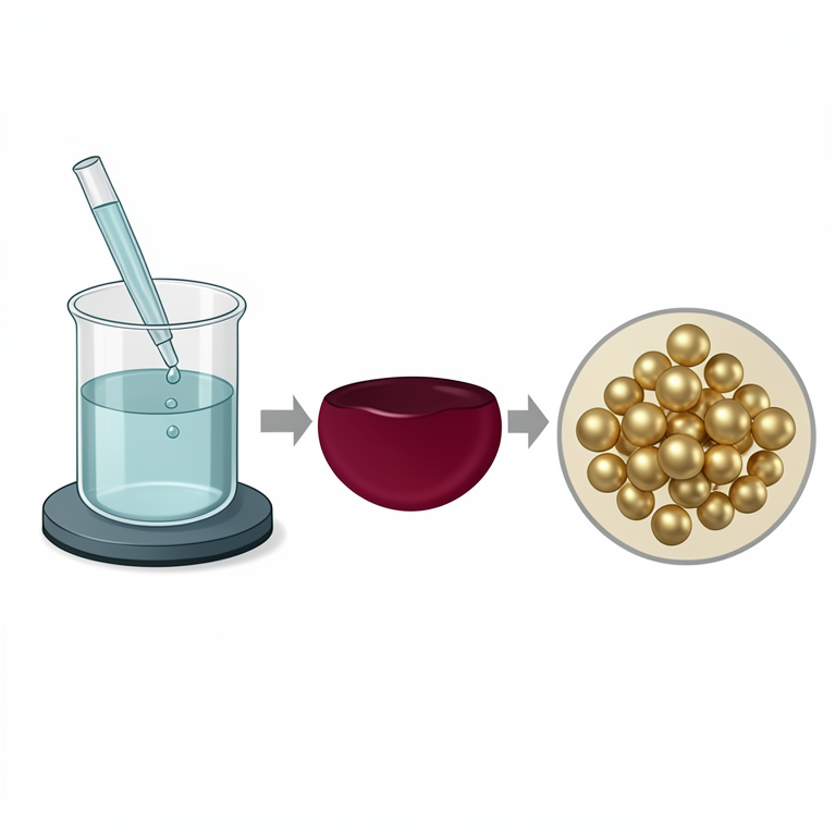
Green Synthesis Process Flow
AI-generated conceptual illustration (Gold nanoparticles synthesized in a laboratory beaker). It explains the idea and does not show study data.
View source evidence ↗Figures & tables
From the original paper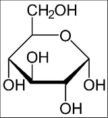 Figure 1
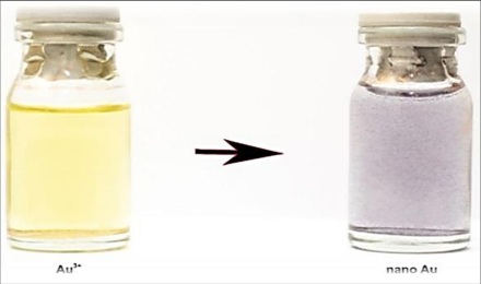 Figure 2
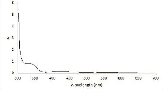 Figure 3
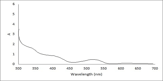 Figure 4
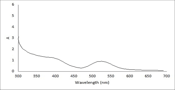 Figure 5
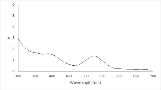 Figure 6
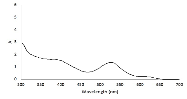 Figure 7
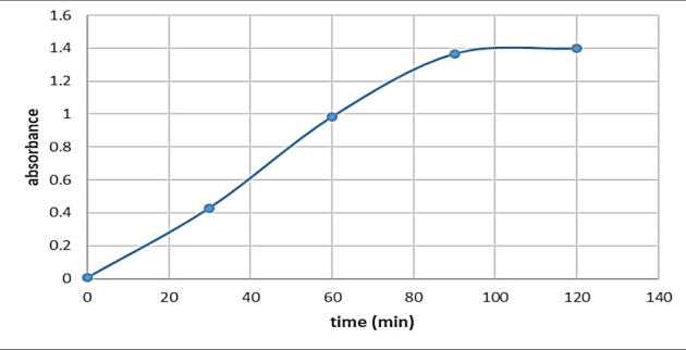 Figure 8
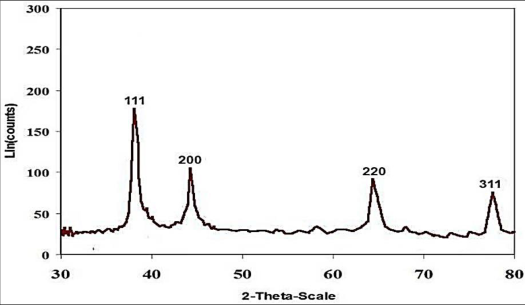 Figure 9
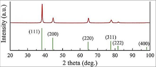 Figure 10
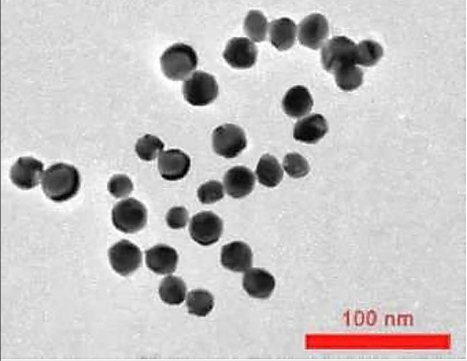 Figure 11
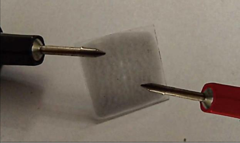 Figure 12
 Figure 13
Introduction
Recent advances in flexible and printed electronics have created a substantial demand for conductive materials that combine high electrical performance with low manufacturing cost, mechanical flexibility, and environmental safety [1] . Conventional electronic devices fabricated from rigid materials such as silicon and glass are increasingly unable to satisfy the requirements of next-generation wearable and portable technologies [2] . Consequently, conductive inks have emerged as a key enabling material for the fabrication of lightweight, flexible, and low-cost electronic devices through various printing techniques including inkjet printing, screen printing, and flexography [4,3] . Conductive inks generally consist of conductive particles, polymeric binders, solvents, and auxiliary additives that collectively determine the rheological, electrical, and mechanical properties of the final printed film [6,5] . Among the various conductive materials used in ink formulation, metallic nanoparticles have attracted significant attention due to their superior electrical conductivity and excellent charge transport characteristics [6] . In particular, gold nanoparticles possess unique physicochemical properties including high chemical stability, tunable optical behavior, excellent biocompatibility, and resistance to oxidation, making them highly suitable for advanced electronic and biomedical applications [7] . Despite the outstanding properties of gold nanoparticles, conventional chemical and physical synthesis methods often involve hazardous reagents, expensive equipment, and environmentally harmful by-products [8] . These limitations have encouraged researchers to develop sustainable and environmentally benign synthesis approaches based on green chemistry principles [9] . Green synthesis methods utilize natural reducing and stabilizing agents derived from biological sources such as sugars, plant extracts, proteins, and microorganisms to produce nanoparticles under mild reaction conditions with minimal environmental impact [10] . Glucose is considered one of the most promising natural reducing agents for nanoparticle synthesis because of its biodegradability, low toxicity, availability, and reducing capability [13,12,11] . Furthermore, the incorporation of stabilizing agents such as polyvinyl pyrrolidone (PVP) can effectively prevent nanoparticle aggregation and improve particle stability and dispersion [14] . The resulting gold nanoparticles can subsequently be incorporated into polymeric matrices to formulate conductive nanocomposite inks with enhanced electrical properties suitable for printed electronic systems [16,15] . Therefore, the present study aimed to develop an environmentally friendly approach for the synthesis of gold nanoparticles using glucose as a reducing agent and PVP as a stabilizer, followed by the preparation of conductive ink based on nitrocellulose-gold nanoparticle nanocomposites. The synthesized nanoparticles and fabricated conductive films were comprehensively characterized to evaluate their structural, morphological, and electrical properties for potential application in flexible and printed electronics.
Materials and Methods
Materials and Methods
Materials
All the chemicals required for this study were of synthetic grade. The necessary ingredients were auric acid tetrachloride trihyrate (with the formula AuCl₄H. 3H₂O and molecular mass of 393.83 g/mol, Merck Germany-4-240-948), acetone (Majlili-Iran), methanol (Merck Germany-106009), glucose (+) German Merck-108337) and ethylene glycol (German Merck-109621).
Equipment
To synthesize and characterize gold nanoparticles and conductive ink, spectroscopic and imaging equipment and devices as well as laboratory equipment were needed, which are: 1. Ultraviolet-visible spectrometer 2. X-ray diffraction spectrometer 3. Fourier transform infrared spectrometer 4. Transmission electron microscope 5. Scanning electron microscope 6. Magnetic stirrer 7. Analytical scale with an accuracy of 0.0001 grams 8. Digital Ohm Meter
Green synthesis of gold nanoparticles
A reducing agent and a stabilizing agent are generally needed for the synthesis of metal nanoparticles. In the basic methods of the green chemistry approach, it is favored to use environmentally friendly compounds as much as possible for this purpose in order to minimize the environmental harm of using chemicals [17] . In this study, glucose (Figure 3-1), which is a reducing monosaccharide [18] , was used as a reducing and stabilizing agent at the same time for the green synthesis of gold nanoparticles. In order to synthesize gold nanoparticles, a 0.001 molar solution of gold salt (auric acid tetrachloride trihydrate) was prepared by dissolving 0.04 gold salt in 100 ml of water. Then, by dissolving 0.7 g of glucose in 250 ml and 0.3 g of polyvinyl pyrrolidone (PVP) as a stabilizer in 250 ml of water and heating it at 80 degrees Celsius, a 1 mM glucose solution was prepared. Glucose and PVP solutions were added dropwise to the gold salt solution which was alkalized by 0.1 M sodium hydroxide solution and was stirred at room temperature at 1500 rpm while adding the solution. After the formation of gold nanoparticles, the formed particles were precipitated by a centrifuge at 13000 rpm and the upper solution was separated. In order to remove unreacted reagents, the sediments were washed with deionized water. For this purpose, after the redistribution of the deposited nanoparticles by ultrasonic bath, centrifuging was performed again and the upper solution was separated. This process was repeated three times and in the third repetition, the sediments were dried by a freeze dryer.
Characterization of the synthesized nanoparticles
Spectroscopy and electron microscopic imaging methods were used to carry out the characterizations. For this purpose, the absorption spectrum of the solution in the range of 300 to 700 nm was used to determine the surface plasmon resonance (SPR) by the UV-VIS spectrometer of Varian company model cary300 in specific time ranges (0 to 120 minutes and every 30 minutes). To determine the crystal structure of the nanoparticles, the obtained gold nanoparticles were dispersed in acetone by an ultrasonic bath and a layer of nanoparticles was placed on a microscope slide. The X-ray diffraction spectrum of the deposited layer was obtained by an XRD spectrometer in the range of 2θ angles between 10 and 80 degrees and using a Germany Seifert XRD device equipped with a copper lamp. The crystal structure of the nanoparticles was determined from the peaks appearing at 2θ angles and their Bragg values. To determine the morphology and size of the particles, transmission electron microscopy (TEM) imaging method was used. For this purpose, the nanoparticles were redispersed in deionized water and were placed on the carbon grid of the TEM device (Philips Company) as a drop and their image was obtained with 100,000 times magnification.
Preparation of the conductive ink
The conductive ink was made using gold nanoparticles according to the method provided by Camargo et al. [19] . According to the mentioned method, 0.5 grams of the gold nanoparticles synthesized by the green method with the help of glucose were mixed with 0.5 grams of nitrocellulose as a polymer material in a centrifuge tube. Acetone was used as a dispersing solvent and lubricant for better dispersion of nanoparticles. Then the mixture was placed in an ultrasonic bath for 20 minutes at a temperature of 25 degrees Celsius so that the gold nanoparticles are homogeneously distributed in the mixture. To investigate the appearance of electrical conductivity, 1 ml of nitrocellulose-gold nanoparticles composite was spread on a microscope glass slide and its electrical resistance was measured using a digital ohmmeter (in the range of microohms).
Results and Discussion
Synthesis of gold nanoparticles Synthesis of nanoparticles was done according to the method presented in the third chapter (3-3). In short, in this method, 0.001 molar solution of gold ions was used as a precursor of nanoparticles. 1 mM glucose solution was used to reduce gold nanoparticles and stabilize them and PVP was used to stabilize nanoparticles. The synthesis steps of gold nanoparticles were carried out at laboratory temperature (25°C) and under stirring.
The color change of the solution from yellow to purple (Figure 4-1) indicates the beginning and progress of the reaction of gold ions and their transformation into gold nanoparticles.
Fig 2:
The gold solution color change from yellow to purple after adding glucose and PVP Next, to check the progress of the reaction, the rate of absorption of the solution in the SPR region of the resulting gold nanoparticles was studied using UV-Vis spectroscopy in the range of 300 to 700 nm from the beginning of the reaction to two hours after the start of the reaction in 30-minute steps (Figures 3 to 7). According to Figure 8, which is the graph of the amount of absorption in terms of time, it was found that the gold ions reduction reaction and their transformation into nanoparticles is completed in about 90 minutes after the start of the reaction.
Fig 8:
Absorption versus the reaction time of gold ion reduction and their conversion to gold nanoparticles X-ray diffraction spectroscopy was used to ensure the formation of gold nanoparticles and to determine their crystal structure. As mentioned earlier, for this purpose, the colloidal mixture of gold nanoparticles formed by glucose was dried by freeze drying method after being precipitated by a centrifuge at 13000 rpm and washed with deionized water. To obtain the XRD spectrum, the resulting gold nanoparticles were redispersed in acetone and a thin layer of nanoparticles was formed on the microscope slide (Figure 45678). Then the XRD spectrum (Figure 9) was obtained in the range of 2θ angles from 10 to 80 degrees with the help of a copper lamp. and 311 crystallographic planes respectively. This indicates that the synthesized gold nanoparticles have an FCC crystal structure, which has also been obtained in previous reports [20] . A single layer of the ink made of the gold nanoparticles, which contained 50 wt% of gold nanoparticles and 50 wt% of nitrocellulose, was fixed as a single layer on a glass microscope slide (Figure 11) at 60 degrees Celsius (Figure 12). The electrical resistance of the nitrocellulose-gold nanocomposite film was measured after drying at 60 Co by a digital ohmmeter (Figure 12). The electrical resistance of the film was 0.00045 Ω, which was lower compared to nitrocellulose without gold nanoparticles (22000 Ω). This indicates that the nanocomposite film is electrically conductive.
Conclusions
In this study, the green synthesis of gold nanoparticles was carried out using glucose as a natural and environmentally friendly material. In the investigations, it was found that glucose alone is not able to cover and stabilize Au particles after the reduction of gold ions (Au³⁺). For this purpose, polyvinyl pyrrolidine compound (PVP) was used as a coating material to prevent the accumulation of gold particles and obtain gold nanoparticles. Observing the color change of the solution from yellow to purple was the first sign of the synthesis of gold nanoparticles. Spectroscopic studies using different methods such as ultraviolet-visible spectroscopy (UV-VIS), Fourier transform infrared spectroscopy (FT-IR) and X-ray diffraction spectroscopy (XRD) as well as electron microscopic studies indicated the synthesis of spherical gold nanoparticles with an average size of 19.42 nm. In addition, the resulting gold nanoparticles had a face-centered cubic structure. The resulting nanoparticles were used to make conductive inks based on previously published methods. For this purpose, a simple method was used by mixing gold nanoparticles and nitrocellulose with a ratio of 1:1. The decrease in electrical resistance compared to the nitrocellulose film without gold nanoparticles indicates the emergence of electrical conductivity due to the addition of gold nanoparticles to the nitrocellulose film.
Figures and tables from the paper
References
19 in the original paper- Yin Z, Huang Y, Bu N, Wang X, Xiong Y. Inkjet printing for flexible electronics: Materials, processes and equipments. Chinese Science Bulletin. 2010;55:33833407.
- KAMYSHNY, Alexander; MAGDASSI, Shlomo. Conductive nanomaterials for printed electronics. small, 2014, 10.17: 3515-3535.
- Tran TS, Dutta NK, Choudhury NR. Graphene inks for printed flexible electronics: Graphene dispersions, ink formulations, printing techniques and applications. Adv Colloid Interface Sci. 2018;261:41-61.
- Li D, Lai WY, Zhang YZ, Huang W. Printable transparent conductive films for flexible electronics. Advanced materials. 2018;30(10):1704738.
- Kamarudin SF, Mustapha M, Kim J-K. Green strategies to printed sensors for healthcare applications. Polymer Reviews. 2021;61(1):116-156.
- Saidina D, Eawwiboonthanakit N, Mariatti M, Fontana S, Hérold C. Recent development of graphene-based ink and other conductive material-based inks for flexible electronics. J Electron Mater. 2019;48:3428-3450.
- Sarfraz N, Khan I. Plasmonic gold nanoparticles (AuNPs): properties, synthesis and their advanced energy, environmental and biomedical applications. ChemistryAn Asian Journal. 2021;16(7):720-742.
- Amina SJ, Guo B. A review on the synthesis and functionalization of gold nanoparticles as a drug delivery vehicle. Int J Nanomedicine. 2020:9823-9857.
- Soltys L, Olkhovyy O, Tatarchuk T, Naushad M. Green synthesis of metal and metal oxide nanoparticles: Principles of green chemistry and raw materials. Magnetochemistry. 2021;7(11):145.
- Tipawan R, Eakkasit P, Jaclyn A, Charles H, Orawon C. Development of Electrochemical Paper-based Glucose Sensor using cellulose-4-aminophenylboric acid modified screen printed electrode. Electroanalysis. 2016;28:462468.
- Na W, Lee J, Jun J, Kim W, Kim YK, Jang J. Highly sensitive copper nanowire conductive electrode for nonenzymatic glucose detection. Journal of Industrial and Engineering Chemistry. 2019;69:358-363.
- Rungsawang T, Punrat E, Adkins J, Henry C, Chailapakul O. Development of Electrochemical Paper-based Glucose Sensor Using Cellulose-4-aminophenylboronic Acid-modified Screen-printed Carbon Electrode. Electroanalysis. 2016;28(3):462-468.
- Mohanraj J, Durgalakshmi D, Rakkesh RA, Balakumar S, Rajendran S, Karimi-Maleh H. Facile synthesis of paper based graphene electrodes for point of care devices: A double stranded DNA (dsDNA) biosensor. J Colloid Interface Sci. 2020;566:463-472.
- Li J, Zhang X, Liu X, Liang Q, Liao G, Tang Z, et al. Conductivity and foldability enhancement of Ag patterns formed by PVAc modified Ag complex inks with lowtemperature and rapid sintering. Mater Design. 2020;185:108255.
- Liang X, Li H, Dou J, Wang Q, He W, Wang C, et al. Stable and biocompatible carbon nanotube ink mediated by silk protein for printed electronics. Adv Mater. 2020;32(31):2000165.
- Salem SS, Fouda A. Green synthesis of metallic nanoparticles and their prospective biotechnological applications: an overview. Biological trace element research. 2021;199:344-370.
- Sedaghat S, Arshadi E, Afshar P, Nafar A, Dabbagh R. Rapid Green Biosynthesis and Characterization of Silver Nanoparticles using Glucose as a Green Route. Rev Roum Chim. 2019;64(5):409-413.
- Camargo JR, Fernandes-Junior WS, Azzi DC, Rocha RG, Faria LV, Richter EM, et al. Development of New Simple Compositions of Silver Inks for the Preparation of Pseudo-Reference Electrodes. Biosensors. 2022;12(9):761.
- Hosny M, Eltaweil AS, Mostafa M, El-Badry YA, Hussein EE, Omer AM, et al. Facile synthesis of gold nanoparticles for anticancer, antioxidant applications, and photocatalytic degradation of toxic organic pollutants. ACS Omega. 2022;7(3):3121-3133.