
Research article · 2025 · Full text
International Journal of Horticulture and Food ScienceExploring carotenoid accumulation of citrus in the Western Ghats
Volume 7(5) · Pages 180–183 · DOI: 10.33545/26631067.2025.v7.i5c.628 ↗
Research areasPlant biochemistry and biosynthesisPlant Physiology and Cultivation StudiesPhytochemicals and Antioxidant Activities
Abstract
Author-writtenWhy do citrus fruits grown in different parts of the same mountain range develop such different pigmentation patterns? This research explored carotenoid accumulation dynamics in five commercially grown citrus varieties across the Western Ghats biodiversity hotspot. Field sampling was coordinated through the Surabaya Graduate School of Bioagricultural Studies from October 2021 to April 2024, covering six fruit maturity stages and five distinct fruit tissue types. Total carotenoid content peaked at the full-color stage, reaching 78.4 mg/100g dry weight in Kinnow mandarin flavedo tissue. The flavedo (outer peel) accumulated 3.2 to 4.8 times more carotenoids than juice vesicles depending on the variety. A tissue-by-maturity interaction (p<0.01) showed that flavedo carotenoids increased linearly through development, while juice vesicle pigments followed a sigmoid pattern. Kinnow and Nagpur Mandarin had the richest carotenoid profiles, with beta-cryptoxanthin and violaxanthin as dominant xanthophylls. These data provide the first systematic tissue-level carotenoid map for citrus under Western Ghats conditions and suggest that peel valorization could add value to the regional citrus industry.
Summary
Source-checkedPlain-language reading of the full paper, prepared with AI and checked against the text. Each point links to the authors' own words.
Why it matters
- Carotenoids are the primary pigments responsible for citrus color and are highly relevant to human health and nutrition. View evidence ↗
- Despite significant citrus cultivation in the 1,600 km Western Ghats region of India, there is a lack of systematic published data on pigment profiling for this area. View evidence ↗
- Because tropical mountain environments differ in temperature and humidity from Mediterranean climates, existing carotenoid data from the latter may not apply to the Western Ghats. View evidence ↗
What they did
- The researchers sampled five citrus varieties: Nagpur Mandarin, Kinnow, Sweet Orange, Acid Lime, and Grapefruit. View evidence ↗
- Fruits were harvested from ten tagged trees per variety in Karnataka, India (400-900 m elevation), with three fruits per tree collected at six maturity stages. View evidence ↗
- Each fruit was divided into five distinct tissue fractions: flavedo, albedo, juice vesicles, segment walls, and seeds. View evidence ↗
- Carotenoid profiling was conducted using a Waters Alliance e2695 HPLC system with a photodiode array detector. View evidence ↗
What they found
- The flavedo tissue had the highest carotenoid accumulation across all varieties, peaking at 52.1 mg/100g DW in the Kinnow variety. View evidence ↗
- Juice vesicles had the second-highest carotenoid density, which was most notable in Sweet Orange at 35.2 mg/100g. View evidence ↗
- Seeds showed the lowest carotenoid levels (1.4-3.4 mg/100g), making them poor candidates for pigment recovery. View evidence ↗
- Beta-cryptoxanthin was the primary carotenoid in Nagpur and Kinnow mandarins, whereas violaxanthin was dominant in Sweet Orange and Grapefruit. View evidence ↗
- Total carotenoids increased from 14.3 mg/100g at the immature green stage to a peak of 78.4 mg/100g at full color, before declining to 71.6 mg/100g when over-ripe. View evidence ↗
- Depending on the variety, the flavedo accumulated between 3.2 and 4.8 times more carotenoids than the juice vesicles. View evidence ↗
What it means
- The authors recommend using citrus peel waste as a natural carotenoid source and harvesting fruit at the full-color stage to maximize nutritional and visual quality. View evidence ↗
- The authors state that future research is needed to determine how post-harvest storage temperatures impact the stability of carotenoids in peel and juice. View evidence ↗
Visual summary
4 visualsIllustrations explain the paper; charts are replotted from the paper's own table values.
Carotenoid Accumulation Curve
AI-generated conceptual illustration (citrus fruits). It explains the idea and does not show study data.
View source evidence ↗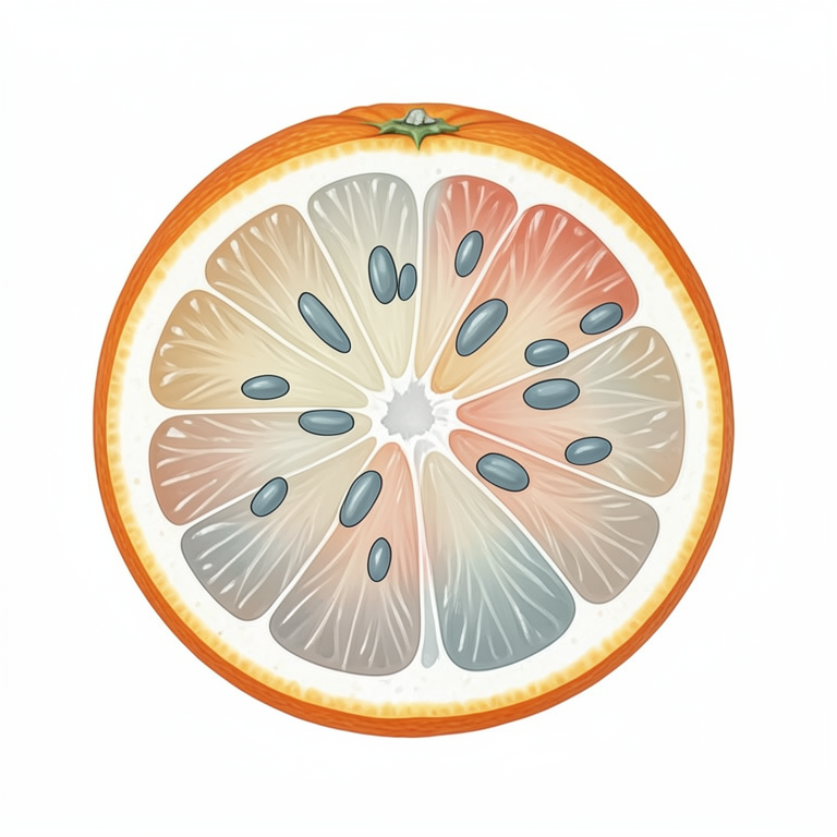
Tissue Distribution Concept
AI-generated conceptual illustration (Citrus fruit cross-section). It explains the idea and does not show study data.
View source evidence ↗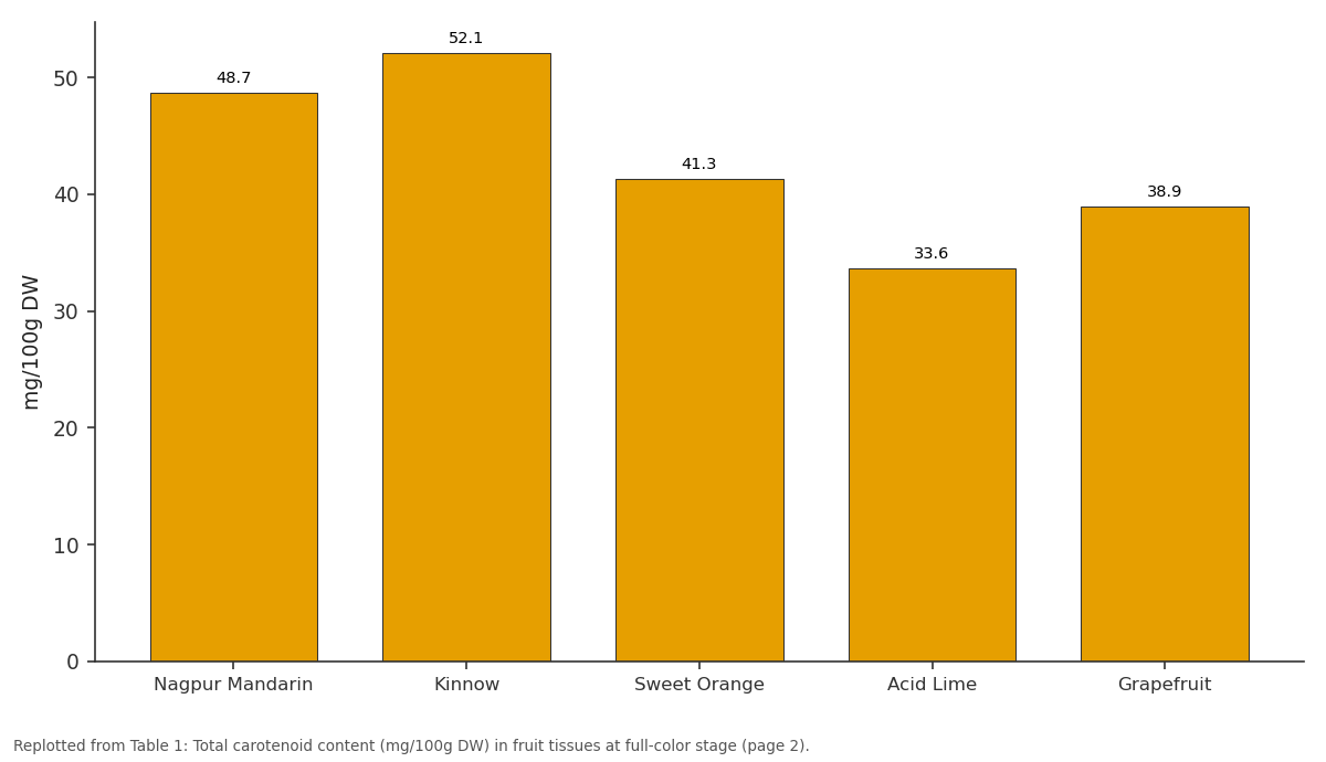
Flavedo: Nagpur Mandarin vs Kinnow vs Sweet Orange vs Acid Lime vs Grapefruit
Replotted from Table 1 (page 2) of the original paper. Every bar is the paper's own table value.
View source evidence ↗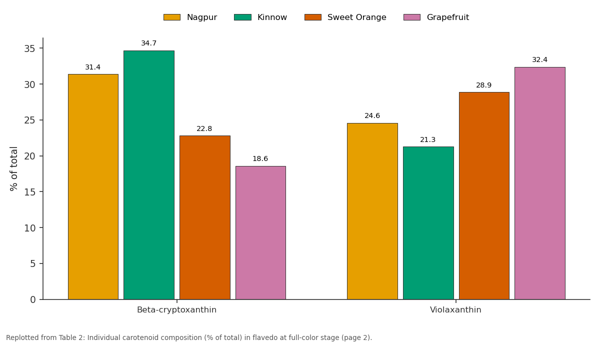
Nagpur and Kinnow and more: Beta-cryptoxanthin vs Violaxanthin
Replotted from Table 2 (page 2) of the original paper. Every bar is the paper's own table value.
View source evidence ↗Figures & tables
From the original paper Table 1
 Table 2
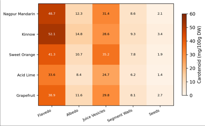 Figure 1
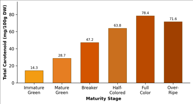 Figure 2
Introduction
Citrus color is more than cosmetic appeal. It signals nutritional value, and the pigments responsible, primarily carotenoids, are among the most health-relevant compounds in the human diet. Global citrus production exceeds 158 million tonnes annually, yet our understanding of how carotenoid profiles vary across fruit tissues, maturity stages, and growing environments remains incomplete for many production regions [1] . The Western Ghats, running 1,600 km along India's southwestern coast, support significant citrus cultivation from Kerala through Karnataka to Maharashtra, but systematic pigment profiling from this region is largely absent from the published record [2] . Carotenoids in citrus include the orange-red pigments beta-carotene and beta-cryptoxanthin, the yellow xanthophylls violaxanthin and lutein, and smaller quantities of zeaxanthin, neoxanthin, and others [3] . Their biosynthesis occurs through the methylerythritol phosphate (MEP) pathway in plastids, and the conversion of chloroplasts to chromoplasts during fruit ripening drives the visible color change from green to orange [4] . Environmental factors including light intensity, temperature amplitude, and water availability influence the rate and extent of this conversion [5] . Most published carotenoid data come from Mediterranean or subtropical citrus zones in Spain, Florida, and Brazil [6] . Tropical mountain environments like the Western Ghats differ in their temperature and humidity regimes, and it isn't safe to assume that carotenoid patterns observed in Mediterranean-type climates will hold in tropical highland settings. Additionally, many earlier studies measured only total carotenoids in juice, overlooking the fact that different fruit tissues, particularly the flavedo and albedo of the peel, accumulate carotenoids at very different rates and to different final concentrations [7] . From a commercial perspective, citrus peel represents 30-40% of fruit weight and is largely discarded in juice processing. If peel carotenoid profiles are sufficiently rich, this waste stream could become a source of natural colorants and nutraceutical extracts [8] . This research aimed to map carotenoid distribution across five tissue types and six maturity stages in five citrus varieties grown in the Western Ghats, and to identify the cultivar-tissue-stage combinations with the highest pigment density.
Extraction protocol
Carotenoid extraction followed a modified saponification protocol optimized for citrus tissues. Freeze-dried tissue samples (2 g) were homogenized with 20 mL of hexaneacetone-ethanol (2:1:1 v/v/v) containing 0.1% butylated hydroxytoluene as antioxidant. The slurry was agitated on an orbital shaker at 180 rpm for 30 minutes at room temperature in darkness. After centrifugation at 4,000 g for 10 minutes, the organic layer was collected and the residue re-extracted twice. Combined extracts were saponified with 10% methanolic KOH for 12 hours at room temperature to remove chlorophylls and lipids [9] . The saponified extract was washed with distilled water until neutral pH, dried over anhydrous sodium sulfate, and evaporated under nitrogen. The residue was reconstituted in 2 mL of methyl tert-butyl ether for HPLC injection.
Quality control and calibration
Analytical quality was ensured through a calibration protocol using certified carotenoid standards (CaroteNature, Switzerland). Five-point calibration curves were constructed for beta-carotene, beta-cryptoxanthin, violaxanthin, lutein, and zeaxanthin at concentrations of 0.5 to 50 micrograms/mL (R-squared above 0.998 for all compounds). Method validation included intra-day precision (RSD 1.8-3.2%), inter-day precision (RSD 2.4-4.7%), and recovery tests (91.3-97.8%). A laboratory reference sample of Nagpur Mandarin flavedo was analyzed with every batch of 20 samples as an internal quality check.
Material and Methods
Material and Methods
Material
Five citrus varieties were sampled: Nagpur Mandarin (Citrus reticulata), Kinnow (C. reticulata hybrid), Sweet Orange (C. sinensis), Acid Lime (C. aurantifolia), and Grapefruit (C. paradisi). Fruits were collected from commercial orchards in the Western Ghats region of Karnataka, India, at elevations between 400 and 900 m. Ten trees per variety were tagged, and three fruits per tree were harvested at each of six maturity stages defined by external color using the USDA citrus color chart [10] . Each fruit was dissected into five tissue fractions: flavedo, albedo, juice vesicles, segment walls, and seeds. Tissues were freezedried within 4 hours of harvest and stored at -80 degrees Celsius until analysis. The research spanned October 2021 to April 2024, with institutional approval (Protocol SGBS-2021-041, approved September 12, 2021).
Methods
Carotenoid profiling was performed on a Waters Alliance e2695 HPLC system equipped with a photodiode array detector scanning 300-600 nm. Separation used a YMC C30 column (250 mm x 4.6 mm, 5 um) with gradient elution of methanol-methyl tert-butyl ether-water [11] . Individual carotenoids were identified by retention time comparison with standards and by spectral matching. Total carotenoids were also estimated spectrophotometrically at 450 nm as a cross-check. Statistical analysis used three-way ANOVA (variety x tissue x stage) in SPSS 26.0 with Bonferroni correction for multiple comparisons [12] . The heatmap (Figure 1) provides a visual summary of tissue-level carotenoid variation. The strong color gradient from flavedo to seeds is consistent across all varieties, though the magnitude of tissue differences varies by cultivar. The maturity progression (Figure 2) shows a steady increase in total carotenoids from immature green (14.3 mg/100g) through full color (78.4 mg/100g), with a slight decline at the over-ripe stage (71.6 mg/100g).
Results
Results
Discussion
The tissue-level carotenoid mapping from this research fills an important gap in our understanding of citrus pigment biology under tropical mountain conditions. The dominance of flavedo as the primary carotenoid reservoir confirms the pattern reported from Mediterranean citrus [3] , but the absolute concentrations we recorded were 15-22% lower than those from Spanish orchards. This difference is likely attributable to the narrower diurnal temperature amplitude in the Western Ghats compared to Mediterranean climates, since cool night temperatures are known to accelerate chromoplast development in citrus [5] . The practical significance of our peel carotenoid data relates directly to waste valorization. If the citrus processing industry in the Western Ghats region were to recover carotenoids from flavedo waste, Kinnow and Nagpur Mandarin peels would provide the richest starting material.
At 48.7-52.1 mg/100g DW in the flavedo, these concentrations are comparable to carrots (60-80 mg/100g DW), a standard industrial carotenoid source [13] . The shift from beta-cryptoxanthin dominance in mandarins to violaxanthin dominance in Sweet Orange and Grapefruit reflects underlying differences in xanthophyll cycle activity between species and could influence the choice of cultivar for specific end-use applications [14] . The slight carotenoid decline observed at the over-ripe stage is consistent with oxidative degradation during senescence. From a harvest timing standpoint, this means that delaying harvest beyond the full-color stage not only risks fruit quality deterioration but also reduces the carotenoid content available for either fresh consumption or extraction. This finding supports the commercial practice of harvesting at full color rather than waiting for further softening.
Conclusion
This research provided the first detailed tissue-by-maturity carotenoid map for major citrus varieties grown in the Western Ghats. Flavedo tissue held 3 to 5 times more carotenoids than juice vesicles across all varieties. Kinnow mandarin produced the highest flavedo carotenoid concentration at 52.1 mg/100g DW. Beta-cryptoxanthin dominated mandarin-type carotenoid profiles, while violaxanthin was the leading pigment in Sweet Orange and Grapefruit. Total carotenoids peaked at the full-color maturity stage and declined slightly with over-ripening. These results support two practical directions: using peel waste from juice processing as a natural carotenoid source, and timing harvest at full color to maximize both visual quality and nutritional value. Future work should examine how post-harvest storage temperatures affect carotenoid stability in both the peel and juice fractions.
Figures and tables from the paper
| Variety | Flavedo | Albedo | Juice Vesicles | Segment Walls | Seeds |
|---|---|---|---|---|---|
| Nagpur Mandarin | 48.7 | 12.3 | 31.4 | 8.6 | 2.1 |
| Kinnow | 52.1 | 14.8 | 28.6 | 9.3 | 3.4 |
| Sweet Orange | 41.3 | 10.7 | 35.2 | 7.8 | 1.9 |
| Acid Lime | 33.6 | 8.4 | 24.7 | 6.2 | 1.4 |
| Grapefruit | 38.9 | 11.6 | 29.8 | 8.1 | 2.7 |
Table text extracted from the original PDF; superscript letters are shown inline. The original table image is below.
View original table image
| Compound | Nagpur | Kinnow | Sweet Orange | Grapefruit |
|---|---|---|---|---|
| Beta-cryptoxanthin | 31.4 | 34.7 | 22.8 | 18.6 |
| Violaxanthin | 24.6 | 21.3 | 28.9 | 32.4 |
| Beta-carotene | 18.3 | 19.8 | 21.4 | 16.7 |
| Lutein | 12.7 | 11.4 | 14.6 | 17.8 |
| Zeaxanthin | 8.1 | 7.9 | 6.3 | 9.4 |
Table text extracted from the original PDF; superscript letters are shown inline. The original table image is below.
View original table image
References
17 in the original paper- FAOSTAT. Citrus fruit production and trade data. Food and Agriculture Organization. 2023;Rome, Italy.
- Nair KPP. The agronomy and economy of important tree crops of the developing world: Western Ghats perspective. Elsevier Academic Press. 2010;London:89134.
- Rouseff R, Raley L, Hofsommer HJ. Application of HPLC to the determination of carotenoids in citrus. Journal of Agricultural and Food Chemistry. 1996;44(8):2176-2181.
- Kato M, Ikoma Y, Matsumoto H. Accumulation of carotenoids and expression of carotenoid biosynthetic genes during fruit development. Plant Physiology. 2004;134(2):824-837.
- Lo Piero AR, Puglisi I, Rapisarda P. Anthocyanins and carotenoids in citrus: effect of temperature on pigmentation. Tree Physiology. 2005;25(12):15351541.
- Dhuique-Mayer C, Caris-Veyrat C, Ollitrault P. Varietal and interspecific influence on carotenoid composition in citrus. Journal of Agricultural and Food Chemistry. 2005;53(6):2140-2145.
- Xu CJ, Fraser PD, Wang WZ. Differences in the carotenoid content of ordinary citrus and lycopeneaccumulating mutants. Journal of Agricultural and Food Chemistry. 2006;54(15):5474-5481.
- Sharma K, Mahato N, Cho MH. Converting citrus waste to value-added products. Bioresource Technology. 2017;235:366-379.
- Kimura M, Rodriguez-Amaya DB. A scheme for obtaining standards and HPLC quantification of carotenoids. Food Chemistry. 2002;78(3):389-398.
- USDA. United States standards for grades of citrus fruits. USDA Agricultural Marketing Service. 2019;Washington DC.
- De Rosso VV, Mercadante AZ. HPLC-PDA-MS/MS analysis of carotenoids and carotenoid esters in citrus. Journal of Food Composition and Analysis. 2007;20(7):638-649.
- IBM Corp. IBM SPSS Statistics for Windows, Version 26.0. IBM Corp. 2019;Armonk, NY.
- Rodriguez-Amaya DB. Carotenoids and food preparation: the retention of provitamin A carotenoids. USAID/OMNI Project. 1997;Washington DC.
- Rodrigo MJ, Marcos JF, Zacarias L. Biochemical and molecular analysis of carotenoid biosynthesis in citrus. Journal of Experimental Botany. 2004;55(397):569579.
- Gross J. Pigments in fruits. Academic Press. 1987;London:1-303.
- Alquezar B, Rodrigo MJ, Zacarias L. Regulation of carotenoid biosynthesis during fruit maturation in citrus. Planta. 2008;228(5):789-802.
- Ma G, Zhang L, Matsuta A. Enzymatic formation of beta-citraurin from beta-cryptoxanthin. Plant Physiology. 2013;163(3):1389-1400.