
Research article · 2026 · Full text
International Journal of Biology SciencesIsolation and molecular identification of Klebsiella spp.in raw meat from Mandla district of Madhya Pradesh
Volume 8(4) · Pages 188–193 · DOI: 10.33545/26649926.2026.v8.i4c.814 ↗
Research areasEnterobacteriaceae and Cronobacter ResearchSalmonella and Campylobacter epidemiologyFood Safety and Hygiene
Abstract
Author-writtenMeat is an essential part of human nutrition; still high nutrient load also makes it a probable carrier for foodborne pathogen. Klebsiella spp., an emerging food borne pathogen alarming public health because of its virulence nature and growing anti-microbial resistant strains. The present study examined 51 retail raw meat samples i.e. poultry meat, pork and goat meat samples collected from retails shops of 12 markets in tribal-dominated Mandla district of Madhya Pradesh. Klebsiella spp. specific morphological characteristics colonies were recovered by bacteriological isolation method and further identified by biochemical tests. Thirty-eight Klebsiella spp. presumptive isolates i.e. 74.5% were identified. Raw goat meat exhibiting the highest exposure to Klebsiella spp. (76.47%) followed by poultry meat (74.07%) and pork (71.4%). Market wise variation in Klebsiella contamination of raw meat was observed with six markets showed 100% exposure rate. Presumptive isolates were confirmed at genomic level by PCR using Klebsiella genus specific gyrA gene primers and Klebsiella pneumonia specific rpoB gene primers. Out of 38 presumptive Klebsiella isolates; 22 and 8 isolates were found positive for gyrA gene and rpoB gene respectively. Most of K.pneumonia isolates recovered from goat meat samples collected from Narayanganj market. Overall retail raw meat samples collected from Mandla district showed higher Klebsiella contamination highlighting need of hygienic slaughtering and trading practices to ensure food safety.
Summary
Source-checkedPlain-language reading of the full paper, prepared with AI and checked against the text. Each point links to the authors' own words.
Why it matters
- Meat is a vital nutritional source for humans, but its high nutrient content makes it susceptible to carrying foodborne pathogens. View evidence ↗
- Klebsiella species are emerging foodborne pathogens that pose a public health risk due to their virulence and increasing antimicrobial resistance. View evidence ↗
- In the Mandla district, retail shops use traditional open-air slaughtering, and there is a lack of public data regarding microbial contamination of raw meat in this region. View evidence ↗
The question
- The study aimed to isolate and identify Klebsiella species in poultry, goat, and pork meat from various markets in the Mandla district using morphological, biochemical, and molecular methods. View evidence ↗
What they did
- Isolation involved pre-enriching 10 grams of each sample in buffered peptone water at 37 °C overnight, followed by plating on Eosin Methylene Blue (EMB) agar. View evidence ↗
- Morphological identification was conducted via Gram and capsule staining, while further identification used a battery of biochemical tests including catalase, oxidase, and TSI. View evidence ↗
- Molecular confirmation was performed using PCR targeting the genus-specific gyrA gene and the K. pneumonia-specific rpoB gene. View evidence ↗
What they found
- Based on culture and phenotypic tests, 38 out of 51 meat samples (74.5%) were identified as presumptive Klebsiella species. View evidence ↗
- Goat meat had the highest contamination rate at 76.47% (13/17), followed by chicken at 74.07% (20/27) and pork at 71.42% (5/7). View evidence ↗
- Six markets, Hirdenagar, Narayanganj, Madhupur, Padmi, Udaipur, and Mandla, showed a 100% contamination rate. View evidence ↗
- Six K. pneumonia isolates were recovered from 8 meat samples (75%) collected specifically from the Narayanganj retail market. View evidence ↗
What it means
- The authors conclude that the high rate of Klebsiella contamination in Mandla district's retail meat underscores the urgent need for more hygienic slaughtering and trading practices. View evidence ↗
- The authors suggest that the lack of proper slaughtering facilities and unhygienic open-market handling likely contribute to the high environmental exposure of meat to Klebsiella. View evidence ↗
- The authors state that further extensive genomic studies are needed on the pure K. pneumonia isolates to identify hypervirulent and multi-drug resistant strains. View evidence ↗
Figures & tables
From the original paper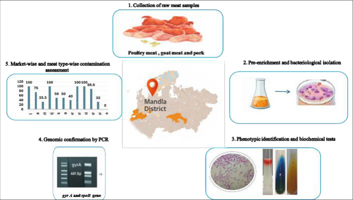 Figure 1
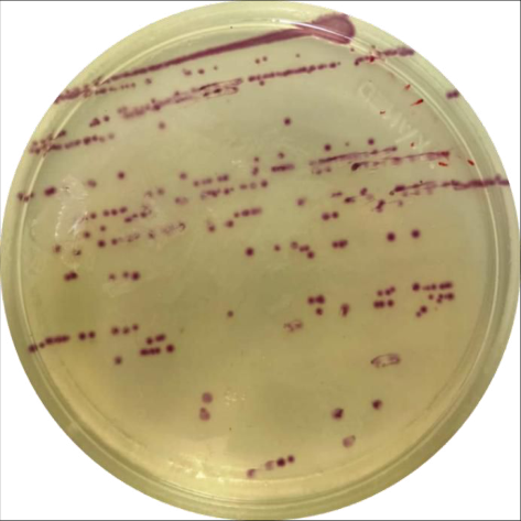 Figure 2
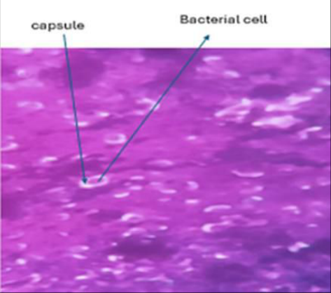 Figure 3
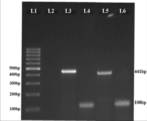 Figure 4
 Table 1
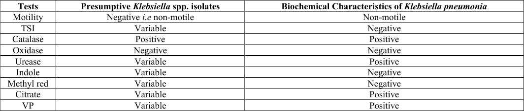 Table 2
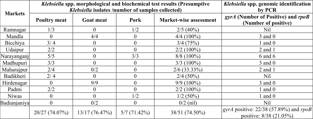 Table 3
Introduction
Meat represents an important component of human food and contamination of raw meat with pathogenic microbes poses high risk of food borne illness [1] . Microbial load of raw meat depends on infrastructure of slaughter house, slaughtering practices, sanitation of meat processing, handling, quality of water sources, availability of cold storage facility etc. [2] . During slaughtering of animals and processing of meat, there are high chances of pathogen contamination of the raw meat from contaminated animal skin, infected internal organs, contaminated faecal material etc. [3] . Monitoring of microbial contamination in raw meat becomes a critical public health-safety requirement. The members of Klebsiella genus are often considered an agent of foodborne illness and reported can be found in seafood, frozen foods, and fresh meat [4] . The members of genus Klebsiella belong to Enterobacteriaceae family are gram negative and encapsulated bacteria, causes severe infections such as septicemia, liver abscesses, urinary-tract infections, and pneumonia in humans. Klebsiella pneumonia (K. pneumonia) is the medically most important member of this genus and leading cause of nosocomial Klebsiella infections in human [5] . Klebsiella spp. are opportunistic pathogens and mostly cause infections in immune-compromised and hospitalized individuals [6,7,8] . The species under Klebsiella genus are known as one of the most critical multi-drug resistance (MDR) organisms globally and increasingly emergence of hyper-virulent strains posing significant public health threat [9] . K. pneumonia is ubiquitous in environment and found in soil, water bodies, sewage, animal bedding material, vegetation etc. and reason for being the common contaminant of food items [10] . In humans and animals, the K. pneumonia localises in upper respiratory tract and gastrointestinal tract and excreted in faeces along with other enterobacteria [11] . Domesticated and commercial farm animals and poultry get K. pneumonia infection when come in contact with contaminated water bodies, soil, vegetation etc. The infected animals and poultry transmit the organism to humans due to cohabitation, routine handling of infected animals, handling of contaminated raw meat, consumption of undercooked meat and all such activities that facilitate exposure possibilities to K. pneumonia. Many studies have reported contamination of K. pneumonia in raw meat and other animal origin foods and genetic relatedness between K. pneumonia isolates recovered from retail meat and human clinical samples [12, 13, 14, 15, and 16] . Therefore, the present study was undertaken to isolate Klebsiella spp. in retail meat sample (poultry meat, goat meat, and pork) collected from different markets of Mandla district of Madhya Pradesh and their identification by morphological, biochemical tests and molecular techniques. The district represents significant tribal population where meat consumption is a daily nutrition and deeply associated to cultural practices and religious beliefs. In this area, the retail shops follow traditional open-air slaughtering of animals. No studies on microbial contamination of raw meat meant for human consumption have been available in public domain.
Materials and Methods
Materials and Methods
Area of study
The present study was conducted in Mandla district, located in the eastern part of Madhya Pradesh, India (22.60°N latitude and 80.37°E longitude). Mandla is a predominantly tribal region characterized by tropical climate with hot summers, moderate rainfall, and mild winters.
Sample Collection
A total of 51 raw meat samples were collected from shops of 12 retail markets of Mandla district including Madhupuri, Mandla, Hirdenagar, Ramnagar, Maharajpur, Udaipur, Badianjaniya, Badikheri, Niwas, Padmi, Bicchiya and Narayanganjbetweendecember 2024 to august 2025. The samples included 27 poultry meat, 17 goat meat, and 7 pork meat. Samples were collected aseptically in sterile tubes and transported to laboratory in thermo-insulated boxes. The meat samples were processed promptly in the laboratory (Animal Biotechnology Centre, NDVSU, Jabalpur).
Bacteriological isolation and identification
The 10 gram of each meat sample was pre-enriched in 90 ml sterile buffered peptone water (HiMedia) and incubated overnight at 37 °C. Diluted pre-enriched culture was spread onto Eosin Methylene Blue (EMB) agar (HiMedia) and incubated overnight. The mucoid pink or purple coloured colonies presumed of Klebsiella spp. were picked up and further streaked on Hichrome Klebsiella selective agar (HiMedia). The purple-magenta coloured colonies were picked up and inoculated nutrient broth for growth. Both agar plates and broth culture were stored for further study. Both Gram staining and capsule staining were performed for morphological identification of Klebsiella isolates. Capsule staining was performed by negative staining method. Briefly, a loopful of the bacterial culture was mixed with a drop of nigrosin on a clean glass slide and a thin smear was made by using edge of a second slide. The thin smear was air dried, followed by flooded with 1% crystal violet for one minute. The excess amount of crystal violet stain was drained off by tilting the slide and the smear was air dried. The slide was then observed under an oil immersion microscope.
Biochemical tests
Presumptive Klebsiella isolates were subjected to a series of biochemical tests including catalase, oxidase, indole, methyl red (MR), Voges-Proskauer (VP), citrate utilization, urease, triple sugar iron (TSI), oxidase and catalase tests for further identification.
Genomic level identification of Klebsiella spp. Genomic DNA extraction
Genomic DNA was extracted using the boiling method [17] with some modifications. A loopful of overnight-grown presumptive Klebsiella colonies were suspended in 500 µL of trypsin-EDTA buffer and centrifuged at 3,000 rpm. The bacteria pellet was resuspended in fresh 100 µL trypsin-EDTA buffer and boiled at 100°C for 10 minutes followed by snap chilling in ice for 5 minute. The bacteria lysate was centrifuged at high speed and the upper clear fraction was transferred to a new micro-centrifuge tube. The clear fraction, contained genomic DNA and plasmid, was then used as DNA templates in subsequent polymerase chain reaction (PCR) experiments.
Molecular identification by PCR
Reported primers for the gyrA and rpoB genes (Table 1) were used to carry out PCR for molecular confirmation of Klebsiella isolates. Fifty microliter of PCR reaction mixture consisted of 25 µL of master mix (Dss Takara Bio India), 0.2µM of each forward and reverse primers, 2.5 µL of template DNA and nuclease-free water. The thermal condition followed for PCR as follows; initial denaturation at 98 ºC for 5 minutes, followed by 30 cycles of denaturation (98ºC / 1 minute), primer annealing (56ºC / 1 minute), and extension (72 ºC / 1 minute) and final extension step (72 ºC / 5 minute). The PCR amplified products were separated on 1.5% agarose gel containing DNA-staining dye and visualized under UV light in gel documentation system.
| Target genes | Primer sequence (5’-3’) | Amplicon size | References |
|---|---|---|---|
| gyrA | F: CGCGTACTATACGCCATGAACGTA R: ACCGTTGATCACTTCGGTCAGG | 441bp | [18] and [19] |
| rpoB | F: CAACGGTGTGGTTACTGACG R: TCTACGAAGTGGCCGTTTT | 108bp |
Table text extracted from the original PDF; superscript letters are shown inline. The original table image is below.
View original table image
Results and Discussion
Isolation, morphological characterization and biochemical characterization A total of 51 raw meat (poultry meat, goat meat, and pork) samples collected from twelve retail markets in Mandla district were subjected to bacterial isolation, morphological and molecular identification to assess the contamination rate of raw meat with Klebsiella spp. (Fig. 1). In microbiological isolation process, included pre-enrichment followed by plating on EMB agar, the mucoid and pink/purple coloured colonies were picked up. The colonies were streaked on Klebsiella selective agar for selective isolation and easy detection of Klebsiella spp. The bacteria colonies exhibited classic round, mucoid and purple to magenta-coloured morphology (Fig. 2) were presumed to represent Klebsiella spp. The morphological identification by capsule staining of the selected colonies confirmed the presence of a thick polysaccharide capsule, observed as a clear halo surrounding violet-stained bacilli (Fig. 3). This capsule is a critical virulence determinant, enhancing environmental persistence and resistance to phagocytosis [20] . The Table 2 represents summary of biochemical tests performed for all presumptive Klebsiella isolates. All isolates were found positive for catalase and oxidase reactions. However, Klebsiella spp. isolates showcased variable results in TSI, urease, citrate, MR-VP and indole tests. As it was already reported that different Klebsiella spp. metabolised a particular substrate differently and produced non-result in biochemical tests. This variation in metabolism exploited as epidemiological tool in differentiation of isolates of Klebsiella spp [21] .
| Tests | Presumptive Klebsiella spp. isolates | Biochemical Characteristics of Klebsiella pneumonia |
|---|---|---|
| Motility | Negative i.e non-motile | Non-motile |
| TSI | Variable | Negative |
| Catalase | Positive | Positive |
| Oxidase | Negative | Negative |
| Urease | Variable | Positive |
| Indole | Variable | Negative |
| Methyl red | Variable | Negative |
| Citrate | Variable | Positive |
| VP | Variable | Positive |
Table text extracted from the original PDF; superscript letters are shown inline. The original table image is below.
View original table image
Molecular confirmation by PCR
Molecular identification of all presumptive isolates at genomic level by PCR further substantiated the preliminary cultural, morphological and biochemical characteristics of pure Klebsiella isolates. The Klebsiella genus specific gene gyrA and K. pneumonia specific gene rpoB were used for screening all presumptive Klebsiella isolates. Observed PCR product bands of approximate size 441bp and 108bp on agarose gel confirmed the amplification of target region of gyrA gene and rpoB gene respectively (Fig. 4).
Market-wise and meat type-wise isolation of Klebsiella spp.
The table 3 represents the summary of bacteria culture isolation and morphological identification, biochemical test results and molecular identification by PCR results. The table 3 also shows the market-wise and meat type-wise Klebsiella contamination in raw meat samples. Results of culture based isolation as well as phenotypic characterization demonstrated that 38 meat samples out of 51 (i.e 74.50%) were presumed to be positive for Klebsiella. Market-wise assessment analysis indicated that highest Klebsiella spp. contamination (i.e. 100%) level was observed in Hirdenagar, Narayanganj, Madhupur, Padmi, Udaipur and Mandla markets. Meat meat samples collected from Ramnagar, Niwas, Bicchiya, Badikheri and Maharajpur showed variable Klebsiella contamination (i.e.0 -75%). The variation in Klebsiella load in raw meat samples demonstrated the influence of market-specific factors such as sanitation, vendor hygiene, and environmental exposure.
| Markets | Klebsiella spp. morphological and biochemical test results (Presumptive Klebsiella isolates /number of samples collected) | — | — | — | Klebsiella spp. genomic identification by PCR |
|---|---|---|---|---|---|
| Poultry meat | Goat meat | Pork | Market-wise assessment | gyrA (Number of Positive) and rpoB (Number of positive) | |
| Ramnagar | 1/3 | 0 | 1/2 | 2/5 (40%) | Nil |
| Mandla | 0 | 4/4 | 0 | 4/4 (100%) | 3 and 0 |
| Bicchiya | 3/ 4 | 0 | 0 | 3/4 (75%) | 1 and 0 |
| Udaipur | 2/2 | 0 | 0 | 2/2 (100%) | 2 and 1 |
| Narayanganj | 5/5 | 0 | 3/3 | 8/8 (100%) | 6 and 6 |
| Madhupuri | 3/3 | 0 | 0 | 3/3 (100%) | 3 and 0 |
| Maharajpur | 2/4 | 0/2 | 0 | 2/6 (33.33%) | 2 and 1 |
| Badikheri | 2/ 4 | 0 | 0 | 2/4 (50%) | Nil |
| Hirdenagar | 0 | 9/9 | 0 | 9/9 (100%) | 3 and 0 |
| Padmi | 2/2 | 0 | 0 | 2/2 (100%) | 1 and 0 |
| Niwas | 0 | 0 | 1/2 | 1/2 (50%) | 1 and 0 |
| Badianjaniya | 0 | 0/2 | 0 | 0/2 (nil) | Nil |
| 20/27 (74.07%) | 13/17 (76.47%) | 5/7 (71.42%) | 38/51 (74.50%) | gyrA positive: 22/38 (57.89%) and rpoB positive: 8/38 (21.05%) |
Table text extracted from the original PDF; superscript letters are shown inline. The original table image is below.
View original table image
Among the different meat types, goat meat exhibited the highest contamination rate (13/17 i.e 76.47%), followed by chicken (20/27 i.e 74.07%) and pork (5/7 i.e 71.42%). Among the 38 presumptive Klebsiella spp. isolates; 22 and 8 isolates were found positive for gyrA gene and rpoB gene respectively. Analysis of gyrA gene PCR-positive Klebsiella spp. isolates across meat types demonstrated that the chicken samples exhibiting the highest positivity (12 out of 20), followed by goat meat (6 out of 13) and pork (4 out of 5). Similarly, analysis of rpoB gene PCR-positive Klebsiella spp. isolates revealed that only 05 chicken and 03 pork raw meat samples were found positive for K. pneumonia. All rpoB positive isolates were showed conformity with results of gyrA gene PCR and standard biochemical tests for K. pneumonia. Out of 05 Klebsiella isolates from pork samples, 03 samples were found K. pneumonia positive (i.e.60%). Out of 20 Klebsiella isolates from chicken samples, 05 samples were found K. pneumonia positive (i.e.25%). Surprisingly, all six K.pneumonia isolates were obtained from 08 meat samples collected from Narayanganj retail market (i.e. 75%). The Narayanganj retail market was surrounded by tribal habitats and the market vendors procured backyard domesticated pigs, poultry and goats from the local tribes for raw meat retailing purpose. Many previous studies done at different locations of India were also reported occurrence of Klebsiella spp. in retail raw meats. One study reported occurrence of Klebsiella spp. in 70.58% (cultural identification) and 11.33% (molecular identification) of poultry meat samples collected from retail markets of Anand, Gujarat [12] . Another study reported presence of Klebsiella spp. in 37.50% of beef samples collected from retail shops at Thrissur and Palakkad districts of Kerala [13] . Mahanti A et al. (2017) isolated Klebsiella spp. from 10.7% of clonal swabs samples collected from indigenous and broilers poultry bird in West Bengal [22] . Many studies undertaken abroad such as Ghana [23] , Germany [24] , Turkey [14] etc. reported higher occurrence of Klebsiella spp. in poultry meat. Collectively, the present study and previous studies demonstrated that the raw meat act as significant reservoirs of Klebsiella spp., and exposure rate may likely be influenced by geographic location, sample type, and hygienic practices followed during raw meat processing.
Acknowledgement
AcknowledgementThe first author is thankful to Department of Microbiology, College of Veterinary Science and Animal Husbandry, Jabalpur for assisting in microbiological isolation process. The necessary financial support was provided by the Nanaji Deshmukh Veterinary Science University.
References
24 in the original paper- Kebede MT, Getu AA. Assessment of bacteriological quality and safety of raw meat at slaughterhouse and butchers' shop (retail outlets) in Assosa Town, Beneshangul Gumuz Regional State, Western Ethiopia. BMC Microbiol. 2023;23(1):403.
- Borch E, Arinder P. Bacteriological safety issues in red meat and ready-to-eat meat products, as well as control measures. Meat Sci. 2002;62(3):381-390.
- Diyantoro, Wardhana DK. Risk factors for bacterial contamination of bovine meat during slaughter in ten Indonesian abattoirs. Vet Med Int. 2019;2019:2707064.
- Riwu KHP, Effendi MH, Rantam FA, Khairullah AR, Widodo A. Virulence factors of Klebsiella pneumoniae as emerging infection on the food chain. Vet World. 2022;15(9):2172-2179.
- Abbas R, Chakkour M, Zein El Dine H, Obaseki EF, Obeid ST, Jezzini A, et al. General overview of Klebsiella pneumoniae: epidemiology and the role of siderophores in its pathogenicity. Biology (Basel). 2024;13(2):78.
- Podschun R, Ullmann U. Klebsiella spp. as nosocomial pathogens: epidemiology, taxonomy, typing methods, and pathogenicity factors. Clin Microbiol Rev. 1998;11(4):589-603.
- Zhang S, Yang G, Ye Q, Wu Q, Zhang J, Huang Y. Phenotypic and genotypic characterization of Klebsiella pneumoniae isolated from retail foods in China. Front Microbiol. 2018;9:289.
- Ndlovu T, Kgosietsile L, Motshwarakgole P, Ndlovu SI. Evaluation of potential factors influencing the dissemination of multidrug-resistant Klebsiella pneumoniae and alternative treatment strategies. Trop Med Infect Dis. 2023;8(8):381.
- Dong N, Li R, Lai Y. Editorial: Klebsiella pneumoniae: antimicrobial resistance, virulence and therapeutic strategies. Front Cell Infect Microbiol. 2022;12:1108817.
- Wareth G, Neubauer H. The animal-foods-environment interface of Klebsiella pneumoniae in Germany: an observational study on pathogenicity, resistance development and the current situation. Vet Res. 2021;52(1):16.
- World Health Organization. Disease outbreak news: antimicrobial resistance, hypervirulent Klebsiella pneumoniae-global situation. 2024.
- Modh KS, Parmar BC, Chaudhary JH, Bhanderi BB, Nayak JB, Thakur S, et al. Molecular detection of Klebsiella spp. from poultry meat. Ind J Pure Appl Biosci. 2021;9(1):511-518.
- Jamal J, Latha C, Mathew B, Vergis J, Gleeja VL. Occurrence of Klebsiella pneumoniae and Salmonella spp. in beef and cow milk from central Kerala. J Vet Anim Sci. 2023;54(4):907-915.
- Gundogan N, Citak S, Yalcin E. Virulence properties of extended spectrum β-lactamase-producing Klebsiella species in meat samples. J Food Prot. 2011;74(4):559564.
- Fielding BC, Mnabisa A, Gouws PA, Morris T. Antimicrobial-resistant Klebsiella species isolated from free-range chicken samples in an informal settlement. Arch Med Sci. 2012;8(1):39-42.
- Kurekci C, Unaldi O, Sahin S, Garcia-Menino I, Hammerl JA. Impact and diversity of ESBL-producing Klebsiella pneumoniae recovered from raw chicken meat samples in Turkiye. Antibiotics (Basel). 2023;13(1):14.
- Dashti A, Jadaon M, Abdulsamad A, Dashti HM. Heat treatment of bacteria: a simple method of DNA extraction for molecular techniques. Kuwait Med J. 2009;41(2):117-122.
- Bobbadi S, Kiranmayi Chinnam B, Nelapati S, et al. Occurrence and genetic diversity of ESBL-producing Klebsiella species isolated from livestock and livestock products. J Food Saf. 2020;40:e12738.
- Liza NA, Hossain H, Chowdhury SR, Naser JA, Lasker RM, Rahman A, et al. Molecular epidemiology and antimicrobial resistance of extended-spectrum βlactamase-producing Klebsiella pneumoniae in retail cattle meat. Vet Med Int. 2024;2024:3952504.
- Xu L, Li J, Wu W, Wu X, Ren J. Klebsiella pneumoniae capsular polysaccharide: mechanism in regulation of synthesis, virulence, and pathogenicity. Virulence. 2024;15(1):2439509.
- Barr JG. Variation in metabolism of biochemical test substrates by Klebsiella species: an epidemiological tool. J Med Microbiol. 1978;11(4):501-511.
- Mahanti A, Ghosh P, Samanta I, Joardar SN, Bandyopadhyay S, Bhattacharyya D, et al. Prevalence of CTX-M-producing Klebsiella spp. in broiler, Kuroiler and indigenous poultry in West Bengal State, India. Microb Drug Resist. 2018;24(3):299-306.
- Eibach D, Dekker D, Boahen KG, Akenten CW, Sarpong N, Campos CB, et al. Extended-spectrum betalactamase-producing Escherichia coli and Klebsiella pneumoniae in local and imported poultry meat in Ghana. Vet Microbiol. 2018;217:7-12.
- von Tippelskirch P, Golz G, Projahn M, Daehre K, Friese A, Roesler U, et al. Prevalence and quantitative analysis of ESBL and AmpC beta-lactamase-producing Enterobacteriaceae in broiler chicken during slaughter in Germany. Int J Food Microbiol. 2018;281:82-89.