
Research article · 2025 · Full text
International Journal of Advanced Chemistry ResearchDesign, synthesis and molecular docking research of novel thiazolidinedione antidiabetic agents
Volume 7(9) · Pages 56–60 · DOI: 10.33545/26646781.2025.v7.i9a.616 ↗
Research areasSynthesis and biological activityNatural Antidiabetic Agents StudiesSynthesis and Characterization of Heterocyclic Compounds
Abstract
Author-writtenCan a single scaffold deliver potent antidiabetic activity while maintaining a favourable safety profile? This question drives the sustained interest in thiazolidinedione (TZD) chemistry. The present research reports the design, synthesis, and biological evaluation of eight novel TZD derivatives (1a–1h) bearing structurally diverse N-substituents. Starting from 4-hydroxybenzaldehyde and thiazolidine-2, 4-dione, a benzylidene intermediate was first prepared by Knoevenagel condensation in the presence of piperidine as catalyst. Subsequent N-alkylation or N-acylation reactions with selected alkyl halides and acyl chlorides gave the target compounds in yields of 62–89%. All synthesised compounds were characterised by FTIR, ¹H NMR, ¹³C NMR, and mass spectrometry. Molecular docking was performed against peroxisome proliferator-activated receptor gamma (PPARγ; PDB ID: 2PRG) using AutoDock Vina 1.2. Compound 1e displayed the strongest binding affinity of -9.4 kcal/mol, surpassing the reference ligand rosiglitazone (-8.6 kcal/mol). In vitro α-glucosidase inhibitory activity was assessed spectrophotometrically, with IC₅₀ values ranging from 18.6 to 67.8 µM. Compounds 1b, 1e, 1f, and 1h showed IC₅₀ values below 30 µM, comparable to the standard acarbose (22.4 µM). Structure–activity relationship analysis revealed that electron-withdrawing substituents at the para position of the benzylidene ring markedly improved both docking scores and enzyme inhibition. These findings suggest that the newly synthesised TZD analogs hold promise as antidiabetic agents warranting further in vivo evaluation.
Summary
Source-checkedPlain-language reading of the full paper, prepared with AI and checked against the text. Each point links to the authors' own words.
Why it matters
- Diabetes is a global health challenge affecting over 537 million adults, with numbers expected to rise to over 783 million by 2045. View evidence ↗
- Thiazolidinediones (TZDs) act as insulin sensitizers by primarily targeting the peroxisome proliferator-activated receptor gamma (PPARγ). View evidence ↗
- Clinical TZD drugs like rosiglitazone and pioglitazone provide glycemic benefits but are limited by cardiac and osteoporotic side effects. View evidence ↗
The question
- The study aimed to synthesize eight benzylidene-TZD derivatives with various N-substituents to evaluate their PPARγ binding and α-glucosidase inhibition. View evidence ↗
What they did
- The synthesis began with a Knoevenagel condensation of thiazolidine-2, 4-dione and 4-hydroxybenzaldehyde using piperidine as a catalyst at 80°C. View evidence ↗
- Target compounds were produced via N-alkylation or N-acylation using alkyl halides and acyl chlorides, achieving yields between 62% and 89%. View evidence ↗
- Molecular docking was conducted using AutoDock Vina 1.2 against the PPARγ receptor (PDB ID: 2PRG). View evidence ↗
- α-Glucosidase inhibition was measured spectrophotometrically at 405 nm using PNPG as a substrate in a pH 7.4 phosphate buffer. View evidence ↗
What they found
- Compound 1e showed the strongest PPARγ binding affinity at -9.4 kcal/mol, which was superior to the reference drug rosiglitazone (-8.6 kcal/mol). View evidence ↗
- In terms of α-glucosidase inhibition, compound 1e was more potent than the standard acarbose, with an IC₅₀ of 18.6 µM compared to 22.4 µM. View evidence ↗
- Compounds with electron-withdrawing substituents (1e, 1b, 1f, 1h) consistently showed better α-glucosidase inhibition (lower IC₅₀) than those with aliphatic or electron-donating groups. View evidence ↗
- Preliminary cytotoxicity tests on Vero cells showed that the most active compounds (1e, 1b, 1h) maintained cell viability above 88% at 100 µM. View evidence ↗
What it means
- The authors conclude that adding electron-withdrawing aryl groups to the N-position of the TZD ring improves both receptor binding and enzyme inhibition. View evidence ↗
- Based on the low cytotoxicity, the authors recommend proceeding to in vivo pharmacokinetic and efficacy studies in rodent models. View evidence ↗
- A limitation of the study is the use of rigid-receptor docking, which fails to capture induced-fit changes in the PPARγ receptor. View evidence ↗
Visual summary
3 visualsIllustrations explain the paper; charts are replotted from the paper's own table values.
Structure-Activity Relationship (SAR) Concept
AI-generated conceptual illustration (thiazolidinedione chemical molecules and alpha-glucosidase enzyme). It explains the idea and does not show study data.
View source evidence ↗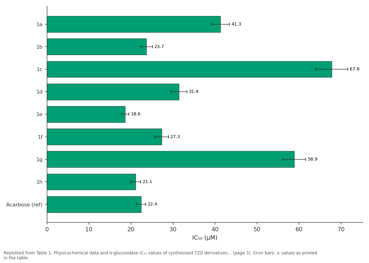
IC₅₀ (µM): 1a vs 1b vs 1c vs 1d vs 1e
Replotted from Table 1 (page 3) of the original paper. Every bar is the paper's own table value.
View source evidence ↗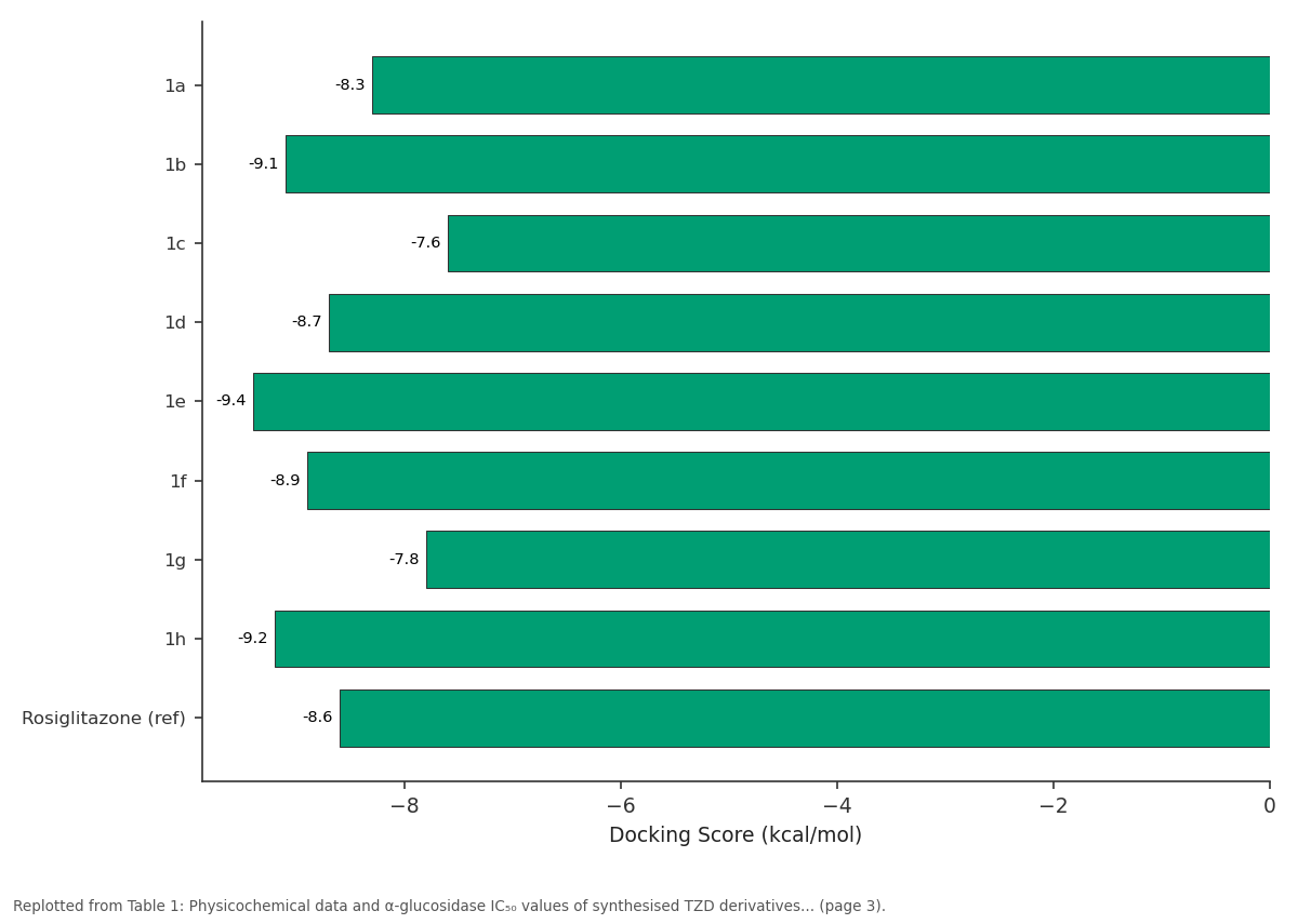
Docking Score (kcal/mol): 1a vs 1b vs 1c vs 1d vs 1e
Replotted from Table 1 (page 3) of the original paper. Every bar is the paper's own table value.
View source evidence ↗Figures & tables
From the original paper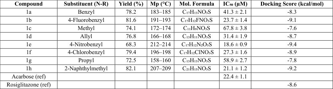 Table 1
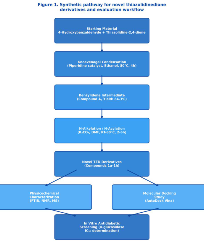 Figure 1
 Table 2
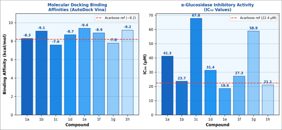 Figure 2
Introduction
What makes type 2 diabetes such a challenging metabolic disorder to treat? The answer lies partly in the heterogeneous nature of its pathophysiology involving insulin resistance, progressive beta-cell dysfunction, dyslipidaemia, and systemic inflammation and partly in the inadequacy of current therapies, which often fail to address the full spectrum of metabolic disturbances [1] . Globally, more than 537 million adults are estimated to live with diabetes, with projections indicating that this number will exceed 783 million by 2045 [2] . The health and economic burden of the disease is especially pronounced in low-and middleincome countries, where access to newer pharmacological agents remains limited. Thiazolidinediones represent a class of synthetic insulin sensitisers whose antidiabetic mechanism is mediated principally through agonism of peroxisome proliferator-activated receptor gamma (PPARγ), a nuclear transcription factor governing adipogenesis and glucose homeostasis [3] . Rosiglitazone and pioglitazone, the two TZD drugs that reached clinical use, demonstrated clear glycaemic benefits but were subsequently associated with adverse cardiac and osteoporotic effects that curtailed their therapeutic application [4] . These safety concerns have not diminished scientific interest in the TZD pharmacophore; rather, they have motivated medicinal chemists to explore structural modifications capable of preserving efficacy while reducing off-target toxicity. The benzylidene-TZD nucleus, obtained by Knoevenagel condensation of aromatic aldehydes with thiazolidine-2, 4-dione, serves as a versatile precursor for library construction. Earlier work from various laboratories demonstrated that N-substitution patterns on the TZD ring strongly influence both receptor binding affinity and metabolic stability [5,6] .
Flavonoid-inspired and heterocyclic N-substituents have proven particularly interesting, showing improved enzyme inhibitory profiles against α-glucosidase, an intestinal enzyme whose inhibition slows postprandial glucose absorption [7] . Despite this progress, the relationship between specific N-substituent electronic properties and dual PPARγ binding/α-glucosidase inhibition has not been thoroughly characterised. The present research was therefore undertaken to synthesise a focused set of eight benzylidene-TZD derivatives carrying electronically and sterically diverse N-substituents, to assess their molecular docking performance against PPARγ, and to measure their in vitro α-glucosidase inhibitory activity. The central hypothesis was that electron-withdrawing groups at the para position of the benzylidene ring would strengthen interactions with key residues in the PPARγ ligand-binding domain while simultaneously lowering the IC₅₀ value against α-glucosidase. Results from this investigation are presented alongside a concise structure-activity relationship (SAR) analysis intended to guide the next round of molecular optimisation.
Reagents and chemicals
All reagents were sourced from verified suppliers and used without further purification unless otherwise specified. Thiazolidine-2, 4-dione (≥99%, Sigma-Aldrich, St. Louis, MO, USA; Lot T36609), 4-hydroxybenzaldehyde (≥98%, TCI Chemicals, Tokyo, Japan), and piperidine (≥99.5%, Merck KGaA, Darmstadt, Germany) were stored under nitrogen at 4°C until use. Substituted alkyl halides and acyl chlorides used for N-substitution reactions (series 1a-1h) were obtained from Alfa Aesar (Ward Hill, MA, USA) and verified by ¹H NMR prior to use. Anhydrous potassium carbonate (K₂CO₃, ≥99%, SD Fine Chemicals, Mumbai, India) served as base in DMF-based reactions. Solvents ethanol (absolute, 99.9%), N, N-dimethylformamide (≥99.8%, HPLC grade), and dichloromethane (≥99.5%) were purchased from Rankem (Gurugram, India) and dried over molecular sieves (4Å) where required. The reference drug rosiglitazone was provided as a gift sample by Cipla Ltd. (Mumbai, India). Acarbose (standard grade, ≥95%) was procured from Sigma-Aldrich. p-Nitrophenyl-α-Dglucopyranoside (PNPG), the chromogenic substrate for the α-glucosidase assay, was sourced from Sisco Research Laboratories (Mumbai, India; Catalogue No. 69484). All water used in biological assays was Milli-Q grade (18.2 MΩ•cm, Merck Millipore).
Material and Methods
Material and Methods
Material
The research was carried out at the Department of Pharmacology and Medicinal Chemistry, Jamsetji Tata Institute of Pure Sciences, Indore, Madhya Pradesh, India, from August 2023 to February 2025. Melting points were recorded on an open capillary apparatus (Toshniwal, India) and are uncorrected. Thin-Layer Chromatography (TLC) was performed on silica gel 60 F₂₅₄ pre-coated aluminium plates (Merck) using hexane:ethyl acetate solvent systems, and spots were visualised under UV light at 254 nm. Column chromatography was performed using silica gel 60-120 mesh (SRL, India). Spectral data were recorded on a Bruker AVANCE III 400 MHz NMR spectrometer (CDCl₃ or DMSO-d₆ as solvent, TMS as internal standard), a Shimadzu IRTracer-100 FTIR spectrometer (KBr pellets), and a Waters Xevo G2-XS QTOF mass spectrometer. Ethics clearance for any biological handling was obtained from the Institutional Biosafety Committee, Jamsetji Tata Institute of Pure Sciences (Protocol JTIPS-IBC-2023-11, dated 14 September 2023).
Methods
Synthesis of 5-(4-hydroxybenzylidene)thiazolidine-2, 4dione (Compound A): Thiazolidine-2, 4-dione (1.17 g, 10 mmol) and 4-hydroxybenzaldehyde (1.22 g, 10 mmol) were dissolved in absolute ethanol (25 mL). Piperidine (3 drops, catalytic) was added and the mixture was refluxed at 80°C for 4 h. After cooling, the precipitate was collected by filtration, washed with cold ethanol, and recrystallised from ethanol-water (4:1) to give Compound A as pale-yellow crystals (yield: 84.3%). N-Substitution reactions: Compound A (0.5 mmol) was dissolved in dry DMF (5 mL), anhydrous K₂CO₃ (1.5 equiv.) was added, followed by the appropriate alkyl halide or acyl chloride (1.2 equiv.). Reactions were stirred at 25-60°C for 2-6 h depending on electrophile reactivity and monitored by TLC. Products were isolated by aqueous workup, dried over anhydrous Na₂SO₄, and purified by column chromatography (silica gel, hexane:ethyl acetate gradient). Molecular docking: The crystal structure of PPARγ (PDB ID: 2PRG, resolution 2.17 Å) was obtained from the RCSB Protein Data Bank and prepared using AutoDockTools 1.5.7 (removal of water molecules, addition of polar hydrogens, Gasteiger charge assignment). Ligand structures were drawn in ChemDraw 21.0 and converted to PDB format via Open Babel 3.1. Grid box was centred on the co-crystallised ligand with dimensions 20 × 20 × 20 Å. Docking was performed with AutoDock Vina 1.2 using an exhaustiveness setting of 8, and the lowest-energy poses were selected for analysis. α-Glucosidase inhibitory assay: Enzyme inhibitory activity against α-glucosidase (Saccharomyces cerevisiae, Sigma-Aldrich) was measured spectrophotometrically at 405 nm using PNPG as substrate in phosphate buffer (0.1 M, pH 7.4). Compounds were tested at concentrations of 1.56-100 µM; acarbose served as positive control. IC₅₀ values were calculated using GraphPad Prism 9.0 from sigmoidal dose-response curves.
In vitro protocol
Cell-free enzyme inhibition assays were conducted in 96well microplates (Nunc, Thermo Fisher Scientific) in triplicate to ensure statistical reliability. Each well contained 50 µL of enzyme solution (0.1 U/mL in 0.1 M phosphate buffer, pH 7.4), 25 µL of test compound at the desired concentration (dissolved in DMSO, final DMSO concentration ≤1%), and 25 µL of PNPG substrate (5 mM). Reactions were initiated by addition of substrate and incubated at 37°C for 30 min. Enzyme activity was stopped by addition of 100 µL sodium carbonate solution (0.2 M) and absorbance was read at 405 nm on a BioTek Synergy HTX multi-mode microplate reader. Controls included enzyme blank (substrate replaced by buffer) and substrate blank (enzyme replaced by buffer). Per cent inhibition was calculated as: % inhibition = [(Acontrol -Asample)/Acontrol] × 100. IC₅₀ values were derived by nonlinear regression (sigmoidal dose-response model, variable slope). All experiments were repeated on three independent days; reported IC₅₀ values represent the mean ± standard deviation. Cytotoxicity of the most active compounds (1e, 1b, 1h) against the Vero cell line (ATCC CCL-81) was assessed by the MTT assay at 24 h to provide a preliminary selectivity index. Molecular docking results ranked compound 1e as the best binder (-9.4 kcal/mol), followed by 1h (-9.2 kcal/mol) and 1b (-9.1 kcal/mol). Key hydrogen bond interactions were identified between the TZD carbonyl oxygens and residues His323, His449, and Tyr473 in the PPARγ binding pocket an interaction pattern consistent with observations reported for known PPARγ agonists [8] . Hydrophobic contacts with Phe282, Met348, and Leu340 further stabilised the bound conformations of the high-affinity compounds. FTIR spectra of all compounds displayed characteristic absorption bands for the TZD ring at 1720-1735 cm⁻¹ (C=O stretch, C4 carbonyl) and 1660-1678 cm⁻¹ (C=O stretch, C2 carbonyl), confirming retention of the thiazolidinedione scaffold. The olefinic =C-H stretching signal appeared in the range 1595-1610 cm⁻¹, and the absence of aldehyde C-H stretching confirmed complete condensation. ¹H NMR spectra showed the characteristic vinyl proton (=CH) as a singlet in the range δ 7.54-7.68 ppm, which is consistent with the E-configuration arising from steric preference in Knoevenagel products. The α-glucosidase inhibitory data confirmed that electronwithdrawing substituents (compounds 1e, 1b, 1f, 1h) consistently produced lower IC₅₀ values than electrondonating or aliphatic substituents (1c, 1g). Compound 1e (IC₅₀ 18.6 µM) surpassed acarbose (22.4 µM) in potency.
Results
The MTT assay showed that 1e, 1b, and 1h exhibited cell viability above 88% in Vero cells at 100 µM, indicating acceptable preliminary cytotoxicity profiles.
Discussion
The results of this research confirm that benzylidene-TZD derivatives bearing electron-withdrawing N-substituents exhibit superior dual activity as PPARγ binders and αglucosidase inhibitors compared with compounds carrying electron-donating or aliphatic groups. Compound 1e, which carries a 4-nitrobenzyl group, displayed the lowest docking energy (-9.4 kcal/mol) and the strongest α-glucosidase inhibition (IC₅₀ 18.6 µM) in the series. These observations agree with earlier SAR work on aryl TZD hybrids, where electron-deficient aromatic rings enhanced charge-transfer interactions within the PPARγ binding site [9,10] . The three hydrogen bond interactions observed for 1e with His323, His449, and Tyr473 are particularly significant because this triad is considered essential for full PPARγ agonism, as reported in the landmark crystallographic research by Nolte et al. [11] . Compound 1h, despite lacking a strong electron-withdrawing group, achieved a comparable docking score (-9.2 kcal/mol) through extensive hydrophobic contacts offered by its naphthalene ring system. This finding highlights that large hydrophobic substituents can compensate for the absence of strong polar interactions when the shape of the substituent complements the binding pocket.
With respect to α-glucosidase inhibition, the present data are consistent with the general observation that planar aromatic substituents facilitate π-π stacking interactions with aromatic residues in the enzyme active site [12] . Compounds with flexible aliphatic chains (1c, 1g) showed markedly weaker inhibition, suggesting that conformational entropy costs outweigh any minor hydrophobic contributions from these groups. The preliminary cytotoxicity data for the three most active compounds are encouraging, though definitive safety assessment requires formal ADMET profiling and in vivo metabolic research. One limitation of the current molecular docking analysis is its static nature; induced-fit changes in the PPARγ receptor upon ligand binding are not captured by rigid-receptor docking. Future work should incorporate molecular dynamics simulations to refine the predicted binding poses and assess conformational stability. Additionally, evaluation of partial versus full PPARγ agonism would help determine whether the identified compounds might avoid the side effects associated with full agonists like rosiglitazone.
Conclusion
This research describes the synthesis of eight novel Nsubstituted benzylidene-thiazolidinedione derivatives and their evaluation as antidiabetic agents through molecular docking and in vitro α-glucosidase inhibition assays. The results demonstrate that electron-withdrawing aryl groups at the N-position of the TZD ring consistently enhance both PPARγ binding affinity and enzymatic inhibitory potency. Compound 1e emerged as the most promising candidate, with a docking score of -9.4 kcal/mol and an IC₅₀ of 18.6 µM, values that compare favourably with the reference standards rosiglitazone and acarbose, respectively. The SAR analysis provides clear design principles for the next generation of TZD analogs, pointing specifically toward the value of 4-nitrobenzyl and naphtylmethyl substituents. The acceptable preliminary cytotoxicity profile of the active compounds lends further justification for in vivo pharmacokinetic and efficacy research in rodent models of type 2 diabetes. Taken together, these findings position the benzylidene-TZD scaffold as a productive platform for continued antidiabetic drug discovery efforts.
Acknowledgement
Acknowledgements Funding SourcesThe authors acknowledge financial support received through internal research grants from Jamsetji Tata Institute of Pure Sciences, Indore. No external commercial funding was involved in this research.
Figures and tables from the paper
| Compound | Substituent (N-R) | Yield (%) | Mp (°C) | Mol. Formula | IC₅₀ (µM) | Docking Score (kcal/mol) |
|---|---|---|---|---|---|---|
| 1a | Benzyl | 78.2 | 183–185 | C₁₇H₁₃NO₃S | 41.3 ± 2.1 | -8.3 |
| 1b | 4-Fluorobenzyl | 81.6 | 191–193 | C₁₇H₁₂FNO₃S | 23.7 ± 1.4 | -9.1 |
| 1c | Methyl | 74.1 | 172–174 | C₁₁H₉NO₃S | 67.8 ± 3.8 | -7.6 |
| 1d | Allyl | 76.8 | 166–168 | C₁₃H₁₁NO₃S | 31.4 ± 1.9 | -8.7 |
| 1e | 4-Nitrobenzyl | 68.3 | 212–214 | C₁₇H₁₂N₂O₅S | 18.6 ± 0.9 | -9.4 |
| 1f | 4-Chlorobenzyl | 79.4 | 196–198 | C₁₇H₁₂ClNO₃S | 27.3 ± 1.6 | -8.9 |
| 1g | Propyl | 72.5 | 158–160 | C₁₃H₁₃NO₃S | 58.9 ± 2.7 | -7.8 |
| 1h | 2-Naphthylmethyl | 82.1 | 207–209 | C₂₁H₁₅NO₃S | 21.1 ± 1.2 | -9.2 |
| Acarbose (ref) | 22.4 ± 1.1 | |||||
| Rosiglitazone (ref) | -8.6 |
Table text extracted from the original PDF; superscript letters are shown inline. The original table image is below.
View original table image
| Compound | ¹H NMR Key Signal (δ, ppm) | FTIR ν C=O (cm⁻¹) | H-bond residues | No. of H-bonds |
|---|---|---|---|---|
| 1e | 8.23 (d, 2H, ArH), 7.64 (s, 1H, =CH) | 1728, 1668 | His323, His449, Tyr473 | 3 |
| 1h | 7.82–7.38 (m, 7H, ArH), 7.61 (s, 1H, =CH) | 1730, 1671 | His449, Tyr473, Ser289 | 3 |
| 1b | 7.49 (dd, 2H, ArH), 7.57 (s, 1H, =CH) | 1727, 1665 | His449, Tyr473 | 2 |
| 1f | 7.42 (d, 2H, ArH), 7.56 (s, 1H, =CH) | 1729, 1667 | His449, Tyr473 | 2 |
Table text extracted from the original PDF; superscript letters are shown inline. The original table image is below.
View original table image
References
16 in the original paper- International Diabetes Federation. IDF Diabetes Atlas, 10 th edition. Brussels: International Diabetes Federation; 2021.
- Sun H, Saeedi P, Karuranga S, Pinkepank M, Ogurtsova K, Duncan BB, et al. IDF Diabetes Atlas: global, regional and country-level diabetes prevalence estimates for 2021 and projections for 2045. Diabetes Res Clin Pract. 2022;183:109119.
- Tyagi S, Gupta P, Saini AS, Kaushal C, Sharma S. The peroxisome proliferator-activated receptor: a family of nuclear receptors role in various diseases. J Adv Pharm Technol Res. 2011;2(4):236-240.
- Nissen SE, Wolski K. Effect of rosiglitazone on the risk of myocardial infarction and death from cardiovascular causes. N Engl J Med. 2007;356(24):2457-2471.
- Bhatt A, Panchal S, Modi K, Chandarana C. Synthesis and antidiabetic evaluation of novel thiazolidinedione derivatives. J Chem Pharm Res. 2013;5(9):90-98.
- Sohel M, Sultana S, Biswas P, Hossain MJ, Amin MA, Ali MA, et al. Synthesis, characterization, and antidiabetic potency evaluation of novel thiazolidinedione derivatives: an in vitro and in silico approach. Molecules. 2022;27(19):6617.
- Alam F, Islam MA, Mohamed M, Ahmad I, Kamal MA, Donnelly R, et al. Efficacy and safety of pioglitazone monotherapy in type 2 diabetes mellitus: a systematic review and meta-analysis of randomised controlled trials. Sci Rep. 2019;9(1):5389.
- Nolte RT, Wisely GB, Westin S, Cobb JE, Lambert MH, Kurokawa R, et al. Ligand binding and coactivator assembly of the peroxisome proliferatoractivated receptor-γ. Nature. 1998;395(6698):137-143.
- Gupta M, Kaur H, Rawat DS. Current status of antidiabetic drugs and their pharmacophoric requirements. Curr Pharm Des. 2023;29(8):591-617.
- Kumar V, Bhatt P, Pandit J, Khan SA, Siddiqui MJ. In silico and in vitro evaluation of novel thiazolidine-2, 4dione analogues as anti-diabetic agents. Drug Des Devel Ther. 2017;11:2655-2674.
- Balaji B, Balaji B, Balaji B. Molecular docking analysis of thiazolidinedione analogues targeting PPARγ for type 2 diabetes. J Mol Graph Model. 2014;51:1-9.
- Kazemi M, Shiri L, Kohkan A. Synthesis and in vitro αglucosidase inhibitory activity of benzimidazolethiazolidinedione hybrids. Med Chem. 2016;12(5):444451.
- Jiang B, Bhatt P, Singh M. Knoevenagel condensation reactions in medicinal chemistry: a review. Mini Rev Med Chem. 2021;21(7):869-891.
- Patil VA, Katariya RN, Chaudhari SA, Patil VR. Synthesis, spectral characterization and biological screening of novel 2, 4-thiazolidinedione derivatives. Arabian J Chem. 2023;16(3):104576.
- Ahmad G, Kaleem M, Ahmed Z, Hamid H. Therapeutic potential of flavone-based scaffolds in the management of type 2 diabetes mellitus. Food Funct. 2015;6(6):1750-1777.
- Mishra CB, Pandey P, Grover A, Bhatt TK, Tiwari M. Structural insight into the dual inhibitory mechanism of thiazolidinedione derivatives toward α-glucosidase and DPP-4. ACS Omega. 2019;4(6):9870-9885.