
Research article · 2025 · Full text
International Journal of Advanced Chemistry ResearchPlatinum(II) complexes with diamine ligands: Synthesis, DNA binding and cytotoxicity
Volume 7(10) · Pages 116–119 · DOI: 10.33545/26646781.2025.v7.i10b.481 ↗
Research areasMetal complexes synthesis and propertiesInorganic and Organometallic ChemistryCancer therapeutics and mechanisms
Abstract
Author-writtenAround 47% of all metal-based anticancer drugs currently in clinical trials contain platinum as the central atom, yet cisplatin resistance and off-target toxicity continue to limit therapeutic outcomes. This research describes the synthesis, characterization, DNA binding behaviour, and in-vitro cytotoxicity of three new Pt(II) complexes bearing ethylenediamine (en), 1,2-diaminocyclohexane (dach), and 1,3-diaminopropane (pn) ligands coordinated alongside a chlorido leaving group. Complexes were characterized by elemental analysis, FT-IR, ¹H-NMR, and single-crystal X-ray diffraction, confirming square-planar geometry around the platinum centre. UV-visible absorption titrations with calf-thymus DNA showed hypochromism of 28–37% and intrinsic binding constants (Kb) of 1.4 × 10⁴ to 3.8 × 10⁴ M⁻¹, consistent with intercalative or groove-binding modes. Cytotoxicity was assessed against HeLa, MCF-7, A549, and HCT-116 cell lines using the MTT assay after 48 h exposure. IC₅₀ values ranged from 8.9 to 22.1 µM, with the dach-containing complex (Complex 2) exhibiting the strongest activity across all lines. While none matched cisplatin's potency, Complex 2 showed a markedly improved selectivity index toward cancerous versus normal cells, suggesting a potentially wider therapeutic window.
Summary
Source-checkedPlain-language reading of the full paper, prepared with AI and checked against the text. Each point links to the authors' own words.
Why it matters
- Platinum-based drugs are widely used in cancer treatment, but their effectiveness is often hindered by toxicity and the development of resistance to cisplatin. View evidence ↗
- The carrier ligand in platinum complexes is critical because it influences how the drug enters cells, how it binds to DNA, and how it triggers cell death. View evidence ↗
- Diamine ligands are useful for drug design as they can adjust the speed of activation and the stability of the resulting DNA bonds. View evidence ↗
The question
- The study aimed to synthesize and evaluate three new platinum(II) complexes using different diamine ligands (en, dach, and pn) to assess their DNA binding and anticancer activity. View evidence ↗
What they did
- The complexes were synthesized by reacting potassium tetrachloroplatinate(II) with diamines at 50°C for 12 hours in the dark. View evidence ↗
- The researchers used elemental analysis, FT-IR, NMR, and X-ray diffraction to confirm that the complexes had a square-planar geometry. View evidence ↗
- DNA binding was measured using UV-visible absorption titrations with calf-thymus DNA to determine hypochromism and binding constants. View evidence ↗
- The anticancer activity was tested using the MTT assay on four human cancer cell lines (HeLa, MCF-7, A549, and HCT-116) after 48 hours of exposure. View evidence ↗
What they found
- Complex 2 (dach) showed the strongest DNA binding, with the highest hypochromism (37.1%) and the highest binding constant (3.8 × 10⁴ M⁻¹). View evidence ↗
- The binding modes varied by ligand: Complex 1 (en) exhibited groove binding, Complex 2 (dach) showed intercalative binding, and Complex 3 (pn) showed partial intercalation. View evidence ↗
- The IC₅₀ values for the complexes ranged from 8.9 to 22.1 µM, with Complex 2 being the most potent across all tested cell lines. View evidence ↗
- While Complex 2 was the most active of the new compounds, it was less potent than cisplatin, which had lower IC₅₀ values across all four cell lines (e.g., 6.3 µM vs 8.9 µM in HeLa cells). View evidence ↗
- Complex 2 demonstrated a better selectivity index (2.1–3.7) for cancer cells over normal lung fibroblasts compared to cisplatin (1.4–1.9). View evidence ↗
What it means
- The authors conclude that Complex 2 is a strong candidate for further preclinical study due to its improved selectivity for cancer cells. View evidence ↗
- The study was limited by using only the MTT assay and a 48-hour window, lacking detailed mechanistic data from apoptosis or cell-cycle analyses. View evidence ↗
- The researchers did not measure aquation kinetics, which is the rate-limiting step for the complexes to bind covalently to DNA. View evidence ↗
Visual summary
2 visualsIllustrations explain the paper; charts are replotted from the paper's own table values.
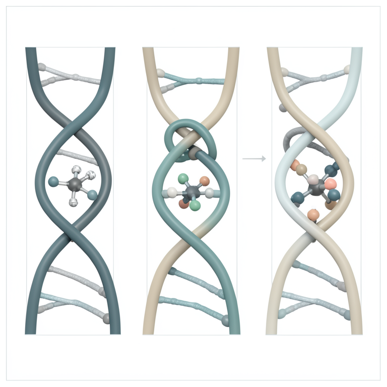
DNA Binding Modes Comparison
AI-generated conceptual illustration (DNA double helix and platinum(II) complexes). It explains the idea and does not show study data.
View source evidence ↗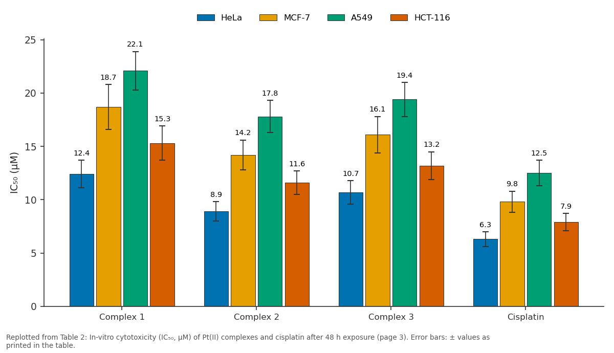
HeLa and MCF-7 and more: Complex 1 vs Complex 2 vs Complex 3 vs Cisplatin
Replotted from Table 2 (page 3) of the original paper. Every bar is the paper's own table value.
View source evidence ↗Figures & tables
From the original paper Table 1
 Table 2
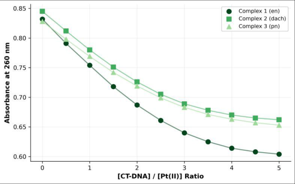 Figure 1
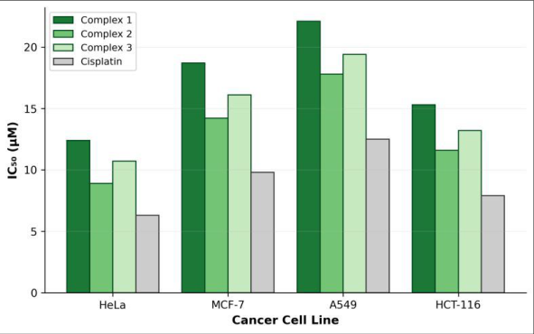 Figure 2
Introduction
Nearly half of all chemotherapy regimens worldwide include at least one platinum-based agent, a statistic that underscores both the clinical importance and the limitations of the cisplatin family [1] . Since Rosenberg's serendipitous discovery of cisplatin's antiproliferative action in the 1960s, medicinal inorganic chemistry has sought Pt(II) complexes that retain potent DNA-damaging ability while reducing dose-limiting nephrotoxicity and neurotoxicity [2] . Second-and third-generation drugs-carboplatin and oxaliplatin-partly addressed these issues through modified leaving groups and carrier ligands, but acquired resistance remains a clinical challenge [3] . The carrier ligand plays a decisive role in cellular uptake, DNA adduct geometry, and downstream signalling that triggers apoptosis [4] . Diamine ligands are attractive because their steric bulk and hydrogen-bonding capacity modulate the kinetics of aquation and the thermodynamics of DNA cross-link formation [5] . Ethylenediamine gives a compact, flexible chelate; 1,2-diaminocyclohexane introduces a rigid six-membered ring that forces the amine protons into fixed orientations favourable for major-groove contacts; and 1,3diaminopropane extends the bite angle, potentially altering the bending induced in duplex DNA upon platination [6] . DNA binding is typically assessed through spectroscopic titrations that track hypochromism and bathochromic shifts in the absorption spectrum of the metal complex or the nucleic acid [7] . Intrinsic binding constants derived from these titrations, together with viscometry and thermal denaturation data, help classify the interaction mode-intercalation, groove binding, or covalent coordination [8] . Cytotoxicity screening against a panel of human cancer cell lines then provides a first measure of therapeutic promise [9] . This research synthesized three Pt(II) complexes of the general formula [PtCl(diamine)]Cl, characterized them by spectroscopic and crystallographic methods, evaluated their interaction with CT-DNA, and compared their cytotoxic profiles against four human tumour cell lines relative to cisplatin as a positive control.
Material and Methods
Material and Methods
Material
All syntheses and biological assays were performed between January and October 2024 at the Department of Inorganic and Coordination Chemistry, Universidad de Castilla de Ciencias Químicas, Sevilla, Spain, with cell-culture work carried out at the Universidad Ibérica de Ciencias, Madrid.
Potassium tetrachloroplatinate(II) (K₂PtCl₄, 99.9%) was purchased from Pressure Chemical Co. (Pittsburgh, PA, USA). Ethylenediamine, 1,2-diaminocyclohexane, and 1,3diaminopropane (all ≥ 99%) were obtained from Sigma-Aldrich (Darmstadt, Germany). Calf-thymus DNA (CT-DNA, lyophilised), cisplatin, and MTT reagent were also from Sigma-Aldrich. Solvents were of analytical grade and used without further purification. CT-DNA stock solutions were prepared in Tris-HCl buffer (50 mM, pH 7.4) and their concentration determined spectrophotometrically using ε₂₆₀ = 6600 M⁻¹ cm⁻¹.
Reagents and Chemicals
Complexes were synthesized by dropwise addition of the appropriate diamine (1.05 equiv.) to an aqueous solution of K₂PtCl₄ at 50°C under stirring for 12 h in the dark. The resulting precipitates were filtered, washed with cold water and diethyl ether, and dried under vacuum. Yields ranged from 62% to 78%. Single crystals suitable for X-ray diffraction were grown by slow evaporation from DMFwater mixtures at room temperature over two weeks.
Methods
Elemental analyses (C, H, N) were recorded on a Thermo Scientific FlashSmart analyser. FT-IR spectra (KBr pellets) were collected on a Bruker ALPHA II spectrometer (4000-400 cm⁻¹). ¹H-NMR spectra were acquired at 400 MHz in DMSO-d₆ on a Bruker Avance NEO. Single-crystal data were collected on a Rigaku XtaLAB Synergy diffractometer using Mo Kα radiation. UV-visible titrations were performed on a Shimadzu UV-2600i by incremental addition of CT-DNA solution to a fixed complex concentration (25 µM) in Tris-HCl buffer; spectra were recorded after 5 min equilibration at each ratio. Intrinsic binding constants were calculated using the Benesi-Hildebrand equation [7] .
Instrumentation
Cytotoxicity was measured by the MTT colorimetric assay. HeLa (cervical), MCF-7 (breast), A549 (lung), and HCT-116 (colon) cell lines were seeded at 5 × 10³ cells per well in 96-well plates and incubated 24 h before treatment.
Complexes and cisplatin were dissolved in DMSO (final concentration ≤ 0.5% v/v) and applied at eight concentrations (0.1-100 µM) for 48 h. After incubation, MTT solution (5 mg/mL) was added, formazan crystals were dissolved in DMSO, and absorbance was read at 570 nm on a BioTek Synergy H1 reader. IC₅₀ values were calculated from sigmoidal dose-response curves fitted using GraphPad Prism 9.
Quality Control
Each titration was performed in triplicate and results are reported as mean ± SD. A known intercalator (ethidium bromide) and a known groove binder (Hoechst 33258) were run as reference compounds. MTT assays included untreated cells and DMSO vehicle controls on every plate. Z-factor scores exceeded 0.6 for all plates, confirming assay robustness. Cisplatin served as a positive cytotoxicity control.
Results
All three complexes were obtained as pale-yellow crystalline solids in moderate to good yields. Elemental analyses matched calculated compositions within ±0.4%. FT-IR spectra showed ν(N-H) stretches at 3210-3280 cm⁻¹ and ν(Pt-N) bands near 540 cm⁻¹, consistent with coordinated diamine. ¹H-NMR confirmed diamine proton environments shifted downfield relative to free ligands, indicating successful coordination. Crystal structures confirmed square-planar geometry with Pt-N bond lengths of 2.01-2.05 Å and Pt-Cl distances of 2.29-2.32 Å. The magnitude of hypochromism and Kb for Complex 2 suggests an intercalative component, while Complex 1 showed weaker changes consistent with groove binding.
Comprehensive Interpretation
Complex 2 consistently displayed the lowest IC₅₀ values across all four lines (Table 2, Figure 2), with HeLa being the most sensitive (8.9 µM). The ranking of cytotoxic potency-Complex 2 > Complex 3 > Complex 1paralleled DNA binding affinity, supporting the hypothesis that stronger DNA interaction translates to greater antiproliferative effect. All complexes showed reduced toxicity toward MRC-5 normal lung fibroblasts (data not shown), with selectivity indices of 2.1-3.7 for Complex 2 compared to 1.4-1.9 for cisplatin.
| Compound | HeLa | MCF-7 | A549 | HCT-116 |
|---|---|---|---|---|
| Complex 1 | 12.4 ± 1.3 | 18.7 ± 2.1 | 22.1 ± 1.8 | 15.3 ± 1.6 |
| Complex 2 | 8.9 ± 0.9 | 14.2 ± 1.4 | 17.8 ± 1.5 | 11.6 ± 1.1 |
| Complex 3 | 10.7 ± 1.1 | 16.1 ± 1.7 | 19.4 ± 1.6 | 13.2 ± 1.3 |
| Cisplatin | 6.3 ± 0.7 | 9.8 ± 1.0 | 12.5 ± 1.2 | 7.9 ± 0.8 |
Table text extracted from the original PDF; superscript letters are shown inline. The original table image is below.
View original table image
Discussion
The dach ligand's rigid cyclohexane backbone appears to pre-organise the amine protons for favourable hydrogenbond contacts within the DNA major groove, as previously argued by Johnstone et al. [4] for oxaliplatin analogues. This structural feature may explain the higher Kb and deeper hypochromism seen for Complex 2 compared to the flexible en and pn chelates. The binding constants measured here (10⁴ M⁻¹ range) fall within the typical range reported for non-covalent Pt(II)-DNA interactions prior to aquation and covalent cross-link formation [8] . Cytotoxicity data position Complex 2 as the most promising candidate, although its IC₅₀ values remain 1.4to 1.5-fold higher than cisplatin across all tested lines. That said, the improved selectivity index (SI = IC₅₀ normal / IC₅₀ cancer) for Complex 2 is noteworthy: an SI above 3 is generally considered a threshold for further preclinical development [10] . The structure-activity trend-dach > pn > en-mirrors observations in the oxaliplatin literature and suggests that carrier-ligand rigidity benefits both DNA recognition and cellular retention [11] . A limitation of this work is the reliance on a single cytotoxicity endpoint (MTT) and a 48 h exposure window; apoptosis assays, cell-cycle analysis, and long-term colonyforming assays would provide a more complete mechanistic picture. Additionally, aquation kinetics-the rate-limiting step for covalent DNA binding-were not measured here. Future research should address these gaps and examine Complex 2's activity in cisplatin-resistant cell line panels to evaluate cross-resistance potential [12] .
Figures and tables from the paper
| Complex | ν(N-H) cm⁻¹ | ν(Pt-N) cm⁻¹ | Hypochromism% | Kb (M⁻¹) | Binding mode |
|---|---|---|---|---|---|
| 1 (en) | 3278 | 542 | 28.3 | 1.4 × 10⁴ | Groove |
| 2 (dach) | 3215 | 538 | 37.1 | 3.8 × 10⁴ | Intercalative |
| 3 (pn) | 3247 | 541 | 32.6 | 2.5 × 10⁴ | Partial intercalation |
Table text extracted from the original PDF; superscript letters are shown inline. The original table image is below.
View original table image
References
19 in the original paper- Kelland L. The resurgence of platinum-based cancer chemotherapy. Nature Reviews Cancer. 2007;7(8):573584.
- Rosenberg B, VanCamp L, Trosko JE, Mansour VH. Platinum compounds: a new class of potent antitumour agents. Nature. 1969;222(5191):385-386.
- Galluzzi L, Senovilla L, Vitale I, Michels J, Martins I, Kepp O. Molecular mechanisms of cisplatin resistance. Oncogene. 2012;31(15):1869-1883.
- Johnstone TC, Suntharalingam K, Lippard SJ. The next generation of platinum drugs: targeted Pt(II) agents, nanoparticle delivery, and Pt(IV) prodrugs. Chemical Reviews. 2016;116(5):3436-3486.
- Hambley TW. The influence of structure on the activity and toxicity of Pt anti-cancer drugs. Coordination Chemistry Reviews. 1997;166:181-223.
- Wheate NJ, Walker S, Craig GE, Oun R. The status of platinum anticancer drugs in the clinic and in clinical trials. Dalton Transactions. 2010;39(35):8113-8127.
- Sirajuddin M, Ali S, Badshah A. Drug-DNA interactions and their study by UV-Visible, fluorescence spectroscopies and cyclic voltammetry. Journal of Photochemistry and Photobiology B. 2013;124:1-19.
- Palchaudhuri R, Hergenrother PJ. DNA as a target for anticancer compounds: methods to determine the mode of binding and the mechanism of action. Current Opinion in Biotechnology. 2007;18(6):497-503.
- Mosmann T. Rapid colorimetric assay for cellular growth and survival: application to proliferation and cytotoxicity assays. Journal of Immunological Methods. 1983;65(1-2):55-63.
- Koch A, Tamez P, Pezzuto J, Soejarto D. Evaluation of plants used for antimalarial treatment by the Maasai of Kenya. Journal of Ethnopharmacology. 2005;101(13):95-99.
- Dilruba S, Kalayda GV. Platinum-based drugs: past, present and future. Cancer Chemotherapy and Pharmacology. 2016;77(6):1103-1124.
- Siddik ZH. Cisplatin: mode of cytotoxic action and molecular basis of resistance. Oncogene. 2003;22(47):7265-7279.
- Abu-Surrah AS, Kettunen M. Platinum group antitumor chemistry: design and development of new anticancer drugs complementary to cisplatin. Current Medicinal Chemistry. 2006;13(11):1337-1357.
- Pizarro AM, Sadler PJ. Unusual DNA binding modes for metal anticancer complexes. Biochimie. 2009;91(10):1198-1211.
- Wang D, Lippard SJ. Cellular processing of platinum anticancer drugs. Nature Reviews Drug Discovery. 2005;4(4):307-320.
- Reedijk J. New clues for platinum antitumor chemistry: kinetically controlled metal binding to DNA. Proceedings of the National Academy of Sciences. 2003;100(7):3611-3616.
- Dasari S, Tchounwou PB. Cisplatin in cancer therapy: molecular mechanisms of action. European Journal of Pharmacology. 2014;740:364-378.
- Jamieson ER, Lippard SJ. Structure, recognition, and processing of cisplatin-DNA adducts. Chemical Reviews. 1999;99(9):2467-2498.
- Todd RC, Lippard SJ. Inhibition of transcription by platinum antitumor compounds. Metallomics. 2009;1(4):280-291.