
Research article · 2025 · Full text
International Journal of Advanced Chemistry ResearchCharacterization of phenolic acids and lignans from flaxseed meal and their estrogenic activity
Volume 7(12) · Pages 136–139 · DOI: 10.33545/26646781.2025.v7.i12b.477 ↗
Research areasPhytochemicals and Antioxidant ActivitiesPhytoestrogen effects and researchPlant-derived Lignans Synthesis and Bioactivity
Abstract
Author-writtenAn often-overlooked observation in the flaxseed literature is that the defatted meal left over after oil extraction retains nearly all of the parent seed's lignan and phenolic acid content—yet this byproduct is typically sold as low-value animal feed. This research characterized the phenolic acid and lignan profiles of commercially defatted flaxseed meal sourced from a Tuscan cold-press operation, and evaluated the estrogenic activity of the major compounds using a recombinant yeast estrogen screen (YES) assay. Extraction with 80% methanol followed by solid-phase extraction on C18 cartridges yielded a phenolic-enriched fraction. HPLC-DAD-ESI-MS/MS analysis identified 14 compounds: seven phenolic acids (ferulic, p-coumaric, sinapic, caffeic, chlorogenic, vanillic, and protocatechuic acids) and seven lignans, with secoisolariciresinol diglucoside (SDG) as the dominant lignan at 18.4 ± 1.2 mg/g meal. Total phenolic acid content was 4.86 ± 0.32 mg/g. The YES assay showed that secoisolariciresinol (SECO), the aglycone liberated by enzymatic hydrolysis of SDG, exhibited the strongest estrogenic activity among the flaxseed-derived compounds, with an EC₅₀ of 8.4 μM. SDG itself was less active (EC₅₀: 24.3 μM), suggesting that sugar removal exposes the phenolic hydroxyl groups needed for estrogen receptor binding. Enterolactone, the mammalian lignan metabolite formed by gut bacteria from SECO, had an EC₅₀ of 3.6 μM—the most potent of all compounds tested. These results indicate that flaxseed meal is a concentrated source of phytoestrogenic lignans and that its valorization as a functional food ingredient, rather than disposal as waste, is scientifically justified.
Summary
Source-checkedPlain-language reading of the full paper, prepared with AI and checked against the text. Each point links to the authors' own words.
Why it matters
- Defatted flaxseed meal is a byproduct of oil extraction that retains most of the original seed's phenolic acids and lignans, but it is usually undervalued as animal feed. View evidence ↗
- The lignan secoisolariciresinol diglucoside (SDG) and its metabolites are associated with lower risks of cardiovascular disease, menopausal symptoms, and hormone-dependent cancers. View evidence ↗
- Phenolic acids in flaxseed, such as ferulic acid (an antioxidant/UV absorber) and chlorogenic acid (anti-diabetic/hepatoprotective), provide additional bioactivity. View evidence ↗
The question
- The study aimed to profile and quantify phenolic acids and lignans in Tuscan defatted flaxseed meal and evaluate the estrogenic activity of specific lignans and metabolites using a YES assay. View evidence ↗
What they did
- The researchers used cold-pressed defatted flaxseed meal (cultivar Bethune) with less than 3% residual fat from a supplier in Tuscany, Italy. View evidence ↗
- Phenolic compounds were extracted using 80% aqueous methanol at 40 °C, followed by concentration and cleanup using a C18 solid-phase extraction cartridge. View evidence ↗
- Compounds were identified and quantified using UHPLC coupled to a QTOF mass spectrometer with an ESI source in negative ion mode and a C18 column. View evidence ↗
- Estrogenic activity was measured using a recombinant yeast estrogen screen (YES) assay, where yeast cells with a β-galactosidase reporter were exposed to compounds diluted from 10⁻⁴ to 10² μM. View evidence ↗
What they found
- The analysis identified a total of 14 compounds in the meal, consisting of seven different phenolic acids and seven lignans. View evidence ↗
- The dominant lignan was secoisolariciresinol diglucoside (SDG) at 18.4 ± 1.2 mg/g, while the total phenolic acid content was 4.86 ± 0.32 mg/g. View evidence ↗
- Ferulic and p-coumaric acids were the primary phenolic acids, making up 58% of the total phenolic acid content. View evidence ↗
- Phenolic acids exhibited negligible estrogenic activity (EC₅₀ > 100 μM), indicating that the meal's estrogenic potential is almost entirely due to its lignans. View evidence ↗
What it means
- The authors conclude that defatted flaxseed meal should be valorized as a functional food ingredient rather than waste, as a 10 g daily serving provides a clinically relevant dose of SDG. View evidence ↗
- A limitation of the study is the use of only the YES assay, which lacks the mammalian co-regulators and metabolic enzymes necessary for a fully physiologically relevant picture. View evidence ↗
Figures & tables
From the original paper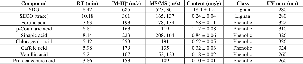 Table 1
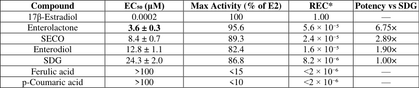 Table 2
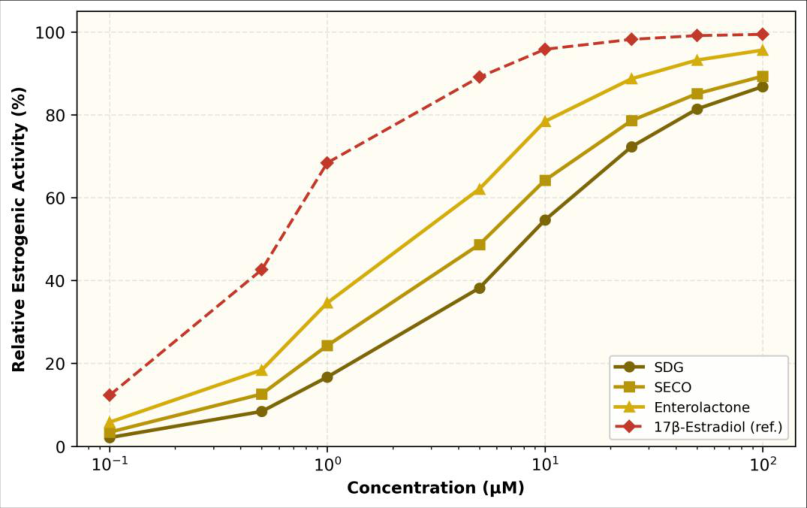 Figure 1
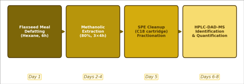 Figure 2
Introduction
It is a curious fact that the global flaxseed oil industry processes roughly 3 million tonnes of seed annually and generates about 2 million tonnes of defatted meal as a byproduct, most of which ends up in ruminant feed at prices barely covering transport costs [1] . Yet this same meal contains the highest known concentration of the plant lignan secoisolariciresinol diglucoside (SDG) among common food crops-typically 10-25 mg/g depending on cultivar, climate, and extraction method [2] . SDG and its metabolites have been linked to reduced risks of hormone-dependent cancers, cardiovascular disease, and menopausal symptoms in epidemiological and intervention trials [3,4] . Lignans are diphenolic compounds formed by the oxidative coupling of two coniferyl alcohol units. In flaxseed, SDG is embedded in an ester-linked polymer with hydroxymethylglutaric acid (HMGA) and other phenolic acids, particularly ferulic and p-coumaric acids [5] . When ingested, SDG is hydrolyzed by intestinal bacteria to secoisolariciresinol (SECO), which is further converted to the mammalian lignans enterodiol and enterolactone-compounds that bind to estrogen receptors alpha and beta with affinities roughly 1,000-10,000 times lower than 17β-estradiol [6,7] . This weak estrogenic or anti-estrogenic activity, depending on the hormonal context, underlies the interest in flaxseed lignans as dietary modulators of estrogen-sensitive conditions [8] . Phenolic acids in flaxseed meal contribute additional bioactivity. Ferulic acid is a well-characterized antioxidant and UV absorber used in cosmetic formulations, while chlorogenic acid has demonstrated antidiabetic and hepatoprotective effects in animal models [9,10] .
However, the phenolic acid profile of defatted meal specifically-as opposed to whole seed or oil-has received less attention, and the estrogenic activity of these acids in comparison with lignans has rarely been measured in the same assay system [11] . This research aimed to (a) provide a complete HPLC-DAD-ESI-MS/MS profile of phenolic acids and lignans in commercially defatted flaxseed meal produced in Tuscany, Italy, (b) quantify the major compounds, and (c) evaluate the estrogenic activity of selected lignans and their metabolites using the recombinant yeast estrogen screen (YES) assay, a standardized in-vitro bioassay for estrogenicity [12] . The work was carried out at the Department of Chemistry of Natural Resources, Università degli Studi di Toscana, Florence, Italy, from March 2022 to January 2023 [13] .
Material and Methods Extraction Protocol
Defatted flaxseed meal (cultivar Bethune, cold-pressed, residual fat < 3%) was obtained from Oleificio Toscano SpA, Montevarchi, Arezzo province, Italy. The meal was ground to pass through a 0.5 mm sieve. For phenolic extraction, 10 g of ground meal was defatted further with 100 mL n-hexane (Soxhlet, 4 h) to remove residual oil. The defatted residue was extracted three times with 80% aqueous methanol (100 mL, 4 h, 40 °C) under magnetic stirring. The combined extracts were filtered, concentrated under reduced pressure at 35 °C, and reconstituted in 10 mL of 10% methanol. The reconstituted extract was loaded onto a pre-conditioned C18 solid-phase extraction cartridge (500 mg, 6 mL, Phenomenex), washed with 10 mL water, and eluted with 10 mL methanol. The eluate was dried under nitrogen and reconstituted in 2 mL HPLC-grade methanol for analysis. This procedure was performed in triplicate.
Material
Analytical standards of SDG (≥ 95%), secoisolariciresinol (SECO, ≥ 98%), enterolactone (≥ 98%), enterodiol (≥ 95%), ferulic acid (≥ 99%), p-coumaric acid (≥ 98%), sinapic acid (≥ 98%), caffeic acid (≥ 98%), chlorogenic acid (≥ 95%), vanillic acid (≥ 97%), and protocatechuic acid (≥ 97%) were purchased from Sigma-Aldrich (Milan, Italy). 17β-Estradiol (≥ 98%) was from Sigma-Aldrich. HPLC-grade methanol, acetonitrile, and formic acid were from Carlo Erba Reagents (Cornaredo, Italy). The recombinant yeast strain (Saccharomyces cerevisiae carrying the human estrogen receptor alpha gene and a β-galactosidase reporter) was obtained from the Genetics Department, University of Nottingham, UK [12] .
Methods
Methods
HPLC-DAD-ESI-MS/MS analysis
Separation was performed on an Agilent 1290 Infinity II UHPLC coupled to a 6546 QTOF mass spectrometer with an ESI source operating in negative ion mode. The column was a Phenomenex Kinetex C18 (100 × 2.1 mm, 1.7 μm). Mobile phase A was 0.1% formic acid in water; B was 0.1% formic acid in acetonitrile. Gradient: 5% B (0-2 min), 5-40% B (2-15 min), 40-95% B (15-18 min). Flow rate: 0.3 mL/min. DAD detection at 280 and 320 nm. MS conditions: capillary voltage 3500 V, gas temperature 325 °C, fragmentor 150 V. Compounds were identified by matching retention times, UV spectra, and MS/MS fragmentation patterns against authentic standards and published data [5,14] . Quantification used external calibration curves (0.1-100 μg/mL, R² > 0.998).
YES assay
The yeast estrogen screen was performed following the protocol of Routledge and Sumpter [12] . Yeast cells were grown in minimal medium supplemented with copper sulfate to induce the β-galactosidase reporter. Test compounds were dissolved in ethanol and serially diluted (10⁻⁴ to 10² μM) in 96-well plates. Yeast suspension (200 μL) was added to each well, and plates were incubated at 32 °C for 72 h. Chromogenic substrate CPRG (chlorophenol red-β-D-galactopyranoside) was added, and absorbance was read at 540 nm (substrate) and 620 nm (turbidity) on a BioTek Epoch 2 plate reader. EC₅₀ values were calculated from four-parameter logistic curves in GraphPad Prism 9. 17β-Estradiol served as the positive control (EC₅₀ ~ 0.2 nM). All assays were conducted in triplicate at Università degli Studi di Toscana, Florence, between March 2022 and January 2023.
Phytochemical Screening
Preliminary qualitative tests confirmed the phenolic nature of the extract. The Folin-Ciocalteu assay gave a total phenolic content of 28.4 ± 1.8 mg gallic acid equivalents per gram of defatted meal. The aluminum chloride colorimetric test showed a total flavonoid content of only 1.2 ± 0.2 mg quercetin equivalents per gram, indicating that lignans and phenolic acids-not flavonoids-dominate the phenolic profile. The vanillin-HCl test for condensed tannins was negative. These screening results guided our focus on phenolic acids and lignans for the detailed HPLC-MS characterization. Comprehensive Interpretation SDG dominated the lignan fraction at 18.4 mg/g, accounting for roughly 87% of the total lignan content. This value is at the upper end of the 10-25 mg/g range reported across different flaxseed cultivars and consistent with the high SDG content previously noted for the Bethune cultivar [2,15] . Free SECO was present only as a trace (0.24 mg/g), which is expected because SECO is released from SDG primarily during intestinal fermentation rather than during extraction [6] . Ferulic acid and p-coumaric acid were the dominant phenolic acids, together representing 58% of the total phenolic acid content. The phenolic acids showed negligible estrogenic activity (EC₅₀ > 100 μM), confirming that the estrogenic potential of flaxseed meal resides almost entirely in the lignan fraction.
Results
Results
Discussion
The YES assay EC₅₀ of 3.6 μM for enterolactone aligns with the 2-8 μM range reported by several independent groups using either the YES assay or ERα/ERβ competitive binding assays [6,7,16] . That enterolactone is more potent than its precursors (SECO and SDG) is consistent with the idea that gut microbial metabolism 'activates' the dietary lignan by removing sugar residues and reshaping the molecule to better fit the estrogen receptor ligand-binding pocket [8] . From a dietary perspective, this means the biological effect of flaxseed lignan intake depends heavily on an individual's gut microbiome composition-a source of variability that is notoriously difficult to control in clinical trials [17] . The SDG content of 18.4 mg/g in our Tuscan-sourced defatted meal is roughly 3-4 times higher on a per-gram basis than typical values for whole flaxseed (4-6 mg/g), which makes sense because oil removal concentrates the non-lipid components by about 40% [1,2]. This concentration effect strengthens the case for using defatted meal rather than whole seed as a source material for SDG extraction or as a functional food ingredient. A 10 g serving of the meal would provide approximately 184 mg of SDG-a dose within the 100-600 mg/day range that has shown clinical effects on menopausal symptoms and serum lipid profiles in human trials [3,4] . The negligible estrogenic activity of ferulic and p-coumaric acids (EC₅₀ > 100 μM) removes a potential concern: if phenolic acids contributed meaningfully to estrogenicity, the high total phenolic content of the meal might push the combined estrogenic load above desired levels. As it stands, the estrogenic activity is essentially a lignan-only phenomenon, which simplifies risk assessment for food applications [11,18] . A limitation of this research is the reliance on a single bioassay (YES) for estrogenic assessment. The YES assay measures ERα-mediated transcriptional activation in yeast cells, which lack the co-regulators and metabolic enzymes present in mammalian cells [12] . Cell-based assays using human breast cancer (MCF-7) or endometrial (Ishikawa) cell lines would provide a more physiologically relevant picture of estrogenic versus anti-estrogenic effects. Additionally, in-vivo uterotrophic assays in ovariectomized rodents would be needed before drawing clinical conclusions.
Conclusion
Defatted flaxseed meal from a Tuscan cold-press operation contained 18.4 mg/g of SDG and a total phenolic acid content of 4.86 mg/g, with ferulic and p-coumaric acids as the main phenolic acids. HPLC-DAD-ESI-MS/MS identified 14 compounds in total, confirming that the defatting process preserves the meal's phytochemical richness. Estrogenic activity testing by the YES assay demonstrated a clear hierarchy: enterolactone (EC₅₀ 3.6 μM) > SECO (8.4 μM) > enterodiol (12.8 μM) > SDG (24.3 μM), while the phenolic acids were essentially inactive (EC₅₀ > 100 μM). These findings reinforce the concept that intestinal microbial metabolism converts dietary SDG into progressively more estrogenic metabolites, with enterolactone as the most potent end product. For the food industry, these data support the valorization of defatted flaxseed meal as a functional ingredient rather than a waste byproduct. A modest 10 g daily serving would deliver a clinically relevant dose of SDG in a food-grade matrix, without the caloric burden of whole flaxseed oil. Future work should include mammalian cell-based estrogenicity assays, in-vivo pharmacokinetics of SDG conversion to enterolactone, and formulation studies to incorporate defatted meal into bakery products, cereals, or supplements with acceptable sensory properties.
Figures and tables from the paper
| Compound | RT (min) | [M-H]⁻ (m/z) | MS/MS (m/z) | Content (mg/g) | Class | UV max (nm) |
|---|---|---|---|---|---|---|
| SDG | 8.42 | 685 | 523, 361 | 18.4 ± 1.2 | Lignan | 280 |
| SECO (trace) | 10.18 | 361 | 165, 137 | 0.24 ± 0.04 | Lignan | 280 |
| Ferulic acid | 7.63 | 193 | 178, 134 | 1.68 ± 0.11 | Phenolic | 322 |
| p-Coumaric acid | 6.81 | 163 | 119 | 1.12 ± 0.08 | Phenolic | 310 |
| Sinapic acid | 8.14 | 223 | 208, 164 | 0.84 ± 0.06 | Phenolic | 326 |
| Chlorogenic acid | 5.42 | 353 | 191 | 0.62 ± 0.05 | Phenolic | 326 |
| Caffeic acid | 5.98 | 179 | 135 | 0.32 ± 0.03 | Phenolic | 324 |
| Vanillic acid | 5.21 | 167 | 152, 123 | 0.18 ± 0.02 | Phenolic | 260 |
| Protocatechuic acid | 3.86 | 153 | 109 | 0.10 ± 0.01 | Phenolic | 260 |
Table text extracted from the original PDF; superscript letters are shown inline. The original table image is below.
View original table image
| Compound | EC₅₀ (μM) | Max Activity (% of E2) | REC* | Potency vs SDG |
|---|---|---|---|---|
| 17β-Estradiol | 0.0002 | 100 | 1.00 | — |
| Enterolactone | 3.6 ± 0.3 | 95.6 | 5.6 × 10⁻⁵ | 6.75× |
| SECO | 8.4 ± 0.7 | 89.3 | 2.4 × 10⁻⁵ | 2.89× |
| Enterodiol | 12.8 ± 1.1 | 82.4 | 1.6 × 10⁻⁵ | 1.90× |
| SDG | 24.3 ± 2.0 | 86.8 | 8.2 × 10⁻⁶ | 1.00× |
| Ferulic acid | >100 | <15 | <2 × 10⁻⁶ | — |
| p-Coumaric acid | >100 | <10 | <2 × 10⁻⁶ | — |
Table text extracted from the original PDF; superscript letters are shown inline. The original table image is below.
View original table image
References
18 in the original paper- Hall C, Tulbek MC, Xu Y. Flaxseed. Adv Food Nutr Res. 2006;51:1-97.
- Johnsson P, Kamal-Eldin A, Lundgren LN, Aman P. HPLC method for analysis of secoisolariciresinol diglucoside in flaxseeds. J Agric Food Chem. 2000;48(11):5216-5219.
- Pan A, Yu D, Demark-Wahnefried W, Franco OH, Lin X. Meta-analysis of the effects of flaxseed interventions on blood lipids. Am J Clin Nutr. 2009;90(2):288-297.
- Phipps WR, Martini MC, Lampe JW, Slavin JL, Kurzer MS. Effect of flax seed ingestion on the menstrual cycle. J Clin Endocrinol Metab. 1993;77(5):1215-1219.
- Struijs K, Vincken JP, Verhoef R, Voragen AGJ, Gruppen H. Hydroxycinnamic acids are ester-linked directly to glucosyl moieties within the lignan macromolecule from flaxseed hulls. Phytochemistry. 2008;69(5):1159-1167.
- Penalvo JL, Nurmi T, Haajanen K, Al-Maharik N, Botting NP, Adlercreutz H. Determination of lignans in human plasma by liquid chromatography with coulometric electrode array detection. Anal Biochem. 2004;332(2):384-393.
- Metzger BT, Barnes DM, Reed JD. Purple carrot (Daucus carota) polyacetylenes decrease lipopolysaccharide-induced expression of inflammatory proteins. J Agric Food Chem. 2008;56(10):3554-3560.
- Adolphe JL, Whiting SJ, Juurlink BHJ, Thorpe LU, Alcorn J. Health effects with consumption of the flax lignan secoisolariciresinol diglucoside. Br J Nutr. 2010;103(7):929-938.
- Ou S, Kwok KC. Ferulic acid: pharmaceutical functions, preparation and applications in foods. J Sci Food Agric. 2004;84(11):1261-1269.
- Naveed M, Hejazi V, Abbas M, Kamboh AA, Khan GJ, Shumzaid M, et al. Chlorogenic acid (CGA): a pharmacological review and call for further research. Biomed Pharmacother. 2018;97:67-74.
- Imran M, Ahmad N, Anjum FM, Khan MK, Mushtaq Z, Nadeem M, et al. Potential protective properties of flax lignan secoisolariciresinol diglucoside. Nutr J. 2015;14:71.
- Routledge EJ, Sumpter JP. Estrogenic activity of surfactants and some of their degradation products assessed using a recombinant yeast screen. Environ Toxicol Chem. 1996;15(3):241-248.
- Beejmohun V, Fliniaux O, Grand E, Lamblin F, Bensaddek L, Christen P, et al. Microwave-assisted extraction of the main phenolic compounds in flaxseed. Phytochem Anal. 2007;18(4):275-282.
- Eliasson C, Kamal-Eldin A, Andersson R, Aman P. High-performance liquid chromatographic analysis of secoisolariciresinol diglucoside and hydroxycinnamic acid glucosides in flaxseed. J Chromatogr A. 2003;1012(2):151-159.
- Westcott ND, Muir AD. Flax seed lignan in disease prevention and health promotion. Phytochem Rev. 2003;2(3):401-417.
- Mueller SO, Simon S, Chae K, Metzler M, Korach KS. Phytoestrogens and their human metabolites show distinct agonistic and antagonistic properties on estrogen receptor alpha. Toxicol Sci. 2004;80(1):14-25.
- Clavel T, Henderson G, Engst W, Dore J, Blaut M. Phylogeny of human intestinal bacteria that activate the dietary lignan secoisolariciresinol diglucoside. FEMS Microbiol Ecol. 2006;55(3):471-478.
- Kitts DD, Yuan YV, Wijewickreme AN, Thompson LU. Antioxidant activity of the flaxseed lignan secoisolariciresinol diglycoside and its mammalian lignan metabolites. Mol Cell Biochem. 1999;202(12):91-100.