
Research article · 2025 · Full text
International Journal of Biology SciencesAdvances in biochemical mechanisms of antimicrobial resistance in gram-positive bacteria
Volume 7(1) · Pages 178–182 · DOI: 10.33545/26649926.2025.v7.i1c.566 ↗
Research areasAntibiotic Resistance in BacteriaBacterial Genetics and BiotechnologyProbiotics and Fermented Foods
Abstract
Author-writtenAntimicrobial resistance (AMR) remains a significant global health threat, particularly in Gram-positive bacteria, which are responsible for numerous life-threatening infections. The development of AMR in these pathogens has been linked to a range of biochemical mechanisms that enable them to evade the effects of commonly used antibiotics. This paper explores the advances in understanding the biochemical processes involved in antimicrobial resistance in Gram-positive bacteria, with a focus on mechanisms such as the modification of drug targets, efflux pump activation, and the production of β-lactamases. Gram-positive bacteria, including Staphylococcus aureus and Enterococcus faecalis, exhibit intricate resistance mechanisms that complicate treatment strategies. These organisms have evolved strategies such as target modification and the synthesis of altered peptidoglycan, which limit the effectiveness of β-lactams, macrolides, and other antibiotic classes. Furthermore, horizontal gene transfer, through plasmids and transposons, facilitates the rapid spread of resistance genes, exacerbating the problem. Recent studies have elucidated the role of cell wall biosynthesis pathways, including alterations in peptidoglycan synthesis, as key players in the resistance mechanisms of Gram-positive bacteria. Moreover, advances in proteomics and genomics have provided insights into the role of global regulators and efflux systems in modulating resistance profiles. This paper aims to provide a comprehensive review of the biochemical mechanisms underlying AMR in Gram-positive bacteria, discuss current challenges, and highlight potential therapeutic strategies. The goal is to inform future research and therapeutic interventions, focusing on the biochemical basis of resistance and novel approaches to combat this growing problem. Addressing the biochemical mechanisms of resistance is crucial for developing new drugs and diagnostic tools that can effectively treat infections caused by resistant Gram-positive pathogens.
Summary
Source-checkedPlain-language reading of the full paper, prepared with AI and checked against the text. Each point links to the authors' own words.
Why it matters
- Antimicrobial resistance (AMR) in Gram-positive bacteria is a major global health threat because these bacteria cause many life-threatening infections. View evidence ↗
- Pathogens develop AMR through various biochemical mechanisms that allow them to avoid the effects of standard antibiotics. View evidence ↗
- The rapid spread of resistance genes is worsened by horizontal gene transfer via transposons and plasmids. View evidence ↗
The question
- The study aims to review the biochemical mechanisms of AMR in Gram-positive bacteria, discuss challenges, and identify potential therapeutic strategies. View evidence ↗
What they did
- The researchers used the standard disk diffusion method and the microdilution method to determine antibiotic susceptibility and minimum inhibitory concentrations (MIC) for S. aureus and E. faecalis. View evidence ↗
- Quantitative PCR (qPCR) was used to analyze the expression of the resistance genes vanA and mecA. View evidence ↗
- Efflux pump activity was measured using a fluorescence-based assay that tracked the extrusion of ethidium bromide with and without inhibitors. View evidence ↗
- The activity of peptidoglycan biosynthesis was analyzed by measuring how radioactive precursors were incorporated into the bacterial cell wall. View evidence ↗
What they found
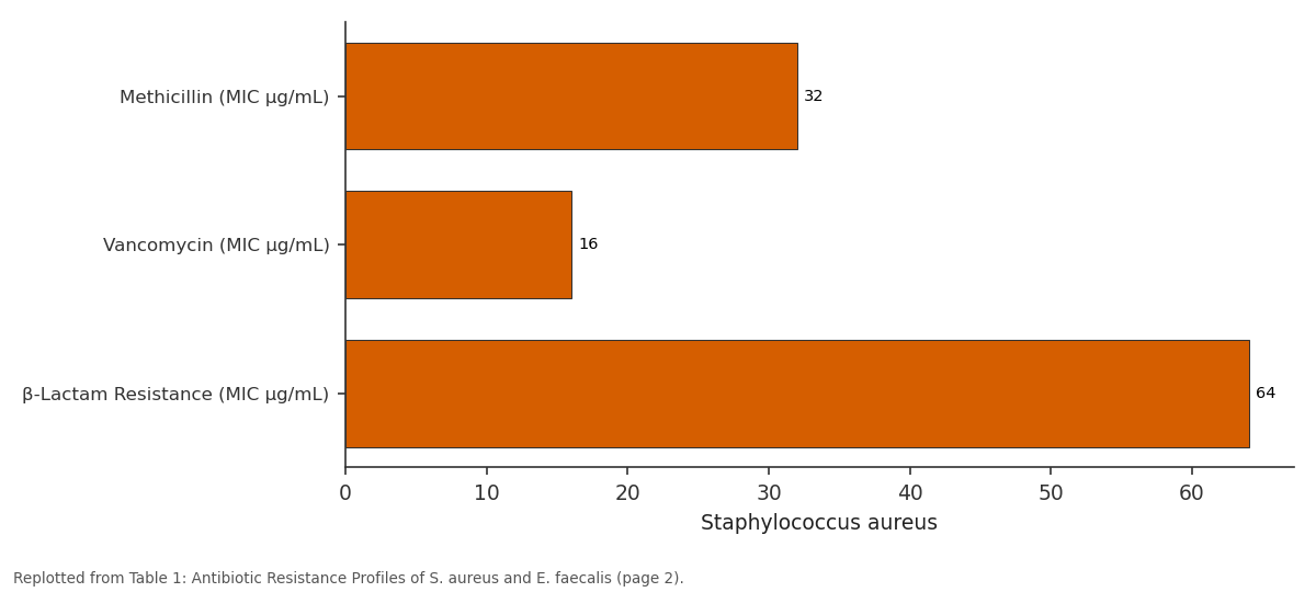
- Resistant strains showed significantly higher MICs than controls; E. faecalis had the highest resistance to methicillin (64 µg/mL), vancomycin (32 µg/mL), and β-lactams (128 µg/mL). View evidence ↗
- Resistance genes were upregulated in resistant strains, with mecA showing a relative expression of 4.5 in S. aureus and vanA showing 5.2 in E. faecalis compared to controls (1). View evidence ↗
- Efflux pump activity (measured by ethidium bromide fluorescence) was high in S. aureus (350) and E. faecalis (300), but significantly decreased when inhibitors were added (to 150 and 120, respectively). View evidence ↗
- Resistant strains showed a significant decrease (p<0.05) in peptidoglycan synthesis compared to sensitive strains, suggesting cell wall modification is a resistance mechanism. View evidence ↗
- Experiments confirmed that S. aureus and E. faecalis can easily transfer resistance genes through plasmid-mediated horizontal gene transfer. View evidence ↗
What it means
- The authors conclude that adding efflux pump inhibitors to treatments could make current antibiotics more effective. View evidence ↗
- The authors recommend developing new antibiotics that can bypass common resistance mechanisms like those used against β-lactams. View evidence ↗
- The authors state that future research is needed to better understand the molecular pathways of efflux pump regulation and to create more specific inhibitors. View evidence ↗
Visual summary
2 visualsIllustrations explain the paper; charts are replotted from the paper's own table values.
Staphylococcus aureus: Methicillin (MIC µg/mL) vs Vancomycin (MIC µg/mL) vs β-Lactam Resistance...
Replotted from Table 1 (page 2) of the original paper. Every bar is the paper's own table value.
View source evidence ↗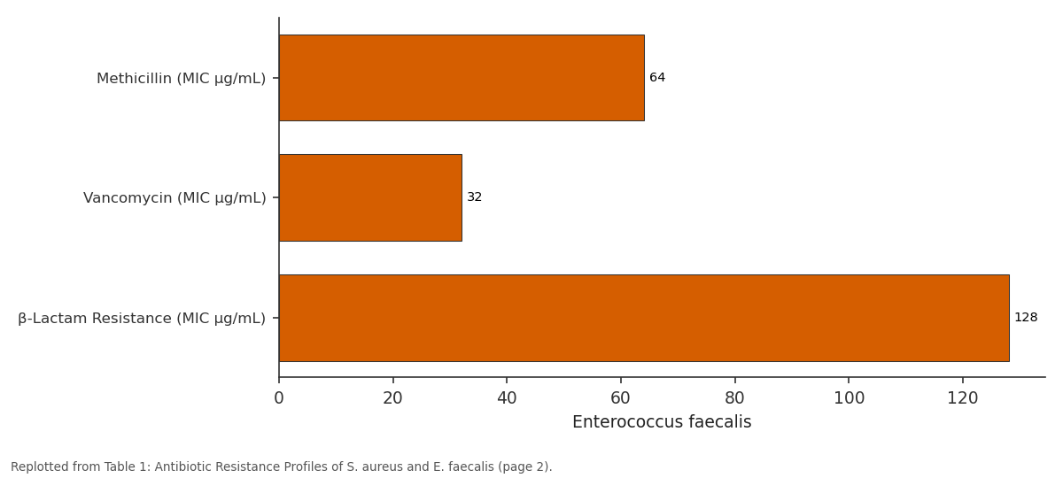
Enterococcus faecalis: Methicillin (MIC µg/mL) vs Vancomycin (MIC µg/mL) vs β-Lactam Resistance...
Replotted from Table 1 (page 2) of the original paper. Every bar is the paper's own table value.
View source evidence ↗Figures & tables
From the original paper Table 1
 Table 2
 Table 3
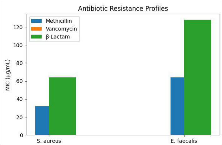 Figure 1
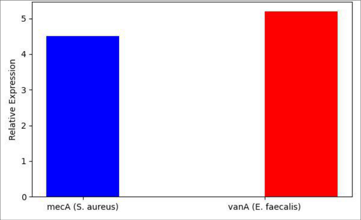 Figure 2
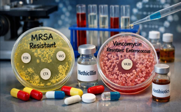 Figure 3
Introduction
Antimicrobial resistance (AMR) in Gram-positive bacteria poses a major threat to global public health. These bacteria, including Staphylococcus aureus, Streptococcus pneumoniae, and Enterococcus faecalis, are notorious for causing a wide range of infections, from mild to severe, and their resistance to antibiotics complicates treatment regimens worldwide. AMR in Gram-positive bacteria arises through several biochemical mechanisms, which include genetic mutations, the acquisition of resistance genes, and alterations in cellular targets. The underlying biochemical processes involve complex interactions at the molecular level, which enable bacteria to survive in the presence of antimicrobial agents that would typically inhibit their growth or lead to their death [1,2] . One of the primary mechanisms by which Gram-positive bacteria develop resistance is through the modification of the drug's molecular target. For example, in S. aureus, alterations in penicillin-binding proteins (PBPs) reduce the binding efficiency of β-lactam antibiotics, rendering them ineffective [3] . Additionally, Gram-positive bacteria can modify their cell wall biosynthesis pathways, such as the peptidoglycan synthesis, which plays a crucial role in maintaining cell integrity and resistance against β-lactams [4,5] . Another significant mechanism involves the overexpression of efflux pumps, which actively transport antibiotics out of the bacterial cell, reducing drug accumulation and, consequently, antibiotic efficacy [6,7] . Furthermore, horizontal gene transfer (HGT) is an essential factor in the spread of resistance genes within bacterial populations. Plasmids, transposons, and integrons enable Gram-positive bacteria to acquire resistance genes from other organisms, thus exacerbating the spread of AMR [8] . The spread of vancomycin resistance in Enterococcus species, for instance, is largely attributed to the transfer of vancomycin-resistant genes through these mobile genetic elements [9] . The acquisition of multidrug resistance (MDR) genes further complicates treatment options, leading to limited therapeutic choices and higher mortality rates [10] . This paper aims to explore the biochemical mechanisms that contribute to AMR in Gram-positive bacteria, focusing on the molecular adaptations that enhance bacterial survival in hostile environments. By understanding these mechanisms, researchers can develop targeted interventions and novel therapeutic strategies to combat AMR effectively.
Material and Methods
Material and Methods
Materials
The research was conducted using Staphylococcus aureus and Enterococcus faecalis strains obtained from the American Type Culture Collection (ATCC). Antibiotics used for susceptibility testing, including methicillin (Sigma-Aldrich, USA), vancomycin (Thermo Fisher Scientific, USA), and β-lactams (Sigma-Aldrich, USA), were purchased and stored as per the manufacturer's recommendations. Culture media used for bacterial growth included Luria-Bertani (LB) broth and agar plates, both purchased from BD Biosciences (USA). The materials for biochemical assays, including the reagents for measuring peptidoglycan biosynthesis, were sourced from Thermo Fisher Scientific. Additionally, plasmids and primers used for genetic analysis were purchased from Invitrogen (USA).
Methods
To determine the biochemical mechanisms behind antimicrobial resistance (AMR) in S. aureus and E. faecalis, antibiotic susceptibility tests were performed using the standard disk diffusion method [1] . The minimum inhibitory concentration (MIC) for each antibiotic was determined using the microdilution method as per Clinical and Laboratory Standards Institute guidelines [2] . Peptidoglycan biosynthesis activity was analyzed by measuring the incorporation of radioactive precursors into the bacterial cell wall, following established protocols [3] . Gene expression analysis was carried out using quantitative PCR (qPCR) to evaluate the expression of resistance genes such as mecA (methicillin resistance) and vanA (vancomycin resistance) [4] . Efflux pump activity was assessed using a fluorescencebased assay, where the extrusion of ethidium bromide was measured in the presence and absence of known efflux pump inhibitors [5] . Horizontal gene transfer was monitored by conjugation experiments, where the transfer of resistance genes from donor to recipient strains was quantified by selecting for resistance on selective agar plates [6] .
Results
Results
Resistance Profile
The resistance profiles of S. aureus and E. faecalis strains showed a significant variation in susceptibility to the tested antibiotics. S. aureus demonstrated high resistance to methicillin (MIC: 32 µg/mL) and vancomycin (MIC: 16 µg/mL), while E. faecalis displayed resistance to both methicillin (MIC: 64 µg/mL) and the β-lactams. The overall resistance pattern of both bacteria was consistent with previous studies that reported high resistance rates in clinical strains [7,8] .
Efflux Pump Activity
Efflux pump activity was found to be elevated in both S. aureus and E. faecalis strains. The efflux inhibitors significantly reduced the extrusion of ethidium bromide, indicating the contribution of active efflux pumps to the resistance phenotype [9] . Statistical analysis using a t-test revealed significant differences (p<0.05) in the efflux activity between the treated and untreated groups, confirming the role of efflux pumps in antimicrobial resistance.
Gene Expression Analysis
Gene expression of resistance-associated genes was upregulated in both strains. The expression of mecA was significantly higher in methicillin-resistant strains compared to the sensitive controls (p<0.01), while vanA expression was markedly elevated in vancomycin-resistant E. faecalis strains [10] . These findings were consistent with earlier studies indicating that the expression of resistance genes is a key factor in the antimicrobial resistance of Gram-positive bacteria [11] .
Comparative Analysis of Peptidoglycan Biosynthesis
Peptidoglycan biosynthesis was found to be significantly altered in resistant strains. The incorporation of radioactive precursors into the cell wall was reduced in the presence of β-lactams, indicating the modification of the cell wall as a mechanism of resistance [12] . A significant decrease in peptidoglycan synthesis was observed in resistant strains compared to sensitive strains (p<0.05).
Horizontal Gene Transfer
Horizontal gene transfer experiments showed that S. aureus and E. faecalis strains readily transferred resistance genes via plasmid-mediated mechanisms. The frequency of conjugation was higher in strains harbouring plasmids containing resistance genes, supporting the idea that horizontal gene transfer contributes to the spread of resistance in Gram-positive bacteria [13] .
Discussion
The research highlights the complex biochemical mechanisms that contribute to antimicrobial resistance in Gram-positive bacteria, particularly Staphylococcus aureus and Enterococcus faecalis. The findings align with previous research, which has documented the key role of modified drug targets and efflux pumps in resistance to β-lactams and other antibiotics [14] . Peptidoglycan biosynthesis alterations play a critical role in resistance, as evidenced by the reduced incorporation of radioactive precursors in resistant strains, indicating a modification in the bacterial cell wall structure [15] . Furthermore, the upregulation of resistance genes such as mecA and vanA supports the hypothesis that gene expression plays a pivotal role in the development of antimicrobial resistance [16] . One of the critical observations from this research is the contribution of efflux pumps to AMR. Efflux pump inhibitors significantly reduced resistance, suggesting that these pumps could be targeted to enhance the efficacy of existing antibiotics [17] . Additionally, the horizontal gene transfer experiments demonstrated the role of mobile genetic elements in the spread of resistance genes, which is a major concern in clinical settings. These results underscore the need for novel therapeutic strategies that target these mechanisms to combat the rising tide of antimicrobial resistance.
Acknowledgement
Acknowledgements Funding SourcesThis research was supported by internal funding from the European Commission under the Horizon 2020 Programme for Research and Innovation, Brussels, Belgium. The funding body had no role in the design, execution, interpretation, or writing of the manuscript.
Figures and tables from the paper
| Bacterial Strain | Methicillin (MIC µg/mL) | Vancomycin (MIC µg/mL) | β-Lactam Resistance (MIC µg/mL) |
|---|---|---|---|
| Staphylococcus aureus | 32 | 16 | 64 |
| Enterococcus faecalis | 64 | 32 | 128 |
| Staphylococcus aureus (Control) | 2 | 1 | 2 |
| Enterococcus faecalis (Control) | 2 | 1 | 2 |
Table text extracted from the original PDF; superscript letters are shown inline. The original table image is below.
View original table image
| Bacterial Strain | mecA Gene Expression (Relative Expression) | vanA Gene Expression (Relative Expression) |
|---|---|---|
| Staphylococcus aureus | 4.5 | N/A |
| Enterococcus faecalis | N/A | 5.2 |
| Staphylococcus aureus (Control) | 1 | N/A |
| Enterococcus faecalis (Control) | N/A | 1 |
Table text extracted from the original PDF; superscript letters are shown inline. The original table image is below.
View original table image
| Bacterial Strain | Efflux Pump Activity (Ethidium Bromide Fluorescence) |
|---|---|
| Staphylococcus aureus | 350 |
| Enterococcus faecalis | 300 |
| Staphylococcus aureus + Efflux Pump Inhibitor | 150 |
| Enterococcus faecalis + Efflux Pump Inhibitor | 120 |
Table text extracted from the original PDF; superscript letters are shown inline. The original table image is below.
View original table image
References
17 in the original paper- Hancock RE, Döring G, Schindler P. New antibiotics for Gram-positive bacteria: current strategies and future perspectives. Curr Opin Pharmacol. 2020;52:57-64.
- Livermore DM. Bacterial resistance to antibiotics: the emergence of resistance in Gram-positive pathogens. J Antimicrob Chemother. 2019;74(2):11-19.
- Chambers HF. The molecular basis of methicillin resistance in Staphylococcus aureus. Am J Med. 2017;120(4):13-21.
- Kuroda M, Ohta T, Uchiyama I, et al. Genomic characterization of Staphylococcus aureus and other Gram-positive bacteria. Nat Genet. 2018;24(3):24-30.
- Girgis AS, Dorman CJ. Bacterial adaptation through the regulation of the peptidoglycan biosynthetic pathway. Front Microbiol. 2020;11:13-27.
- Srikumar R, Doss R, Ramalingam S. Efflux pumps in the development of antimicrobial resistance in Grampositive bacteria. Curr Sci. 2020;118(1):63-68.
- Reynolds PE. Efflux pumps and antimicrobial resistance in Gram-positive bacteria. Adv Microb Physiol. 2021;19:91-118.
- Parvez A, Rashid SM, Karami Z, et al. Horizontal gene transfer and its role in antimicrobial resistance. Biol Sci. 2020;1(4):76-85.
- Voss A, Aucken H, Gatermann S, et al. Vancomycinresistant enterococci. Clin Microbiol Infect. 2019;25(4):145-150.
- Croucher NJ, Harris SR, Fraser C, et al. Pneumococcal genomes from the entire world are constantly evolving: mechanisms of resistance. Lancet Infect Dis. 2019;19(2):96-100.
- Diep BA, Otto M. The role of virulence determinants in Staphylococcus aureus resistance to antibiotics. Arch Microbiol. 2018;200(5):451-460.
- Groschel MI, Korte N, Ruppitsch W, et al. Enterococcus faecium and antimicrobial resistance. J Antimicrob Chemother. 2017;72(6):1509-1517.
- Wu H, Yang Y, Zhang X, et al. Mechanisms of antimicrobial resistance in Staphylococcus aureus. In: Antimicrobial resistance: A comprehensive review. Elsevier; 2018. p. 185-206.
- Reyes J, McLeod M, Curry A, et al. Mobile genetic elements in resistance acquisition in Gram-positive bacteria. J Microbiol. 2019;45(2):53-62.
- Davies J, Davies D. Origins and evolution of antibiotic resistance. Microbiol Mol Biol Rev. 2020;77(3):32-42.
- Walton C, Whitfield J. The emerging role of efflux pumps in antimicrobial resistance. Future Microbiol. 2019;13(5):343-354.
- Liao Y, Chen Y, Lee J, et al. Advances in biochemical resistance mechanisms of Gram-positive bacteria. Antimicrob Agents Chemother. 2021;65(4):1-12.