
Research article · 2026 · Full text
International Journal of Horticulture and Food SciencePathogenicity test of major fungal pathogens in vanilla (Vanilla planifolia Andrews)
Volume 8(1) · Pages 41–43 · DOI: 10.33545/26631067.2026.v8.i1a.454 ↗
Research areasBiochemical and biochemical processesAllelopathy and phytotoxic interactionsBiological and pharmacological studies of plants
Abstract
Author-writtenVanilla (Vanilla planifolia Andrews) is an economically important spice crop affected by several fungal diseases that significantly reduce yield and quality. The present investigation was undertaken to isolate, identify, and prove the pathogenicity of major fungal pathogens associated with vanilla under Konkan conditions. Diseased samples of leaves, stems, roots, and pods were collected and subjected to tissue isolation on Potato Dextrose Agar (PDA). The isolated fungi were purified, mass multiplied, and tested for pathogenicity on healthy vanilla plants and pods following Koch’s postulates. Five major fungal pathogens, namely Colletotrichum gloeosporioides Penz., Fusarium oxysporum F. sp. vanillae Schlecht, Rhizoctonia bataticola (Taub.) Butler, Sclerotium rolfsii Sacc and Aspergillus niger van Tieghem, were isolated and confirmed. Pathogenicity tests revealed characteristic symptoms such as stem rot, leaf spot, basal rot, wilting, and pod rot. Reisolation of pathogens confirmed their causal role. The study highlights the importance of these fungal pathogens in vanilla cultivation and provides a basis for developing effective disease management strategies.
Summary
Source-checkedPlain-language reading of the full paper, prepared with AI and checked against the text. Each point links to the authors' own words.
Why it matters
- Vanilla is a commercially grown climbing orchid valued for its vanillin-rich cured beans, which provide a distinct aroma and flavor. View evidence ↗
- The vanilla crop is highly vulnerable to various viral and fungal diseases, leading to significant losses in yield. View evidence ↗
- As vanilla cultivation has intensified, the frequency of disease has risen, creating a need for systematic research into the organisms causing these diseases. View evidence ↗
The question
- The study aimed to isolate and identify major fungal pathogens of vanilla using morphological traits and confirm their pathogenicity using Koch's postulates. View evidence ↗
What they did
- The researchers collected diseased samples from the roots, stems, leaves, and pods of vanilla plants. View evidence ↗
- Infected tissues were sterilized with 0.1% mercuric chloride for two minutes, washed, and then incubated on Potato Dextrose Agar (PDA) at 28 ± 1 °C for seven days. View evidence ↗
- Pathogenicity tests were conducted using healthy, one-month-old vanilla vines grown in pots. View evidence ↗
- Inoculum was applied either to the soil near the collar region or to slightly injured plant parts, with non-inoculated plants serving as controls. View evidence ↗
What they found
- The study isolated and confirmed five major fungal pathogens: Colletotrichum gloeosporioides, Fusarium oxysporum F. sp. vanillae, Rhizoctonia bataticola, Sclerotium rolfsii, and Aspergillus niger. View evidence ↗
- Colletotrichum gloeosporioides caused water-soaked lesions at the collar region after 18 days, leading to stem and shoot tip rot and complete wilting within 45 days. View evidence ↗
- Fusarium oxysporum F. sp. vanillae produced water-soaked lesions near the soil line after 11 days, resulting in stem rot and complete wilting within 20 days. View evidence ↗
- Sclerotium rolfsii caused collar rot 12 days after inoculation, characterized by white mycelia and sclerotia on the stem and soil, leading to plant death. View evidence ↗
- Aspergillus niger inoculation of fresh green vanilla pods caused yellowing, wrinkling, foul odor, and rotting at the distal end. View evidence ↗
What it means
- The authors concluded that the five identified fungi are major pathogens of vanilla, as their pathogenic potential was demonstrated through artificial inoculation and reisolation. View evidence ↗
- The authors state that understanding these fungi is essential for creating integrated disease management strategies to reduce yield losses. View evidence ↗
Visual summary
1 visualsIllustrations explain the paper; they do not show study data.
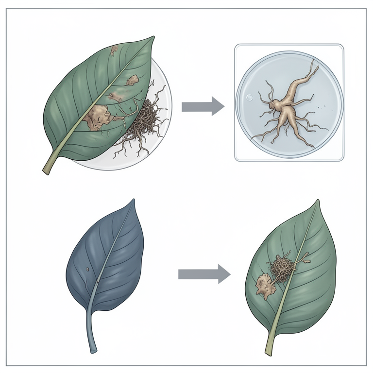
Pathogenicity Verification Process
AI-generated conceptual illustration (Vanilla planifolia plants and fungal cultures). It explains the idea and does not show study data.
View source evidence ↗Introduction
Vanilla (Vanilla planifolia Andrews), a perennial climbing orchid belonging to the family Orchidaceae, is cultivated commercially for its cured beans rich in vanillin, which imparts characteristic aroma and flavour. Vanilla is widely used in food, beverage, pharmaceutical, perfumery, and tobacco industries and is popularly known as the "Prince of Spices". In India, vanilla cultivation has expanded to states such as Karnataka, Kerala, Maharashtra, Andhra Pradesh, and parts of the Konkan region due to its high economic returns. Vanilla thrives under warm and humid climatic conditions with well-distributed rainfall and moderate shade. However, the crop is highly susceptible to a range of fungal and viral diseases that cause considerable yield losses. Important fungal diseases include stem rot, root rot, basal rot, leaf spot, anthracnose, bean rot, and shoot tip rot. With the intensification of vanilla cultivation, disease incidence has increased, necessitating systematic studies on causal organisms. The present investigation was therefore carried out to isolate major fungal pathogens associated with vanilla, identify them based on morphological characteristics, and confirm their pathogenicity through Koch's postulates.
Materials and Methods
The present investigation was undertaken to isolate, identify, and prove the pathogenicity of major fungal pathogens associated with vanilla under Konkan conditions.
Isolation of fungal pathogens
Diseased samples of vanilla leaves, stems, roots, and pods showing typical symptoms were collected. Small infected tissue bits were surface sterilized using 0.1% mercuric chloride for two minutes, followed by three washings with sterile distilled water. The tissue bits were placed on Potato Dextrose Agar (PDA) plates and incubated at 28 ± 1 °C for seven days. Fungal colonies emerging from the tissues were examined microscopically and sub-cultured.
Purification and Maintenance of cultures
Pure cultures were obtained using the single hyphal tip method. The purified isolates were maintained on PDA slants under aseptic conditions and stored at optimum temperature for further studies.
Mass Multiplication of Inoculum
Each fungal isolate was mass multiplied on Potato Dextrose Broth (PDB). Sterilized PDB in 500 ml conical flasks was inoculated with 5 mm mycelial discs from 7-day-old cultures and incubated at room temperature for 10 days.
Pathogenicity Test
One-month-old healthy vanilla vines grown in pots were used for pathogenicity tests. The mycelial mat and spores from PDB cultures were blended with sterile water to obtain a uniform inoculum. The inoculum was applied to the soil near the collar region or on slightly injured plant parts, depending on the pathogen. Control plants were maintained without inoculation. Inoculated plants were covered with polythene bags to maintain high humidity and observed regularly for symptom development.
Re-isolation and Identification
Pathogens were reisolated from artificially infected plants showing typical symptoms and cultured on PDA to confirm Koch's postulates. Identification of pathogens was carried out based on cultural and morphological characteristics and confirmed at NIKU Bio-Research Laboratory, Pune.
Results and Discussion
Results and Discussion
Isolation and Identification of Pathogens
Five fungal pathogens were consistently isolated from diseased vanilla samples. These included Colletotrichum gloeosporioides, Fusarium oxysporum F. sp. vanillae, Rhizoctonia bataticola, Sclerotium rolfsii, and Aspergillus Niger. Identification was based on colony morphology, mycelial characteristics, and spore structures, which were in agreement with standard descriptions reported in the literature.
Pathogenicity Tests
Pathogenicity Tests
Colletotrichum gloeosporioides
Inoculated vanilla plants developed water-soaked lesions at the collar region 18 days after inoculation, followed by chlorosis, stem rot, and shoot tip rotting. Complete wilting occurred within 45 days. Similar symptoms have been reported earlier for anthracnose and stem rot of vanilla by Amaresh et al. (2005) [2] . Also similar results for Colletotrichum sp. were obtained on inoculation by Thakare and Patil (1995) [6] , Mertely and Legard (2004) [4] when associated with chrysanthemum and strawberry, respectively.
Fusarium oxysporum F. sp. vanillae
Symptoms appeared 11 days after inoculation as watersoaked lesions near the soil line, progressing rapidly to stem rot and die-back of the shoot tip. Complete wilting occurred within 20 days, confirming its aggressive pathogenic nature. Alconero (1968) [1] , also proved the pathogenicity of species of Fusarium and Rhizoctonia on vanilla.
Rhizoctonia bataticola
Water-soaked lesions appeared within six days of inoculation, followed by basal stem discoloration, shredding, and complete wilting within 30 days. The symptoms closely resembled natural infection observed in the field. Proved the pathogenicity of Rhizoctonia bataticola on cowpea. The inoculated plants showed typical symptoms of leaf blight after 8-10 days of inoculation. Patil (2003) [6] proved the pathogenicity of R. bataticola causing stem and root rot of sunflower in field condition and also in pot experiment.
Sclerotium rolfsii
Infected plants showed collar rot symptoms 12 days after inoculation, with white radiating mycelia and sclerotial formation on soil and stem surfaces, leading to girdling and plant death. Shredding of stem and roots was observed only in infection of Rhizoctonia sp. In support of these findings, Ashoka et al. (2006) [3] similarly reported for root rot of vanilla caused by Rhizoctonia bataticola (Taub.) Butler. Thomas et al. (2002) [9] also proved pathoginicity of S. rolfsii causing bean and stem rot of vanilla.
Aspergillus Niger
Inoculation of fresh green vanilla pods resulted in yellowing, distal-end rotting, wrinkling, and development of foul odour. Reisolated cultures were identical to the original isolate, confirming pathogenicity. The successful reproduction of disease symptoms and reisolation of pathogens fulfilled Koch's postulates, establishing the causal role of these fungi in major vanilla diseases. Reddy et al. (1991) [7] proved that groundnut seed germination was affected by pathogens like A. Niger, A. terreus, A. Niger. Suryawanshi (2005) [8] proved the pathogenicity of Aspergillus Niger on vanilla pods.
Conclusion
The study confirmed that Colletotrichum gloeosporioides, Fusarium oxysporum F. sp. vanillae, Rhizoctonia bataticola, Sclerotium rolfsii, and Aspergillus Niger are major fungal pathogens associated with vanilla. Their pathogenic potential was conclusively demonstrated through artificial inoculation and reisolation. Understanding the pathogenic behaviour of these fungi is essential for developing integrated disease management strategies to minimize yield losses in vanilla cultivation.
References
9 in the original paper- Alconero R. Natural and artificial infection of vanilla roots by Fusarium oxysporum F. sp. vanillae and Rhizoctonia sp. Phytopathology. 1968;58:553.
- Amaresh YS, Raveendra BH, Negalur RB, Teggi MY, Lokesh MS. Scenario of diseases of vanilla in Uttar Kannada district. In: National Seminar on ETPPSR, Annamalai University; 2005 Mar 7-8, p. 82.
- Ashoka S, Hegde YR. Scenario of fungal diseases of Vanilla planifolia in Karnataka. Int J Plant Sci. 2006;1(2):269-272.
- Mertley JC, Legard DE. Detection, isolation and pathogenicity of Colletotrichum spp. from strawberry petioles. Plant Dis. 2004;88(4):407-412.
- Nurawan, Nazurudin SB, Nikil M, Matsumoto K. Experiments on the introduction of biological and cultural control of stem rot diseases of vanilla. Ind Crop Res J. 1992;4:20-26.
- Patil PP. Bioefficacy of antagonists against seed/root/stem rot of sunflower. Akola: Dr. Punjabrao Deshmukh Krishi Vidyapeeth; 2003.
- Reddy GR, Reddy AGR, Rao KC. Effect of different seed dressing fungicides against certain seed-borne fungi of groundnut. J Oilseed Res. 1991;8(1):79-83.
- Suryawanshi BB. Studies on major diseases of vanilla and their management. Dapoli: Dr. B.S. Konkan Krishi Vidyapeeth; 2005.
- Thomas J, Vijayan AK, Suseela Bahi R. Vanilla diseases in India and their management. Indian J Arecanut Spices Med Plants. 2002;4:143-149.