
Research article · 2025 · Full text
International Journal of Advanced Chemistry ResearchTargeting the sars-cov-2 main protease: A mini-review of warheads, p-site design, and lead optimization
Volume 7(11) · Pages 90–98 · DOI: 10.33545/26646781.2025.v7.i11b.336 ↗
Research areasComputational Drug Discovery MethodsClick Chemistry and ApplicationsSARS-CoV-2 and COVID-19 Research
Abstract
Author-writtenSARS-CoV-2 main protease (Mpro / 3CLpro) is an essential viral enzyme responsible for polyprotein processing and maturation of the replication complex. Its strict preference for a Leu-Gln↓(Ser/Ala/Gly) motif and lack of close human homologs make it a high-selectivity antiviral target. Following early release of high-resolution crystal structures, global discovery programs rapidly applied structure-based design, converging on three non-negotiable interaction principles: (i) a P1 glutamine surrogate anchoring the S1 pocket, (ii) hydrophobic P2 bulk occupying S2, and (iii) tunable P3/P4 groups to balance solubility and pharmacokinetics. This mini-review summarizes the major chemotype classes advanced in 2020-2022: peptidomimetic aldehydes (11a/11b), ketoamides (13b), repurposed covalent inhibitors (N3, GC-376, boceprevir), nitrile-based reversible covalent inhibitors culminating in nirmatrelvir (Paxlovid), and emerging non-peptidyl scaffolds and metal complexes. We highlight design rules that consistently produced potent leads and caution against scaffolds that showed biochemical activity but failed under reducing or orthogonal validation assays. Collectively, these efforts illustrate how structural biology and computer-aided drug design accelerated the transition from concept to a clinically approved oral antiviral.
Summary
Source-checkedPlain-language reading of the full paper, prepared with AI and checked against the text. Each point links to the authors' own words.
Why it matters
- The SARS-CoV-2 main protease (Mpro) is critical for the virus because it processes polyproteins into functional proteins needed for replication. View evidence ↗
- Mpro is an ideal drug target because it recognizes a specific protein sequence not found in human proteases, reducing the risk of off-target toxicity. View evidence ↗
- Small-molecule antivirals are necessary because the virus evolves to escape vaccines and antibody-based therapies. View evidence ↗
The question
- The review aims to summarize the design evolution of four classes of Mpro inhibitors and identify the rules that lead to clinical success. View evidence ↗
What they did
- Inhibitor design focused on three key structural requirements: a P1 glutamine surrogate for the S1 pocket, a hydrophobic P2 group for S2, and modifiable P3/P4 groups for solubility. View evidence ↗
- The design of these inhibitors targets the catalytic dyad consisting of Cys145 and His41. View evidence ↗
What they found
- Peptidomimetic aldehydes 11a and 11b showed nanomolar inhibitory activity (IC₅₀ of 0.053 µM and 0.040 µM respectively) and were non-toxic above 100 µM. View evidence ↗
- The repurposed Michael acceptor N3 demonstrated antiviral activity in Vero cells with an EC₅₀ of 16.77 µM. View evidence ↗
- Ketoamide inhibitor 13b showed an IC₅₀ of 0.67 µM and blocked replication in Calu-3 cells with an EC₅₀ of 4-5 µM. View evidence ↗
- Nirmatrelvir, a nitrile-based reversible inhibitor, became the first FDA-approved oral antiviral targeting Mpro. View evidence ↗
- Ebselen was identified as an Mpro inhibitor with an IC₅₀ of 0.67 µM. View evidence ↗
- The natural products neoechinulin A and echinulin A inhibited Mpro with IC₅₀ values of 0.47 µM and 3.90 µM, respectively. View evidence ↗
- Covalent inhibitors 14a (vinyl sulfone) and 16a (chloroketone) were 10 times more potent than their parent compound X77. View evidence ↗
What it means
- The authors recommend that future development against new variants should integrate computer-aided drug design (CADD), crystallography, and fragment optimization. View evidence ↗
Visual summary
1 visualsIllustrations explain the paper; they do not show study data.
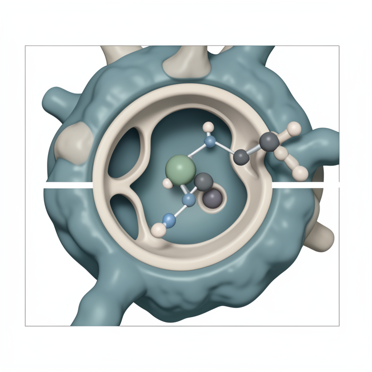
Mpro Inhibitor Design Framework
AI-generated conceptual illustration (SARS-CoV-2 main protease (Mpro) protein structure and a small molecule inhibitor). It explains the idea and does not show study data.
View source evidence ↗Figures & tables
From the original paper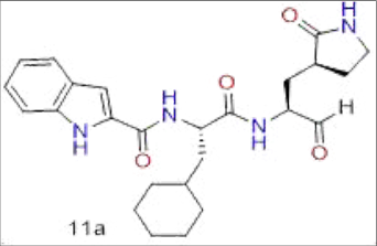 Figure 1Page 2
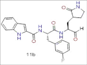 Figure 1Page 2
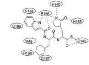 Figure 1Page 2
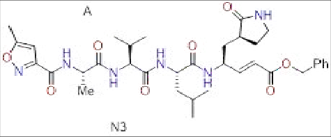 Figure 2Page 3
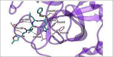 Figure 2Page 3
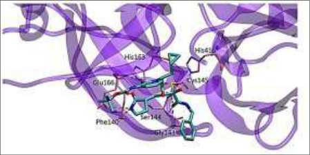 Figure 3Page 3
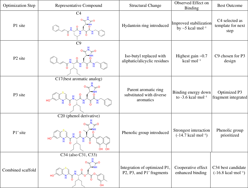 Table 1Page 4
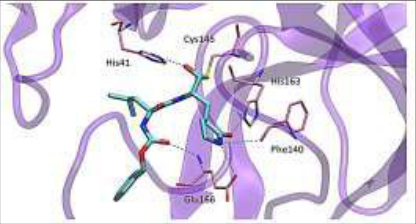 Figure 4Page 4
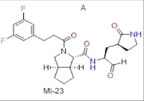 Figure 5Page 5
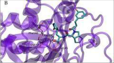 Figure 5Page 5
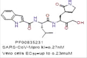 Figure 6Page 5
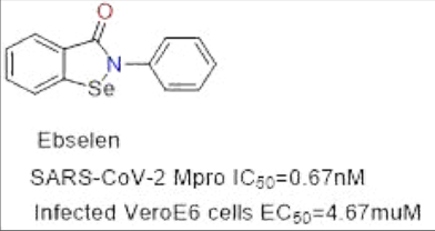 Figure 7Page 5
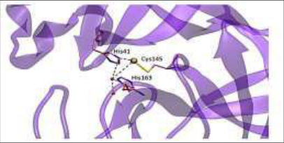 Figure 8Page 5
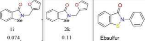 Figure 9Page 5
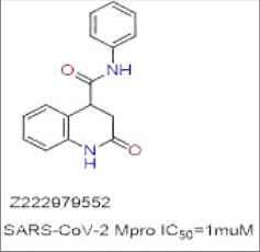 Figure 10Page 6
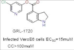 Figure 11Page 6
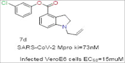 Figure 11Page 6
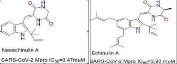 Figure 12Page 6
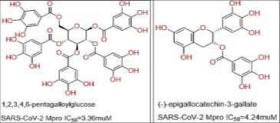 Figure 13Page 6
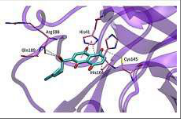 Figure 14Page 7
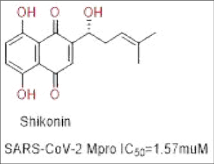 Figure 14Page 7
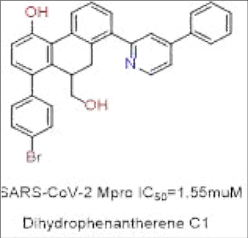 Figure 15Page 7
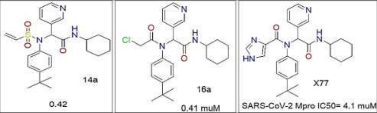 Figure 16Page 7
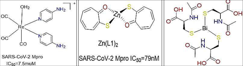 Figure 2Page 7
Introduction
"The COVID-19 pandemic exposed the limitations of relying solely on vaccines and immune-based therapeutics. SARS-CoV-2 continues to evolve, generating variants with altered transmissibility and partial immune escape, reducing the sustained effectiveness of antibody-based therapies and first-generation vaccines. In contrast, small-molecule antivirals that target conserved viral mechanisms remain resilient to viral evolution and are essential for outpatient treatment, early intervention, and protection of immunocompromised patients. Among the viral proteins required for replication, two cysteine proteases such as the papainlike protease (PLpro) and the main protease (Mpro / 3CLpro) cleave the viral polyprotein into functional non-structural proteins (nsps). Mpro performs more than 11 out of 14 proteolytic cleavages, making it indispensable for viral maturation. Unlike host proteases, Mpro requires a unique Leu-Gln↓(Ser/Ala/Gly) motif, which does not occur in human proteases.
This gives Mpro two critical properties from a drug-discovery standpoint 1. High target selectivity: low risk of off-target protease toxicity 2. Conserved substrate recognition: low likelihood of resistance development With the first Mpro crystal structures released just weeks into the pandemic (PDB 6LU7), global research rapidly converged on structure-guided inhibitor design. Early medicinal chemistry campaigns discovered three non-negotiable interaction principles: A P1 glutamine surrogate (e.g., (S)-γ-lactam or hydantoin) to anchor the S1 pocket via His163/Glu166, A hydrophobic P2 group to occupy the deep S2 subsite, A modifiable P3/P4 cap, affecting solubility and pharmacokinetics more than potency.
Warhead choice defined the direction of each scaffold like aldehydes and ketoamides (covalent), Michael acceptors (irreversible), and nitriles (reversible covalent). The earliest leads (11a/11b and 13b) validated the design logic, while repurposing efforts identified N3, boceprevir, and GC-376. Optimization of polarity, metabolic stability, and warhead reactivity ultimately produced nirmatrelvir (Paxlovid), the first FDA-approved oral Mpro inhibitor and a clinical proof that rational covalent design works. This mini-review summarizes the evolution of Mpro inhibitor design across four chemotype classes peptidomimetic aldehydes/ketoamides, repurposed covalent inhibitors, non-peptidyl small molecules, and metal-based complexes and extracts practical design rules: which decisions led to clinical success, and which scaffolds looked promising but collapsed under biochemical scrutiny (redox sensitivity, PAINS behavior).
Target and Pocket Primer
The SARS-CoV-2 main protease (Mpro) contains a catalytic dyad Cys145-His41, responsible for peptide bond cleavage during viral polyprotein processing. The active site is organized into well-defined subsites: S1, S2, S3, and S4, each accommodating corresponding positions on the inhibitor (P1, P2, P3, P4) according to Schechter-Berger notation. The S1 pocket forms a highly conserved hydrogenbonding network (His163, Glu166, Phe140/oxyanion hole), which requires a glutamine-mimicking group at P1 typically an (S)-γ-lactam or hydantoin making it the most critical determinant of potency and selectivity. The S2 pocket is deep and hydrophobic, favoring bulky aliphatic or bicyclic residues at P2, while S3/S4 are more solvent exposed and allow chemical variation to tune solubility and pharmacokinetics rather than intrinsic binding energy. Together, these structural features define the "anchor points" that guide rational inhibitor design.
Peptidomimetic aldehydes were the first chemotype to validate covalent inhibition of Mpro through rational structure-guided design. They exploit the anchoring interactions in the S1 (P1 glutamine mimic) and S2 (hydrophobic P2) pockets while presenting an electrophilic aldehyde warhead that forms a covalent adduct with Cys145. Early optimization focused on identifying the optimal P2 bulky group and a solubility-improving P3 cap. Dai et al. reported the first low-nanomolar leads (11a/11b), establishing aldehydes as credible antiviral candidates and setting the SAR template for subsequent scaffolds.
Peptidomimetic Aldehyde Inhibitors (11a and 11b, Dai et al., 2020)
One of the earliest studies applying structure-based drug design to SARS-CoV-2 Mpro was carried out by Dai and colleagues. Building on knowledge of SARS-CoV inhibitors, particularly those containing an (S)-γ-lactam ring for the S1 pocket, the group designed two new compounds, 11a and 11b. Substituents at P2 (cyclohexyl or 3fluorophenyl) and an indole group at P3 were introduced to enhance interactions. Both inhibitors covalently bound to Cys145 in the protease active site. Crystallographic analysis revealed hydrogen bonding of the lactam oxygen with His163 and Phe140, as hydrophobic stabilization by the P2 group. Additionally, there were contacts from the P3 indole with Glu166 and Pro168. Compound 11a exhibited a half-life of 7.6 hours in rats and 5.5 hours in dogs, with low clearance, making it a stronger candidate than 11b. Both compounds exhibited nanomolar inhibitory activity (IC₅₀ = 0.053 µM for 11a; 0.040 µM for 11b) and sub micromolar EC₅₀ values in infected cells, with no observed toxicity (CC₅₀ > 100 µM). These findings established aldehyde-based peptidomimetics as promising leads for COVID-19 therapy [1] .
A B . This study provided the first structural framework for SARS-CoV-2 protease inhibition, guiding subsequent design efforts [2] .
A B µM) [3] . The crystal structure (PDB 6Y2F) revealed the formation of a thiohemiketal at Cys145, stabilized by hydrogen bonds within the oxyanion hole. A γ-lactam occupied the S1 pocket, while cyclopropyl at P2 gave better fit than cyclohexyl. These results confirmed ketoamides as viable protease inhibitors [2] . 100 µM) [4] .
The crystal structure of GC-376 bound to Mpro (PDB 6WTT) showed hemithioketal formation with Cys145, a γlactam interacting with His163 and Glu166, and benzyl groups filling the S4 pocket. These findings highlighted the potential of repurposing existing protease-targeting drugs [2,5] .
A In 2020, Frecer and colleagues reported a rational design campaign targeting the SARS-CoV-2 main protease (Mpro) using peptidomimetic ketoamide scaffolds closely related to earlier SARS-CoV inhibitors. Beginning with the parent compound 11a, the team performed systematic fragment optimization at the P1, P2, P3, and P1′ positions. [6] .
The crystal structure showed covalent bonding to Cys145 and multiple stabilizing hydrogen bonds within the oxyanion hole and S1 pocket. Many compounds exhibited favorable pharmacokinetics and oral bioavailability of greater than 10%, supporting their development as candidates [2] . A phosphate prodrug (PF-07304814) improved solubility for IV administration. Both compounds showed strong in vitro activity and good safety [7,8] .
Later, oral bioavailability issues with PF-00835231 prompted the design of PF-07321332, a nitrile-based reversible inhibitor. Substitutions at P2 and P3 improved solubility and potency. This compound, now known as nirmatrelvir (marketed as Paxlovid with ritonavir), became the first FDA-approved oral antiviral targeting Mpro [9] .
Organoselenium compounds: Ebselen and derivatives
Screening identified ebselen, a seleno-organic drug with antioxidant and anti-inflammatory activity, as a SARS-CoV-2 Mpro inhibitor (IC₅₀ = 0.67 µM). It also inhibited PLpro (IC₅₀ = 2.26 µM). Crystal structures revealed binding via selenium-sulfur interaction with Cys145 and a secondary domain interface site important for dimerization [10][11][12][13][14][15][16] . Further analogues such as ebsulfur derivatives (compounds 1i, 2k) and hydantoin-based analogs improved potency (IC₅₀ = 0.077 µM). However, activity was diminished in reducing conditions (e.g., DTT), raising concerns of non-specific inhibition [17][18][19] .
Fig 9: Ebselen derivatives and Ebsulfur
Non-peptidyl small molecules Several non-peptidyl scaffolds emerged to address limitations of peptidomimetics (large size, polarity, limited oral exposure), multiple groups explored non-peptidyl chemotypes indole esters, dihydroquinolinones, natural products, and organoselenium compounds. These scaffolds engage Mpro through π-stacking, hydrophobic interactions, or alternative covalent chemistries. However, several hits showed redox sensitivity or promiscuous inhibition, highlighting the need for stringent validation beyond enzymatic IC₅₀. Dihydroquinolinone derivative Z222979552, from large library screening, showed antiviral activity in cells and favorable binding (PDB 7P2G) [20] . Indole chloropyridinyl esters such as GRL-1720 and optimized analog 7d acted as covalent inhibitors, acylating Cys145. Crystal structures confirmed indolemediated π-π interactions (PDB 7RC0) [21,22,2] . stabilized by hydrogen bonds with active-site residues [23] . Polyphenols PGG and EGCG showed micromolar inhibition of Mpro through hydrogen bonding and hydrophobic contacts [24] . Shikonin, a naphthoquinone, induced conformational changes at the catalytic dyad but lost activity in reducing conditions [2,25] .
A B Fig 14 : (A) Crystal structure of the complex formed between shikonin and SARS.CoV-2 Mpro Chemical structure of shikonin.
9, 10-Dihydrophenanthrene derivatives also inhibited Mpro by binding near the dimer interface [26] . and chloroketone (16a) were 10-fold more potent than the parent compound [27] .
Metal-based inhibitors
Metal complexes also demonstrated inhibitory activity. Rhenium tricarbonyl complex 34 inhibited Mpro [28] , while zinc [29] and zinc-thiotropilone complexes showed nanomolar potency [30] . Bismuth drugs such as colloidal bismuth subcitrate (CBS), alone or combined with N-acetyl-L-cysteine, also reduced protease activity [31] .
Conclusion
The rapid discovery of SARS-CoV-2 Mpro inhibitors demonstrated how structural biology and rational design can compress the antiviral discovery timeline. Early peptidomimetic aldehydes and ketoamides established the essential binding rules a P1 glutamine mimic, hydrophobic P2 bulk, and a warhead positioned toward Cys145. Repurposed inhibitors validated covalent engagement, and systematic optimization ultimately produced nirmatrelvir, the first oral clinical Mpro inhibitor. Although many nonpeptidyl and metal-based molecules displayed biochemical activity, only those guided by structure-activity logic and pharmacokinetic feasibility translated into drug-like candidates. Future antiviral development against emerging variants must continue to integrate CADD, crystallography, and fragment-wise optimization to rapidly identify leads with genuine translational potential.
Figures and tables from the paper
| Optimization Step | Representative Compound | Structural Change | Observed Effect on Binding | Best Outcome |
|---|---|---|---|---|
| P1 site | C4 | Hydantoin ring introduced | Improved stabilization by ~5 kcal·mol⁻¹ | C4 selected as template for next step |
| P2 site | C9 | Iso-butyl replaced with aliphatic/alicyclic residues | Highest gain ~0.7 kcal·mol⁻¹ | C9 chosen for P3 design |
| P3 site | C17(best aromatic analog) | Parent aromatic ring substituted with diverse aromatics | Binding energy down to -3.6 kcal·mol⁻¹ | Optimized P3 fragment integrated |
| P1′ site | C20 (phenol derivative) | Phenolic group introduced | Strongest interaction (-14.7 kcal·mol⁻¹) | Phenolic group prioritized |
| Combined scaffold | C34 (also C31, C33) | Integration of optimized P1, P2, P3, and P1′ fragments | Cooperative effect enhanced binding | C34 best candidate (-16.8 kcal·mol⁻¹) |
Table text extracted from the original PDF; superscript letters are shown inline. The original table image is below.
View original table image
References
29 in the original paper- Dai W, Zhang B, Jiang XM, Su H, Li J, Zhao Y, et al. Structure-based design of antiviral drug candidates targeting the SARS-CoV-2 main protease. Science. 2020;368(6497):1331-1335.
- Humphrey W, Dalke A, Schulten K. VMD: visual molecular dynamics. J Mol Graph. 1996;14(1):33-38.
- Zhang L, Lin D, Sun X, Curth U, Drosten C, Sauerhering L, et al. Crystal structure of SARS-CoV-2 main protease provides a basis for design of improved α-ketoamide inhibitors. Science. 2020;368(6489):409412.
- Ma C, Sacco MD, Hurst B, Townsend JA, Hu Y, Szeto T, et al. Boceprevir, GC-376, and calpain inhibitors II, XII inhibit SARS-CoV-2 viral replication by targeting the viral main protease. Cell Res. 2020;30(8):678-692.
- Cáceres JC, Cardenas-Garcia S, Carnaccini S, Seibert B, Rajao DS, Wang J, et al. Efficacy of GC-376 against SARS-CoV-2 virus infection in the K18 hACE2 transgenic mouse model. Sci Rep. 2021;11:9609.
- Qiao J, Li YS, Zeng R, Liu FL, Luo RH, Huang C, et al. SARS-CoV-2 Mpro inhibitors with antiviral activity in a transgenic mouse model. Science. 2021;371(6536):1374-1378.
- Hoffman RL, Kania RS, Brothers MA, Davies JF, Ferre RA, Gajiwala KS, et al. Discovery of ketone-based covalent inhibitors of coronavirus 3CL proteases for the potential therapeutic treatment of COVID-19. J Med Chem. 2020;63(21):12725-12747.
- Boras B, Jones RM, Anson BJ, Arenson D, Aschenbrenner L, Bakowski MA, et al. Discovery of a novel inhibitor of coronavirus 3CL protease as a clinical candidate for the potential treatment of COVID19. bioRxiv. 2020.
- Owen DR, Allerton CMN, Anderson AS, Aschenbrenner L, Avery M, Berriff S, et al. An oral SARS-CoV-2 Mpro inhibitor clinical candidate for the treatment of COVID-19. Science. 2021;374(6575):1586-1593.
- Jin Z, Du X, Xu Y, Deng Y, Liu M, Zhao Y, et al. Structure of Mpro from SARS-CoV-2 and discovery of its inhibitors. Nature. 2020;582(7811):289-293.
- Qiao Z, Wei N, Jin L, Zhang H, Luo J, Zhang Y, et al. The Mpro structure-based modifications of ebselen derivatives for improved antiviral activity against SARS-CoV-2 virus. Bioorg Chem. 2021;117:105455. 12. Węglarz-Tomczak E, Tomczak JM, Talma M, Brul S. Ebselen as a highly active inhibitor of PLProCoV2. bioRxiv. 2020.
- Menéndez CA, Byléh F, Perz-Lemus GR, Alvarado W, De Pablo JJ. Molecular characterization of ebselen binding activity to SARS-CoV-2 main protease. Sci Adv. 2020;6(23):eabd0345.
- Humphrey W, Dalke A, Schulten K. VMD: visual molecular dynamics. J Mol Graph. 1996;14(1):33-38.
- Amporndanai K, Meng X, Shang W, Jin Z, Rogers M, Zhao Y, et al. Inhibition mechanism of SARS-CoV-2 main protease by ebselen and its derivatives. Nat Commun. 2021;12:3061.
- Madabeni A, Nogara PA, Omage FB, Teixeida Rocha JB, Orian L. Mechanistic insight into SARS-CoV-2 Mpro inhibition by organoselenides: the ebselen case study. Appl Sci. 2021;11(13):6291.
- Sun LY, Chen C, Su J, Li JQ, Jiang Z, Gao H, et al. Ebsulfur and ebselen as highly potent scaffolds for the development of potential SARS-CoV-2 antivirals. Bioorg Chem. 2021;112:104889.
- Ma C, Hu Y, Townsend JA, Lagarias PI, Marty MT, Kolocouris A, et al. Ebselen, disulfiram, carmofur, PX12, tideglusib, and shikonin are non-specific promiscuous SARS-CoV-2 main protease inhibitors. ACS Pharmacol Transl Sci. 2020;3(6):1265-1277.
- Luttens A, Gullberg H, Abdurakhmanov E, Vo DD, Akaberi D, Talibov VO, et al. Ultralarge virtual screening identifies SARS-CoV-2 main protease inhibitors with broad-spectrum activity against coronaviruses. J Am Chem Soc. 2022;147:2905-2920.
- Rossetti GG, Ossorio MA, Rempel S, Kratzel A, Dionellis VS, Barriot S, et al. Non-covalent SARSCoV-2 Mpro inhibitors developed from in silico screen hits. Sci Rep. 2022;12:2505.
- Hattori SI, Higashi-Kuwata N, Hayashi H, Rao Allu S, Raghavaiah J, Bulut H, et al. A small molecule compound with an indole moiety inhibits the main protease of SARS-CoV-2 and blocks virus replication. Nat Commun. 2021;12:668.
- Ghosh AK, Raghavaiah J, Shahabi D, Yadav M, Anson BJ, Lendy EK, et al. Indole chloropyridinyl esterderived SARS-CoV-2 3CLpro inhibitors: enzyme inhibition, antiviral efficacy, structure-activity relationship, and X-ray structural studies. J Med Chem. 2021;64(19):14702-14714.
- Alhadrami HA, Burgio G, Thissera B, Orfali R, Jiffri SE, Yaseen M, et al. Neoechinulin A as a promising SARS-CoV-2 Mpro inhibitor: in vitro and in silico study showing the ability of simulations in discerning active from inactive enzyme inhibitors. Mar Drugs. 2022;20:163.
- Chiou WC, Chen JC, Chen YT, Yang JM, Hwang LH, Lyu YS, et al. The inhibitory effects of PGG and EGCG against the SARS-CoV-2 3C-like protease. Biochem Biophys Res Commun. 2022;591:130-136.
- Zhang Y, Gao H, Hu X, Wang Q, Zhou F, Zhou X, et al. Structure-based discovery and structural basis of a novel broad-spectrum natural product against the main protease of coronavirus. J Virol. 2022;96(6):e01253-21.
- Stille JK, Tjutrins J, Wang G, Venegas FA, Hennecker C, Rueda AM, et al. Design, synthesis and in vitro evaluation of novel SARS-CoV-2 3CLpro covalent inhibitors. Eur J Med Chem. 2022;229:114046.
- Karges J, Kalaj M, Gembicky M, Cohen SM. Re(I) tricarbonyl complexes as coordinate covalent inhibitors for the SARS-CoV-2 main cysteine protease. Angew Chem Int Ed. 2021;60(21):10716-10723.
- Panchariya L, Khan WA, Kuila S, Sonkar K, Sahoo S, Ghoshal A, et al. Zn2+ ion inhibits SARS-CoV-2 main protease and viral replication in vitro. Chem Commun. 2021;57(79):10083-10086.
- DeLaney C, Sheng Y, Pectol DC, Vantansever E, Zhang H, Bhuvanesh N, et al. Zinc thiotropolone combinations as inhibitors of the SARS-CoV-2 main protease. Dalton Trans. 2021;50(33):12226-12235.
- Wang R, Chan JF, Wang S, Li H, Zhao J, Ip TK, et al. Orally administered bismuth drug together with Nacetyl cysteine as a broad-spectrum anti-coronavirus cocktail therapy. Chem Sci. 2022;13(7):2238-2251.