
Research article · 2025 · Full text
International Journal of Agriculture and NutritionHistological changes in albino rats fed with germinated green gram protein isolate based diets
Volume 7(12) · Pages 01–06 · DOI: 10.33545/26646064.2025.v7.i12a.317 ↗
Research areasMuscle metabolism and nutritionAnimal Nutrition and PhysiologyProtein Hydrolysis and Bioactive Peptides
Abstract
Author-writtenProtein intake and specific amino acids (AAs) roles in the body limited information available, histological studies of rats while consuming green gram were poorly known. Varying germination hours of differently treated green gram protein isolate were introduced in the diets to examine the hepatic histological changes of albino rats. Thirty-five weaned male albino rats were used to determine the histological response in liver, kidney, heart, stomach, small intestine of Albino rats fed with green gram germinated for different hours. There were 7 treatments, with five rats per treatment arranged experimental layout with seven processing methods (Control (casein protein), un-germinated green gram, 12 hr soaked green gram, 12 hr germinated green gram, 24 hr germinated green gram, 36 hr germinated green gram, 48 hr germinated green gram) protein isolate. Histological studies showed that changes in tissue of liver, kidney, heart, stomach, small intestine. The changes observed ranges from mild congestion, cellular infiltration and necrosis of the cell in the liver with methionine deficiency was observed. Methionine in the diet caused histopathological changes in liver, kidneys, heart, stomach and small intestine. This study suggests that protein consumption should be upon recommendations since it has deleterious effects on vital organs, nutrition education program should be carried out to the society for proper consumption of complete protein diet.
Summary
Source-checkedPlain-language reading of the full paper, prepared with AI and checked against the text. Each point links to the authors' own words.
Why it matters
- Legume seeds are highly important for nutrition because their protein content (20-50%) is twice as high as that found in cereal grains. View evidence ↗
- Germination is used to improve the nutritional quality of bean seeds by breaking down complex macromolecules into digestible ones and reducing antinutritional factors. View evidence ↗
- Mung beans are a rich source of protein and lysine, making them a useful supplement for cereal-based diets. View evidence ↗
The question
- The study aimed to examine how different germination durations of green gram protein isolate affect the histological changes in the livers of albino rats. View evidence ↗
What they did
- Rats were divided into seven groups of five: a casein control, un-germinated green gram, 12-hour soaked, and germinated green gram for 12, 24, 36, and 48 hours. View evidence ↗
- The experimental diets were formulated to provide a 10% protein content by replacing part of the corn starch with either casein or the green gram samples. View evidence ↗
- Histological analysis involved cutting 5 µm tissue sections and staining them with hematoxylin and eosin. View evidence ↗
What they found
- In the liver, the hepatic portal vein shrank in rats fed un-germinated or soaked green gram, whereas those fed germinated green gram or casein maintained a prominent architecture. View evidence ↗
- Kidney damage, specifically to the convoluted tubes and arterioles, was observed in rats fed un-germinated or soaked green gram protein. View evidence ↗
- The protein supplementation treatments did not cause any significant histological changes in the heart tissue. View evidence ↗
- In the stomach, gastric glands appeared as vacuoles in rats fed un-germinated or soaked green gram. View evidence ↗
- The muscularis mucosa was completely absent in rats fed un-germinated green gram, while a thin layer remained in those fed soaked green gram. View evidence ↗
- Rats fed un-germinated green gram showed a reduction in the number of goblet cells and lymphocyte nuclei, as well as a decrease in mucosa in the small intestine. View evidence ↗
- Rats fed the un-germinated protein isolate consumed less food compared to those in the control and germinated groups. View evidence ↗
What it means
- The authors conclude that legume proteins like green gram are lower in methionine than animal proteins, and methionine deficiency in the diet leads to histopathological changes across multiple vital organs. View evidence ↗
- The authors recommend that protein consumption follow professional guidelines and suggest nutrition education programs to ensure the consumption of complete protein diets. View evidence ↗
Visual summary
3 visualsIllustrations explain the paper; charts are replotted from the paper's own table values.
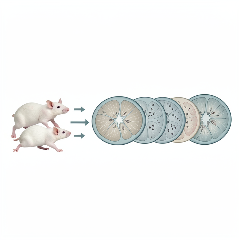
Experimental Group Layout
AI-generated conceptual illustration (albino rats and rat liver tissue). It explains the idea and does not show study data.
View source evidence ↗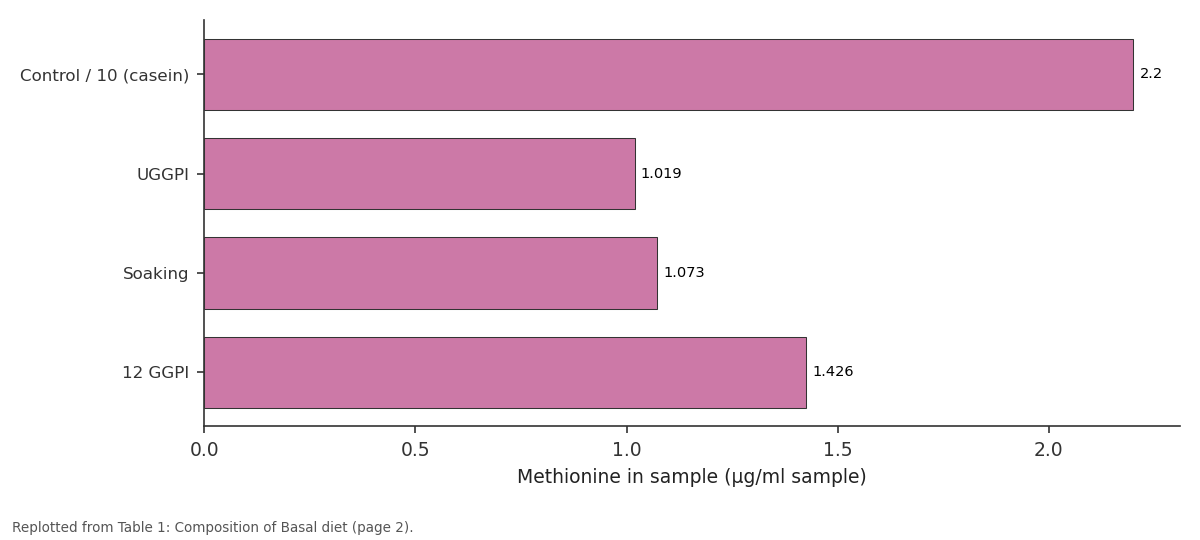
Methionine in sample (µg/ml sample): Control / 10 (casein) vs UGGPI vs Soaking vs 12 GGPI
Replotted from Table 1 (page 2) of the original paper. Every bar is the paper's own table value.
View source evidence ↗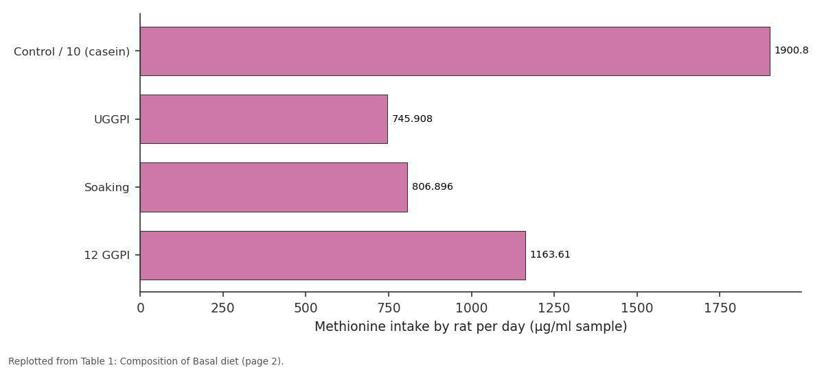
Methionine intake by rat per day (µg/ml...: Control / 10 (casein) vs UGGPI vs Soaking vs 12 GGPI
Replotted from Table 1 (page 2) of the original paper. Every bar is the paper's own table value.
View source evidence ↗Figures & tables
From the original paper Table 1Page 2
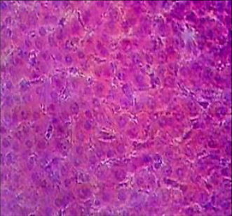 Figure 1APage 3
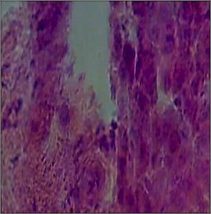 Figure 1BPage 3
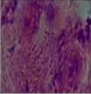 Figure 1CPage 3
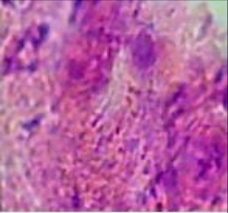 Figure 1DPage 3
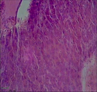 Figure 1EPage 3
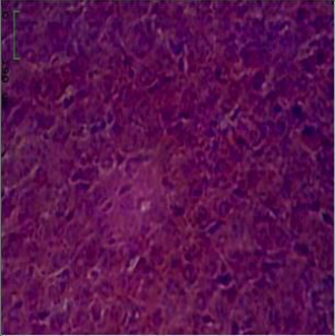 Figure 1FPage 3
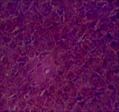 Figure 1GPage 3
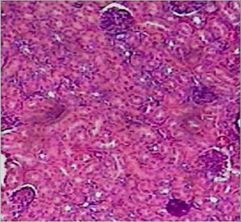 Figure 2APage 3
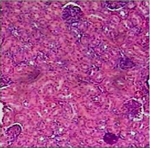 Figure 2BPage 4
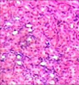 Figure 2BPage 4
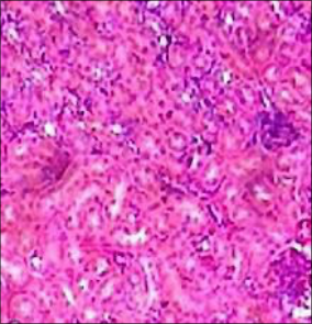 Figure 2CPage 4
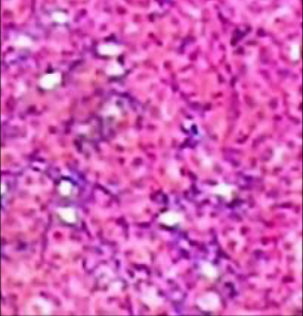 Figure 2EPage 4
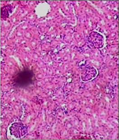 Figure 2FPage 4
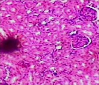 Figure 2GPage 4
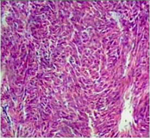 Figure 3APage 4
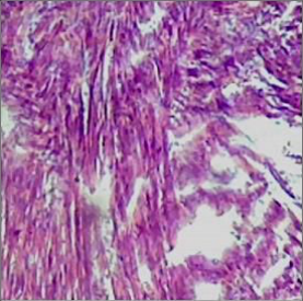 Figure 3BPage 4
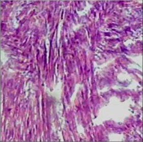 Figure 3CPage 4
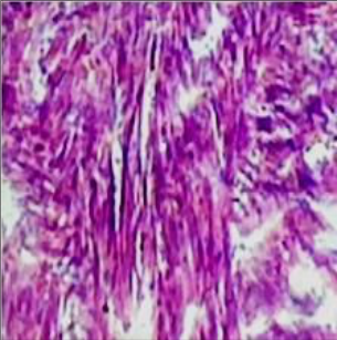 Figure 3DPage 4
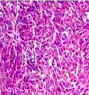 Figure 3EPage 4
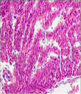 Figure 3FPage 4
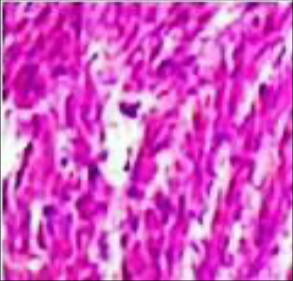 Figure 3GPage 5
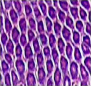 Figure 4APage 5
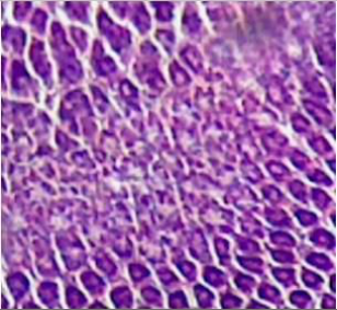 Figure 4BPage 5
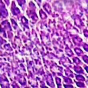 Figure 4CPage 5
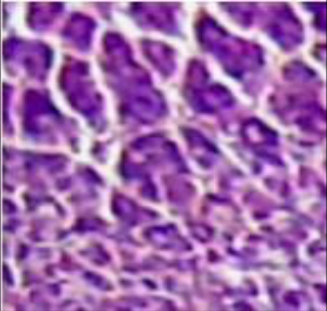 Figure 4DPage 5
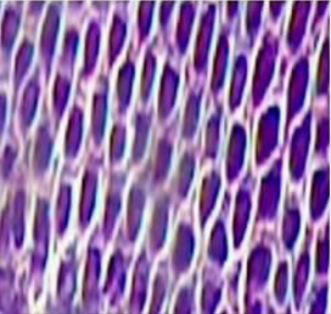 Figure 4EPage 5
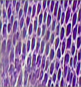 Figure 4FPage 5
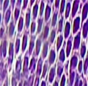 Figure 4GPage 5
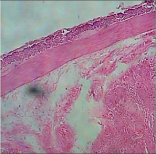 Figure 5APage 5
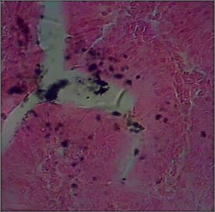 Figure 5BPage 6
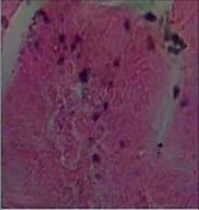 Figure 5CPage 6
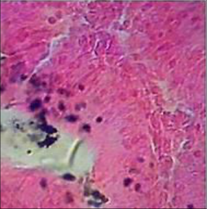 Figure 5DPage 6
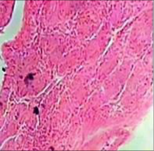 Figure 5EPage 6
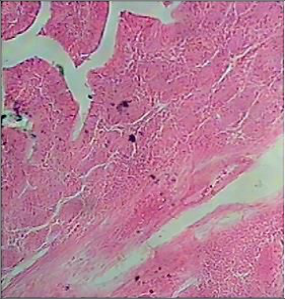 Figure 5FPage 6
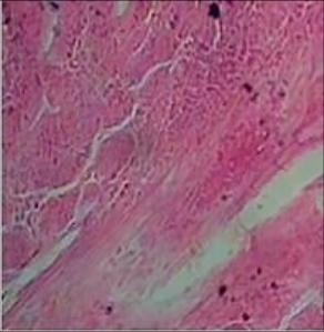 Figure 5GPage 6
Introduction
Legume seeds are of prime importance in human and animal nutrition due to their high protein content (20-50%), which is twofold higher than the level found in cereal grains and is significantly greater than that reported for conventional root crops (Ustimenko-Bakumovsky 1983) [14] . Mung bean (Vigna radiata L.) is one of the most commonly consumed food legumes in India. The production of plant proteins is of growing interest to developing and developed countries alike because of its increasing food and non-food applications (Marcello and Gius 1997) [8] . Plant protein isolates are utilized in foods to improve nutritional quality and functionality. Germination has often been proposed as a means by which the nutritional quality of bean seeds might be improved. This improvement is usually a result of breakdown of complex macromolecules such as starch and proteins into smaller and more digestible molecules, while at the same time lowering the amounts of antinutritional factors (Chang & Harrold, 1988 [2] ; Labaneiah & Luh, 1981) [5] . Isolated proteins often have improved appearance and taste compared with the original meal; therefore, they can better be used as nutritional and functional ingredients in many food products (Mizrahi et al., 1967) [9] . Mung bean is a rich source of protein and amino acid especially lysine and thus can supplement cereal-based human diets (Hussain & Burhanddin 2011) [4] . There is need for further research into processing procedure that will ensure maximum utilization of the nutrients to prepare an acceptable product from green gram for livestock use. Albino rats were used in this experiment due to its tractable nature and sensitivity to diverse diets. Varying germination levels of differently treated green gram protein isolate were introduced in the diets to examine the growth response and hepatic histological changes of albino rats.
Materials and Methods
Materials and Methods
Preparation of Germinated Green Gram Flour
The hybrid variety of Green gram purchased from Salem Super market, Tamil Nadu. Green gram seed were washed well to remove dust and stones, soaked for 12 hrs (soaking) and germinated in a germination container for 12 hrs (12 hr GG), 24 hrs (12 hr GG), 36 hrs (36 hr GG) and 48 hrs (48 hr GG). Germinated seeds were sundried made in to flour and passed through 60 mesh (BSS unit) test sieve to get enhance the uniform in particle size and packed in an airtight container.
Preparation of Germinated Green Gram Protein Isolate
The un germinated, soaking, 12 hr, 24 hr, 36 hr and 48 hr germinated green gram flour was defatted using hexane as solvent ratio of 1:3 at 250 rpm in a lab stirrer for 30 min and centrifuged at 5000 g for 10 min, at room temperature and was air dried overnight, packed and stored at 5 ºC (Wang et al 1999) [7] . Then protein extraction carried out Trichloro acetic acid (TCA) method (Bhardwaj & Yadav 2013) [1] .
Feeding Experiment to Albino Rats
Thirty-five male albino rat (Wistar strain) aged four weeks were obtained from Bangalore. The rats were fed on commercial diet for 3 days before the feeding experiment. The un-germinated, soaked and germinated protein isolate samples were used to formulate diets. The composition of control diet was as follows (Hegisted 1941 [3] ) Corn starch 80%, sunflower Oil 10%, Cellulose 5%, Salt mixture 4%, Vitamin mixture 1%, Protein. Protein was added by replacing part of corn starch with casein or with the tested sample to provide 10% protein diet for the experimental animals. In the protein methionine were supplemented as µg/1 ml sample. Feed water was supplied ad libitum daily for a period of four weeks.
Histopathological examinations
Histopathological examinations were done on liver, kidney, heart, stomach and small intestine samples. In the procedure, organs were fixed in formaldehyde (10%) and the tissues subsequently dehydrated in upgraded concentrations of ethanol (10-90%), cleaned in xylene, impregnated and embedded in paraffin. Sections of 5 µm were cut using a microtome, stained with hematoxylin and eosin stains. Light microscopic examination of multiple tissue sections from each organ in all groups was performed and image representatives of the typical histological profile were examined (Silva et al., 1999) [12] .
Data Collection
At the end of the experiments two rats per treatment were sacrificed and sample of liver, kidney, heart, stomach, small intestine were collected, the organs were preserved in 10% formalin on treatments basis in a small bottle, before taken to laboratory for the micrograph.
Statistical Analysis:
The experiment was arranged in a 5 x 7 experimental layout with seven processing methods (Casien protein, un germination, soaking and germination at 12, 24, 36 and 48 hr). Completely randomized design of SAS 2000 was used to analyze the data. The control was common to all treatments and was taken care in the package used. Significant means were separated using Duncan Multiple Range portion of the statistical package.
Results and Discussion
Pathological Observation of Albino Rats Fed Differently Treated Green gram protein isolate Diets. Albino rats on Green gram protein isolate diets physically showed some clinical aberration ranging from constriction of the eyes, cuddling, anorexia, pale color skin and hair loss particularly those on un-germinated and soaked green gram compared to germinated green gram protein isolate. Similar results were agreed with (Parcell et al., 2002 [11] ; Levine et al., 1996 [6] ) Methionine deficiency can cause liver damage, muscle loss weakness, slow growth and skin lesions.
Histological Changes in Albino Rats Fed Differently Treated green gram protein isolates Diets
The comparative analysis of the animal treated with germinated and un-germinated green gram showed remarkable changes in the metabolic processes regulated through the vital organs such as liver, kidney, heart, stomach and small intestine. The histological analysis of liver clearly demonstrated (Fig 1 (a-g)) that there is no change in the histology of hepatocytes in the treated animals however the hepatic portal vein was found to be shrinking in animals treated both un-germinated and soaked green gram protein isolate when compared with treated with germinated green gram and control (casein) found have prominent histo architecture of liver with hepatic portal vein. These parts of the study clearly authenticated that the histological development of organs and its functions may be regulated through the protein supplements as the liver is reported to be play a vital role in major metabolic processes. [10] . The histology of myocardium cells stains with Hematoxylin and Eosin (Fig. 3a-g), did not showed any prominent were noticeable variation between the control (casein) and treated animals. The mesoepithelium, cardiac muscle fiber and intercalated discs were found to be excellent in the control, germinated and un-germinated protein supplemented animals. Thus the present study concluded that the protein supplementation did not have any remarkable changes in the histology of heart. However the gastric gland was somewhat better when compared to animals treated with un-germinated green gram. Interestingly the muscularis mucosa was found to be totally absent in animal treated with un-germinated green gram, where as a thin mucosa layer was noticed in the inner wall of stomach in the animal treated with soaked green gram. Thus the present investigations scientifically proves that the supplemented protein substances not only digested in stomach but it also support to develop the vital organs like stomach.
The protein supplemented rats such as rats treated with exogenous protein casein and germinated green gram found have a well-developed brush border around the microvelli which was supported the columnar epithelial goblet cells and occurrence of lymphocytes nuclei decides that the rat treated with un-germinated green gram cells found have less number of lymphocytes nuclei and goblet cells with less amount of mucosa. Nevertheless the animal treated with soaking green gram found to have a small size of brush border around the microvelli and lumen of the small intestine was found to be very much reduced. Furthermore it is very difficult to notice the gastric mucosa and intestinal mucosa in animal treated with un-germinated green gram proteins. Thus the present study clearly pointed out that the protein supplemented nutrition on the protein metabolism and small intestinal mucosal metabolism which have been well proved (Fig. 5 a-g). The rat treated with ungerminated protein isolate showed less intake of diet compared to germinated and control treatments. This study indicated that methionine supplementation in the diet caused an increase in food intake. This result may be explained on the basis of that body stores many of the nutrients absorbed from the intestine which are necessary for body functions. Wretlind (2008) [14] has been reported that maximal growth was obtained when rats were given methionine at concentration of 0.25% D-methionine in the diet.
Conclusion:
According to the results in the present study concluded that Plant proteins especially those derived from legumes like green gram tend to be lower in methionine than animal proteins and milk proteins. Methionine in the diet caused histopathological changes in liver, kidneys, heart, stomach and small intestine. Hence proper consumption of methionine in diet has to be recommended.
Figures and tables from the paper
| Treatments | Control | UGGPI | Soaking | 12 GGPI | 24 GGPI | 36 GGPI | 48 GGPI |
|---|---|---|---|---|---|---|---|
| Protein % | 10 (casein) | 10 | 10 | 10 | 10 | 10 | 10 |
| Methionine in sample (µg/ml sample) | 2.2 | 1.019 | 1.073 | 1.426 | 1.0526 | 1.227 | 1.3047 |
| Methionine intake by rat per day (µg/ml sample) | 1900.8 | 745.908 | 806.896 | 1163.61 | 867.34 | 1011.04 | 1077.68 |
| Vitamin premix % | 1 | 1 | 1 | 1 | 1 | 1 | 1 |
| Mineral premix % | 4 | 4 | 4 | 4 | 4 | 4 | 4 |
| Vegetable Oil % | 10 | 10 | 10 | 10 | 10 | 10 | 10 |
| Corn starch % | 70 | 70 | 70 | 70 | 70 | 70 | 70 |
| Cellulose % | 5 | 5 | 5 | 5 | 5 | 5 | 5 |
Table text extracted from the original PDF; superscript letters are shown inline. The original table image is below.
View original table image
References
14 in the original paper- Bhardwaj J, Yadav SK. A common protein extraction protocol for proteomic analysis: Horse gram as a case study. American Journal of Agricultural and Biological Sciences. 2013;8(4):293-301.
- Chang KC, Harrold RL. Changes in biochemical components, in vitro protein digestibility and amino acids in two bean cultivars during germination. Journal of Food Science. 1988;53:783-787.
- Hegested HD, Bork JK, Mohr KL. Amer J Clin Nutr. 1941;26:1251-1251.
- Hussain I, Burhanddin M. Optimization of germination conditions for germinated mungbean flour by response surface methodology. African Journal of Food Science and Technology. 2011;2(10):232-239.
- Labaneiah MEO, Luh BS. Changes of starch, crude fiber and oligosaccharides in germinating dry beans. Cereal Chemistry. 1981;58:135-138.
- Levine RL, Mosoni L, Berlett BS, Stadtman ER. Methionine residues as endogenous antioxidants in proteins. Proc Natl Acad Sci. 1996;93:15036-15040.
- Wang M, Hettiarachchy NS, Qi M, Burks W, Siebenmorgen T. Preparation and functional properties of rice bran protein isolate. J Agric Food Chem. 1999;47:411-416.
- Marcello D, Gius C. Legume seeds: Protein content and nutritional value. Field Crops Research. 1997;53:31-45.
- Mizrahi S, Zimmerman G, Berk Z, Cogan D. The use of isolated proteins in bread. Cereal Chemistry. 1967;44:193-193.
- Nada AAEA, Hoda SI, Reham SR. Does methionine supplementation alter beta-amyloid levels in brain cells, liver and kidney functions? Nat Sci. 2012;10(7):26-36.
- Parcell SW, Candidate ND. Sulfur in human nutrition and applications in medicine. Altern Med Rev. 2002;7(1):22-41.
- Silva AM, Bambirra EA, Oliveira AL, Sousa PP, Gomes OA, Viera EE, Nicoli JR. Protective effect of bifidus milk on the experimental infection with Salmonella enteritidis subsp. typhimurium in mice. J Appl Microbiol. 1999;86:331-336.
- Ustimenko-Bakumovsky GV. Plant Growing in the Tropics and Subtropics. New York: Macmillan; 1983.
- Wretllnd. The effect on growth and toxicity of the two isomers of methionine. Basic Medical Sciences. 2008;20:1-12.