
Research article · 2025 · Full text
International Journal of Biology SciencesBacterial motility in solidified agar medium for an Apple and Rosogolla
Volume 7(4) · Pages 91–92 · DOI: 10.33545/26649926.2025.v7.i4b.332 ↗
Research areasMicrobial Metabolites in Food BiotechnologyMycorrhizal Fungi and Plant Interactions
Abstract
Author-writtenBacterial Motility test is used for checking whether the bacteria motile or not, there are numerous media to identify the motile bacteria. This paper gives a basic overview that from solidified agar medium motility can be detected easily and an overview of the shape of bacteria that depends how much they can survive on the motile test.
Summary
Source-checkedPlain-language reading of the full paper, prepared with AI and checked against the text. Each point links to the authors' own words.
Why it matters
- Bacteria use flagella, which are thread-like appendages extending from the cell wall and plasma membrane, to move across agar media. View evidence ↗
- Motility can be visually identified in different media: motile bacteria 'swarm' in semi-solid agar and create a 'spreader' structure in solid agar. View evidence ↗
- Testing for motility is important for determining pathogenicity, as some motile coliforms can act as pathogens in humans. View evidence ↗
The question
- The study aims to demonstrate that bacterial motility can be easily detected from solidified agar medium and to examine how bacterial shape relates to survival during the test. View evidence ↗
What they did
- The researchers used the 'Hanging Drop Method' to test for bacterial motility. View evidence ↗
- The samples tested for motility included apple, water, and rosogolla (a sweetmeat product). View evidence ↗
- Bacterial cultures were taken from spreader colonies on Nutrient Agar and Violet Red Bile Agar plates. View evidence ↗
- Samples were observed under a 100X microscope using immersion oil. View evidence ↗
What they found
- In the rosogolla sample, both rod-shaped and cocci-shaped bacteria were found to be motile on Nutrient agar and Violet red bile agar. View evidence ↗
- Bacterial motility was observed most extensively in the apple sample and least in the water sample. View evidence ↗
- Bacteria moved less rapidly on the cavity slide compared to their movement in broth or semi-solid agar media. View evidence ↗
- Bacterial movement decreased over time; cocci-shaped bacteria lost survivability after 5-6 minutes, while rod-shaped bacteria survived an average of 2 to 3 minutes longer. View evidence ↗
What it means
- The authors conclude that because motility increases host-pathogen interactions, it may serve as a virulence factor that warrants further investigation. View evidence ↗
Visual summary
1 visualsIllustrations explain the paper; they do not show study data.

Bacterial Survival Comparison
AI-generated conceptual illustration (cocci-shaped and rod-shaped bacteria in solidified agar medium). It explains the idea and does not show study data.
View source evidence ↗Figures & tables
From the original paper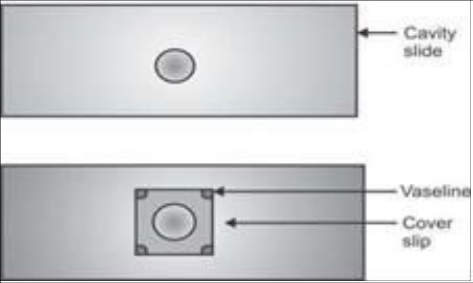 Figure 1
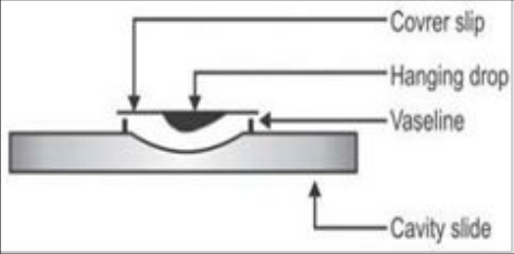 Figure 2
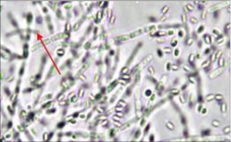 Figure 3
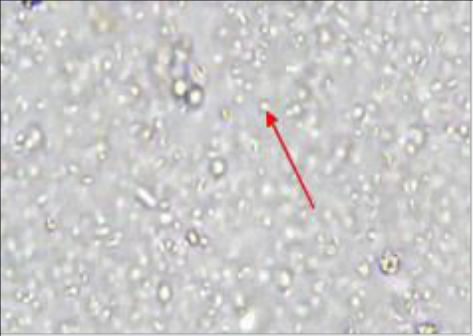 Figure 4
Introduction
A bacterium has their flagella which has thread like locomotor appendages extending outward from the plasma membrane and cell wall either single flagellum or multiple flagella used to move or spread forward on any agar medium. Agar medium maybe the liquid in the form of broth, or semi solid or maybe solidified form. If a bacterium is towards any direction, it is considered as motile bacterium, if there is no movement then it is considered as non-motile bacterium. Some Bacillus, cocci or some coliforms are motile in nature, and the motility test which we have done that is completely based upon these bacteria. But in case of other bacteria which are motile in nature they can be easily identified by the same methodology which was discussed. So, the motility test is performed directly without any smear form or any stain induced in it, and this is the principal criteria when motility was tested. In semi-solid agar media, motile bacteria "swarm" and give a diffuse spreading growth that is easily recognized by the naked eye; In solid media like in agar medium motile bacteria gives a structure of a spreader which can also be detected by naked eye, and the spreader may be bacillus, cocci or a coliform. In case of broth media, it is a distinctive media to move the bacterium very swiftly, in the microscopic test broth media gives always a better swim effect as compared to other media. For this study "Hanging Drop Method" for motility test was performed.
Materials and Method
In terms of materials required for solidified media were cover slips, cavity slides, inoculating loops, Bunsen burner, compound microscope, Sample and Sticky item like Vaseline for adherence of cover slips to cavity slides. The Motile test from solidified agar medium only can be done when the bacteria are in log or in stationary phase. The bacteria should be active and the test was undertaken after taking out the petri-plates from the incubator. The samples used were an apple, water, and rosogolla (Sweetmeat Product), where the motile bacillus as well motile coliform and motile cocci were observed. In the laminar air flow system, the Vaseline/gelly should be given on the four-corner side of the cover slips as well as cavity slide so that the cover slip can adhere to the cavity slide (Fig. 1). Inoculating loop should be red heated in the Bunsen burner and then one loop taken from a dilution blank (0.9% saline solution) one to two drops was taken to the cover slips. Then loop should be further red heated and a loop of culture from the spreader into the petri-plates is taken out (i.e. taken from Nutrient Agar & Violet Red Bile Agar plates spreader bacteria). A loop of culture can be taken out in any solidified agar medium. A clean cavity slide is taken and placed it over the cover slip i.e. concavity facing the drop, gently over the cover slip (Fig. 2). After placing it, a drop of immersion oil is given and observed under 100X Microscope as quick as possible.
Results
On 100X Microscopic view the motile bacteria were certainly seen, which were rapidly moving towards either direction. If the bacteria are non-motile in nature there is no movement is observed under the microscope and seen as immobile, clustered or non-clustered form of bacteria. In case of rosogolla sample (sweetmeat product), from standard plate count (Nutrient agar) and from coliform count (Violet red bile agar) some bacteria are motile whether it was rods or cocci. In Apple sample the bacterial motility is massively seen. In water sample motility was less observed. Although motility of bacteria can only be seen either live in microscope or recorded video format, but some figures which are attached below (Fig. 3, Fig. 4) that identify the motile bacterium, where maximum bacteria are motile in nature and for better understanding red arrow marked bacterium is specifically identified as a distinctive motile bacterium.
Discussion
The motility test was mainly required in terms of application whether it has pathogenicity or not, because some coliforms which are motile in nature they may produce pathogen inside the human body, for example, Escherichia Coli, Strain O157:H7 which expresses somatic (O) antigen 157 and flagella (H) antigen 7. In our study, the cocci shaped bacteria are motile in nature but not stable for long time as compared to rod shaped bacteria, observed mainly in standard plate count plates. In case of the motility test there is no specific direction of movement of the bacteria, they can move from any of the portion amongst the dip surface of the cavity slides. In the cavity slide the bacteria were not moving rapidly as compared to broth or semisolid agar medium. The movement time is very less on either direction. Under compound microscope the bacteria movement rapidly decreased with respect to time, i.e. 5-6 minutes the cocci shaped bacteria losses their survivability and rod-shaped bacteria can survive more 2 to 3 minutes than cocci on an average. Since motility increases the occurrence of host-pathogen interactions hence the role of motility can be a virulence factor in bacteria which can be further investigated.
References
2 in the original paper- Tille PM, Forbes BA. Bailey & Scott's diagnostic microbiology. 13th ed. St. Louis (MO): Elsevier; 2014.
- Cappuccino JG, Sherman N. Microbiology: a laboratory manual. 8th ed. San Francisco (CA): Pearson Benjamin Cummings; 2008.