Scinovex

Research article · 2025 · Full text

International Journal of Veterinary Sciences and Animal Husbandry

Physical and pathological assessment of cardio-hepatobiliary system with salivary gland in buffalo infected with FMD

Muna A. Al-khafaji

Volume 10(2) · Pages 107–113 · DOI: 10.22271/veterinary.2025.v10.i2b.2062 ↗

Research areasAnimal Virus Infections StudiesMeat and Animal Product Quality

↓ Read full-text PDF0 citations

Abstract

Author-written

This study achieved assessing clinical manifestation with salivary gland, liver, heart, gallbladder histopathological changes in buffalo infected with Foot and mouth disease. Out of 50 buffalo herd in Al-Qasim district, Babil/Iraq, the positive cases previously detected with FMD screening kit (VDRG® FMDV 3Diff/PAN Ag Rapid kit) examined clinically, compared with 10 clinically healthy animal used as control, furthermore Gross lesions in mouth dental pads, gums and hoofs and histopathological study done on salivary gland, gall bladder, liver and heart. Clinical examination revealed recumbency, rope salivation, fever, polypnea, tachycardia, depression, anorexia and lameness, tow calves with primary lactation failure died within 1-3 days. Gross pathology exhibit vesicles, erosion and ulceration in its early stages by vesicles in the plamim nasale, in the oral cavity, tongue and hoof. Histopathological examination of infected calves revealed extensive necrotic myocarditis, necrotic hepatitis with ulcerative cholecystitis although no pathological lesion in salivary glands. this study emphasise the exceeding mortality and morbidity rate of young buffalo calves infected with FMD due to sever myocardial necrosis with outlining the associated pathological lesions in non-traditional organs as salivary glands, gall bladder and human consuming organs liver along with heart.

Summary

Source-checked

Plain-language reading of the full paper, prepared with AI and checked against the text. Each point links to the authors' own words.

  1. Why it matters

    • Foot-and-mouth disease (FMD) is a highly contagious viral disease that causes significant annual economic losses for livestock owners. View evidence ↗

      Why it matters

      Foot-and-mouth disease (FMD) is a highly contagious viral disease that causes significant annual economic losses for livestock owners.

      In the paper's own words

      “FMD is a highly contagious, modifiable viral disease that can cross the species barrier causes annual massive economic losses to owner’s stock”

      Page 1

      Found in: Introduction

      Read it in the full textOpen PDF at page 1 ↗
    • FMD has become an endemic transboundary emerging disease across the Middle East, Asia, and Africa. View evidence ↗

      Why it matters

      FMD has become an endemic transboundary emerging disease across the Middle East, Asia, and Africa.

      In the paper's own words

      “The disease has expanded its geographical distribution and become endemic across extensive areas in Africa, Asia, and the Middle East, making FMD a trans boundary emerging disease of ruminants”

      Page 1

      Found in: Introduction

      Read it in the full textOpen PDF at page 1 ↗
    • While adult animals typically have low mortality rates, young calves can experience sudden death from FMD. View evidence ↗

      Why it matters

      While adult animals typically have low mortality rates, young calves can experience sudden death from FMD.

      In the paper's own words

      “The disease-related morbidity rate reaches 100% while mortalities are low in adult animals as well as (Knight-Jones et al., 2017) [14] except sudden death in young calves”

      Page 1

      Found in: Introduction

      Read it in the full textOpen PDF at page 1 ↗
  2. The question

    • The study aimed to identify the clinical manifestations of FMD and assess histopathological changes in the liver, heart, gall bladder, and salivary glands of buffalo calves. View evidence ↗

      The question

      The study aimed to identify the clinical manifestations of FMD and assess histopathological changes in the liver, heart, gall bladder, and salivary glands of buffalo calves.

      In the paper's own words

      “this study focused to figure clinical manifestations of the disease as well as assessing the salivary gland, myocardial, gall bladder and liver histopathological changes in buffalo calves.”

      Page 2

      Found in: Introduction

      Read it in the full textOpen PDF at page 2 ↗
  3. What they did

    • The study examined 50 unvaccinated buffalo calves of both sexes, aged between ≤ 6 months and 2 years, in the Al-Qasim district of Babil, Iraq, in February 2024. View evidence ↗

      What they did

      The study examined 50 unvaccinated buffalo calves of both sexes, aged between ≤ 6 months and 2 years, in the Al-Qasim district of Babil, Iraq, in February 2024.

      In the paper's own words

      “This study examined a total of 50 buffalos (Syncerus caffer) calves were obtained in Al-Qasim district, Babil/Iraq at February, 2024. Both sexes aged between (≤ 6 months and 2 years), all analyzed buffalo calves have no history of FMDV vaccination.”

      Page 2

      Found in: Methodology Animals and Samples Collection

      Read it in the full textOpen PDF at page 2 ↗
    • Histopathological analysis was performed on 1cm³ tissue samples from the heart, liver, gall bladder, and salivary glands using Hematoxylin & Eosin staining. View evidence ↗

      What they did

      Histopathological analysis was performed on 1cm³ tissue samples from the heart, liver, gall bladder, and salivary glands using Hematoxylin & Eosin staining.

      In the paper's own words

      “The specimens of 1cm³ taken from salivary gland heart, liver and gall bladder, specimens were processed through the routine tissue fixation, clearing, mounting and stained by (Hematoxylin & Eosin stain) procedure”

      Page 2

      Found in: Pathological studies Histopathological Techniques

      Read it in the full textOpen PDF at page 2 ↗
  4. What they found

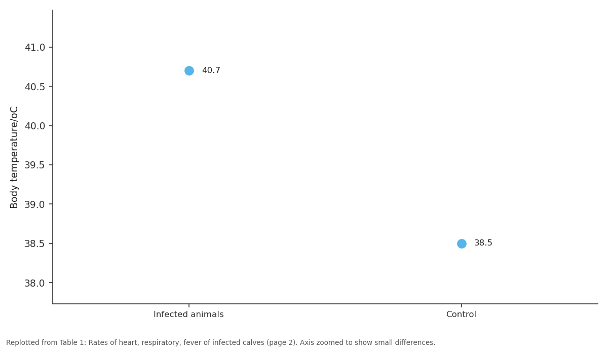
    • Infected calves exhibited significant hyperthermia with a body temperature of 40.7°C compared to 38.5°C in the control group. View evidence ↗

      What they found

      Infected calves exhibited significant hyperthermia with a body temperature of 40.7°C compared to 38.5°C in the control group.

      In the paper's own words

      “Body temperature/oC | 40.7 | 38.5”

      Page 2

      Found in: Table 1

      Read it in the full textOpen PDF at page 2 ↗
    • The heart rate of infected calves was elevated (116-119 min) compared to the control group (91-101 min). View evidence ↗

      What they found

      The heart rate of infected calves was elevated (116-119 min) compared to the control group (91-101 min).

      In the paper's own words

      “Heart rate / min | 116-119 | 91-101”

      Page 2

      Found in: Table 1

      Read it in the full textOpen PDF at page 2 ↗
    • The respiratory rate was higher in infected calves (41-45 min) than in the control group (29-33 min). View evidence ↗

      What they found

      The respiratory rate was higher in infected calves (41-45 min) than in the control group (29-33 min).

      In the paper's own words

      “Respiratory rate / min | 41-45 | 29-33”

      Page 2

      Found in: Table 1

      Read it in the full textOpen PDF at page 2 ↗
    • The study observed a 4% mortality rate in calves that were recumbent and unable to suck their dams, dying within 3 days. View evidence ↗

      What they found

      The study observed a 4% mortality rate in calves that were recumbent and unable to suck their dams, dying within 3 days.

      In the paper's own words

      “all calves that unable to suck their dams, recumbent and died within 3 days, at 4%.of mortality rate”

      Page 2

      Found in: Clinical examination and gross lesions

      Read it in the full textOpen PDF at page 2 ↗
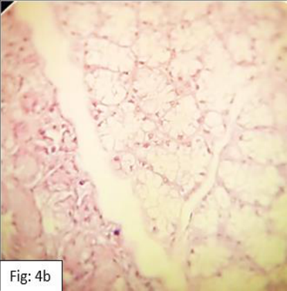
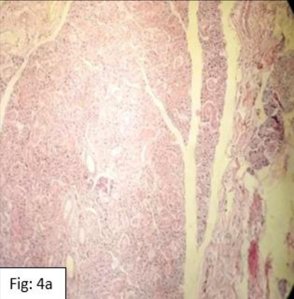
    • Histopathological examination revealed no pathological lesions in the sublingual and mandibular salivary glands of infected calves. View evidence ↗

      What they found

      Histopathological examination revealed no pathological lesions in the sublingual and mandibular salivary glands of infected calves.

      In the paper's own words

      “Histopathological examination of the mandibular and sublingual salivary glands of calves died from infection with FMD showed no pathological lesions”

      Page 4

      Found in: Histopathological examination of internal organs:

      Read it in the full textOpen PDF at page 4 ↗
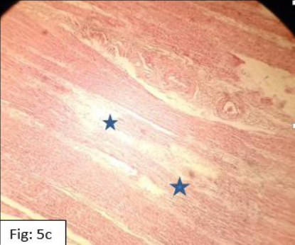
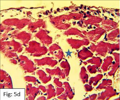
    • The heart of infected calves showed endomyocardium edema and focal coagulative necrosis (zinker necrosis) of the muscle fibers. View evidence ↗

      What they found

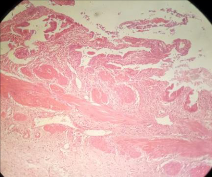
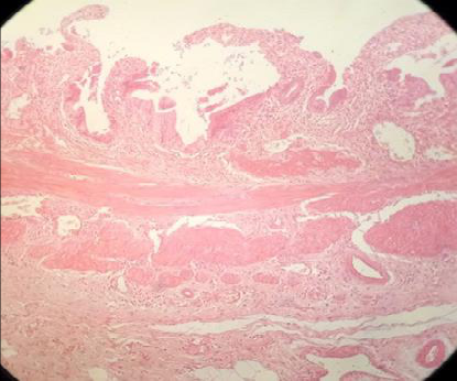
      The heart of infected calves showed endomyocardium edema and focal coagulative necrosis (zinker necrosis) of the muscle fibers.

      In the paper's own words

      “heart histopathological examination of heart shows focal coagulative necrosis (zinker necrosis) of muscle fibers (Figure 5 a, b, c and d) with endomyocardium edema between myocardium muscles”

      Page 4

      Found in: Histopathological examination of internal organs:

      Read it in the full textOpen PDF at page 4 ↗
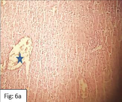
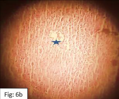
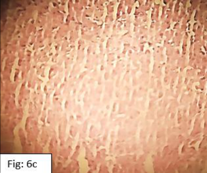
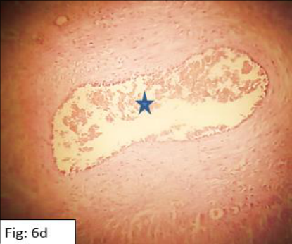
    • Liver examination of infected calves showed hepatocyte necrosis, hemorrhage in sinusoids, and congestion of the portal and central veins. View evidence ↗

      What they found

      Liver examination of infected calves showed hepatocyte necrosis, hemorrhage in sinusoids, and congestion of the portal and central veins.

      In the paper's own words

      “histopathological examination of infected calf liver died from FMD infection revealed congestion of central and portal vein( Figure 6 a, b and d) with necrosis of hepatocytes and hemorrhage in sinusoids between hepatocytes”

      Page 4

      Found in: Histopathological examination of internal organs:

      Read it in the full textOpen PDF at page 4 ↗
    • The gall bladder of infected calves exhibited mucosal erosions and acute cholecystitis. View evidence ↗

      What they found

      The gall bladder of infected calves exhibited mucosal erosions and acute cholecystitis.

      In the paper's own words

      “histopathological examination of gall bladder shows acute cholecystitis with mucosal erosions”

      Page 4

      Found in: Introduction

      Read it in the full textOpen PDF at page 4 ↗
  5. What it means

    • The authors conclude that necrotic myocardiopathy is the likely cause of death in young buffalo calves infected with FMD. View evidence ↗

      What it means

      The authors conclude that necrotic myocardiopathy is the likely cause of death in young buffalo calves infected with FMD.

      In the paper's own words

      “Necrotic myocardiopathy assumes the etiology of death in young buffalo calves.”

      Page 6

      Found in: Conclusion

      Read it in the full textOpen PDF at page 6 ↗
    • The authors recommend the application of active prevention and control measures to manage FMD in Iraq. View evidence ↗

      What it means

      The authors recommend the application of active prevention and control measures to manage FMD in Iraq.

      In the paper's own words

      “FMD in Iraq still requires the application of active measures that aids in its prevention and control.”

      Page 6

      Found in: Conclusion

      Read it in the full textOpen PDF at page 6 ↗

Visual summary

2 visuals

Illustrations explain the paper; charts are replotted from the paper's own table values.
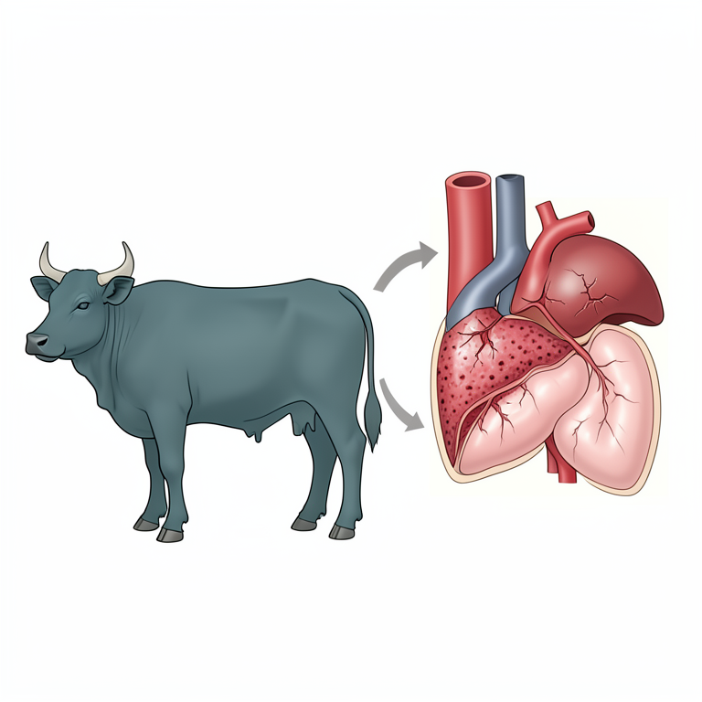
AI-generated illustration
AI-generated conceptual illustration (water buffalo calf). It explains the idea and does not show study data.↗ Enlarge
Systemic Pathological Effects of FMD
AI-generated conceptual illustration (water buffalo calf). It explains the idea and does not show study data.

AI-generated conceptual illustration (water buffalo calf). It explains the idea and does not show study data.

Open full-size image ↗

Systemic Pathological Effects of FMD

AI-generated conceptual illustration (water buffalo calf). It explains the idea and does not show study data.

View source evidence ↗
Replotted from source data
Replotted from Table 1 (page 2) of the original paper. Every bar is the paper's own table value.↗ Enlarge
Body temperature/oC: Infected animals vs Control
Replotted from Table 1 (page 2) of the original paper. Every bar is the paper's own table value.

Replotted from Table 1 (page 2) of the original paper. Every bar is the paper's own table value.

Open full-size image ↗

Body temperature/oC: Infected animals vs Control

Replotted from Table 1 (page 2) of the original paper. Every bar is the paper's own table value.

View source evidence ↗

Figures & tables

From the original paper

Rates of heart, respiratory, fever of infected calvesTable 1Page 2Frequency clinical symptoms in infected animals (%)Figure 1Page 2Gross lesions of calves infected with FMD showing vesculo-ulcerative lesions in dental pads, gums and hoof respectivelyFigure 2Page 3Dead calves with emaciationFigure 3Page 4Dead calves with emaciationFigure 3Page 4Histopathological section of infected calf with salivary gland, showing no pathological lesions, (H&E, 400x)Figure 4Page 4Histopathological section of infected calf with salivary gland, showing no pathological lesions, (H&E, 400x)Figure 4Page 4Histopathological section of FMD infected calf heart, showing endomyocardial focal coagulative necrosis with edema (star), (H&E, 400x)Figure 5Page 5Histopathological section of FMD infected calf heart, showing endomyocardial focal coagulative necrosis with edema (star), (H&E, 400x)Figure 5Page 5Histopathological section of FMD infected calf heart, showing endomyocardial focal coagulative necrosis with edema (star), (H&E, 400x)Figure 5Page 5Histopathological section of FMD infected calf heart, showing endomyocardial focal coagulative necrosis with edema (star), (H&E, 400x)Figure 5Page 5Histopathological section of liver of infected calf with FMD Shows congestion of central and portal vein (star) with necrosis ofFigure 6Page 5Histopathological section of liver of infected calf with FMD Shows congestion of central and portal vein (star) with necrosis ofFigure 6Page 5Histopathological section of gall bladder of infected calf with FMD shows acute cholecystitis with mucosal erosions, (H&E, 200X)Figure 7Page 6Histopathological section of gall bladder of infected calf with FMD shows acute cholecystitis with mucosal erosions, (H&E, 200X)Figure 7Page 6
FULL TEXT Original article · reflowed for reading

Introduction

FMD is a highly contagious, modifiable viral disease that can cross the species barrier causes annual massive economic losses to owner's stock (Al-Salihi KA, 2019) [6] , it is an infectious disease that affects wide range of cloven-hoofed mammals of small and large ruminants and swine (Sameer and Jarullah, 2014) [20] . FMDV is non-enveloped RNA virus in the genus Aphthovirus with four structural proteins surrounded the icosahedral capsid genome of approximately 8500 bases. There are seven immunologically, distinct, serotypes (O, A, C, Asia 1, and Southern African Territories (Peng et al., 2020) [16] , with over 70 subtypes with no cross protection Antigenic variation and instability contribute to persistent nature. The disease has expanded its geographical distribution and become endemic across extensive areas in Africa, Asia, and the Middle East, making FMD a trans boundary emerging disease of ruminants, Disease spread via movement of infected animals, contaminated animal products, and fomites; can be transmitted up to 250 km by wind (Jamal and Belsham, 2015) [11] . Clinical manifestation of excessive salivation and lameness due to vesicles and blisters of oral cavity, nostrils, teats, udder, lethargy, fever, decreased milk production, and blanching of coronary bands may precede vesicles, all with hyperthermia occur in infected calves while signs in small ruminant including sheep and goat were mild to nil making disease difficult in diagnosis (Knight-Jones et al., 2017) [14] . The pharyngeal mucosa is thought to be hostile site for primary FMD virus replication, then it is transferred through lymphatics and blood vessels by viremia to oral mucosa which is secondary sites of replications (El-Karim, et al., 2022) [7] . The diseaserelated morbidity rate reaches 100% while mortalities are low in adult animals as well as (Knight-Jones et al., 2017) [14] except sudden death in young calves (Admassu et al., 2015) [1] . clinical signs demarks huge economic damages due to loss of milk production post 2 months of observation (Arzt et al., 2024) [4] , uncontrolled open Iraqi borders, substantial economic losses due to control measures to attain disease-free status severely weakened the disease control making introduction of exotic FMDV to Iraq as well as creates a threat to the neighboring countries and worldwide, and vice versa (Al-Salihi KA, 2019) [6] . However, this study focused to figure clinical manifestations of the disease as well as assessing the salivary gland, myocardial, gall bladder and liver histopathological changes in buffalo calves.

Methodology Animals and Samples Collection

This study examined a total of 50 buffalos (Syncerus caffer) calves were obtained in Al-Qasim district, Babil/Iraq at February, 2024. Both sexes aged between (≤ 6 months and 2 years), all analyzed buffalo calves have no history of FMDV vaccination. All the calves examined physically after being diagnosed with screening FMD diagnostic kit VDRG® FMDV 3Diff/PAN Ag Rapid kit by using saliva. Ten clinically healthy calves were served as the control group.

Clinical examination

General examinations performed on detectable changes in animal hair, gait and muscle (Radostits et al., 2000) [18] . Vital signs of pulse and respiratory rate, body temperature (rectal and axial), appetite were monitored. Examinations of the buccal cavity and claws were also performed.

Pathological studies Histopathological Techniques

The specimens of 1cm³ taken from salivary gland heart, liver and gall bladder, specimens were processed through the routine tissue fixation, clearing, mounting and stained by (Hematoxylin & Eosin stain) procedure according to (Ahmed, 2016) [5] .

Results and Discussion

Results and Discussion

Clinical examination and gross lesions

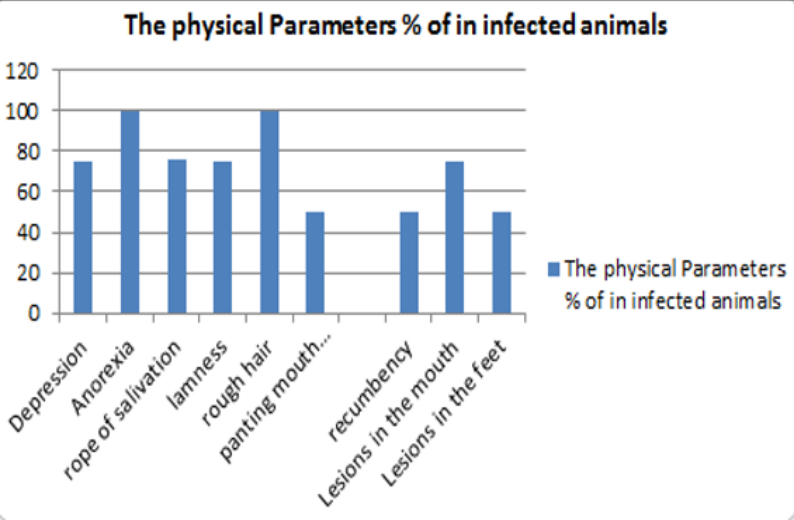
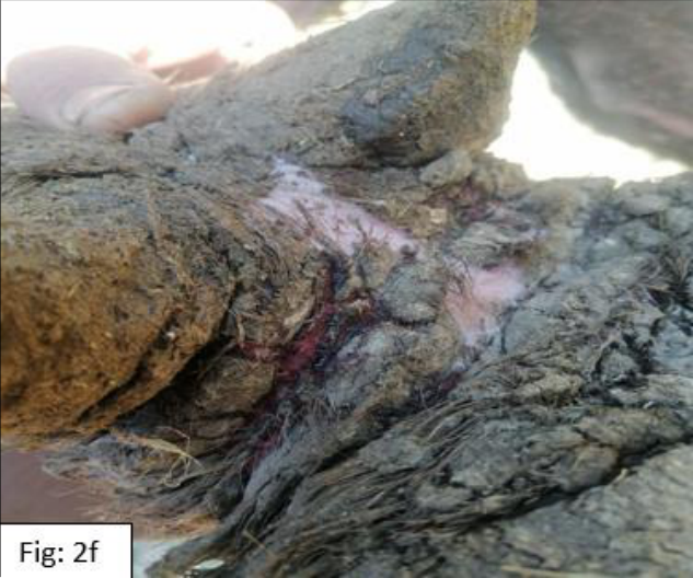
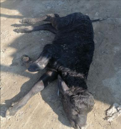
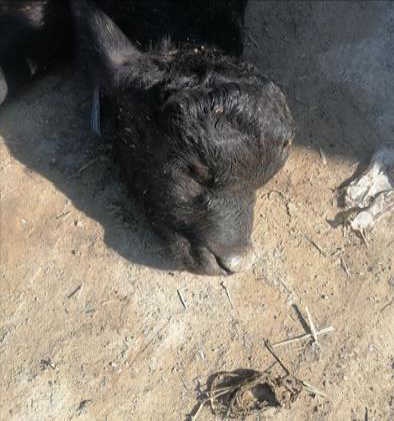
The infected examined calves revealed that all animals suffering from vesicles on the nasal planum, oral cavity, oral mucosa with rope salivation, anorexia depression, panting mouth breathing, fever ≥ 40.7°C, lameness, rough hair., tachycardia, all calves that unable to suck their dams, recumbent and died within 3 days, at 4%.of mortality rate (Figure 1) shows frequencies and rates of each clinical sign. Moreover the infected calves have indicative elevation in heart rate and respiratory rate with significant hyperthermia (Table 1) compared with control group. Gross pathology of calves infected calves dental pads exhibit vesicular lesion, multiple areas of ulceration and erosion (Figure 2a,b,c), upper surface of the gum (Figure 2d) and hoof (Figure 2e), dead calves appear emaciated (Figure 3). [19] ; Aktas et al., (2015) [3] findings, whom displaced that the mortality rate was 9.90% and 11.34% respectively in the acute myocardial form of the FMD-diseased calves, although this study showed that the diseased calves of clinical form were have, depression, feverish, accompanying in appetence, vesicles that may coalesce to form bullae which rupture leaving raw ulcers. Fluid-filled vesicles are present in the oral cavity, lips, rostral plate, and tongue accompanied with ptyalism. The same signs indicated by (Sarry et al., 2022) [17] . Sudden death conceder conspicuous sign of 1-2 months old calves. These findings totally agreed with (Ali et al., 2016) [5] who registered in young calves (2) months of age just before death they have tachypnea, arrhythmia, fever, with tachycardia; while with aged calves, peculiar signs are mouth and feet vaculation lesions. Elhaig and Elsheery (2014) [9] recorded cardiac arrhythmia besides labored breathing just before death. Meantime, less than 1-month old buffalo calves were found dead with indistinguishable clinically signs. This study showed presence of vesicular lesions dispend on dental pad, gum and hoof erosion and ulceration with inflamed borders frequently appear at the site of raptured vesicles, the same lesions mentioned by (Jubb et al., 2016) [

Figure 3. Dead calves with emaciation
Dead calves with emaciation↗ Enlarge
Figure 3
Dead calves with emaciation

Dead calves with emaciation

Open full-size image ↗
Original paper figureSource PDF · page 4 ↗
Figure 3. Dead calves with emaciation
Dead calves with emaciation↗ Enlarge
Figure 3
Dead calves with emaciation

Dead calves with emaciation

Open full-size image ↗
Original paper figureSource PDF · page 4 ↗
Figure 1. Frequency clinical symptoms in infected animals (%)
Frequency clinical symptoms in infected animals (%)↗ Enlarge
Figure 1
Frequency clinical symptoms in infected animals (%)

Frequency clinical symptoms in infected animals (%)

Open full-size image ↗
Original paper figureSource PDF · page 2 ↗
Table 1. Rates of heart, respiratory, fever of infected calves
ParametersInfected animalsControl
Body temperature/oC40.738.5
Heart rate / min116-11991-101
Respiratory rate / min41-4529-33

Table text extracted from the original PDF; superscript letters are shown inline. The original table image is below.

View original table image
Rates of heart, respiratory, fever of infected calves↗ Enlarge
Table 1
Rates of heart, respiratory, fever of infected calves

Rates of heart, respiratory, fever of infected calves

Open full-size image ↗
Original paper tableSource PDF · page 2 ↗

Histopathological examination of internal organs:

Histopathological examination of the mandibular and sublingual salivary glands of calves died from infection with FMD showed no pathological lesions (Figure 4a The histological changes in the myocardium of infected calves revealed myocardial muscle cells coagulative necrosis with myocarditis associated with interstitial edema, which are compatible with (Karapinar et al., 2010) [13] . In the acute, form, of FMD, degeneration and necrosis of myocardial fibers occur due to viral replication (Stalker, 2007) [15] , laterally focal myocardiac ischemic infraction developed, ending in complete heart failure due to stoppage of electrical signal transport to other parts of the cardiac muscle (Islam et al., 2017) [10] . In this study, liver lesion were represented by congestion of central and portal vein with necrosis of hepatocytes; a result totally agreed with (Ali et al., 2016) [6] that assisted hepatic cells degeneration as well as thickening hepatic capsule in addition to infiltration of lymphocytes and macrophages in fibrotic portal areas; these findings were in a partial agreement with (El-Amir et al., 2014) [8] , that conducted presence multi-focal hepatocellular necrobiosis and necrosis areas due to secondary clostridial infection within the necrotic area.

Conclusion

FMD in Iraq still requires the application of active measures that aids in its prevention and control. This work emphasis the importance of FMD as a viral disease causing significant morbidity and mortality in buffalo, and this work denotes pathological lesions with full description of non-traditional organs as salivary glands, gall bladder and liver along with heart. Necrotic myocardiopathy assumes the etiology of death in young buffalo calves.

Acknowledgement

AcknowledgmentsNot applicable.

Figures and tables from the paper

Figure 2. Gross lesions of calves infected with FMD showing vesculo-ulcerative lesions in dental pads, gums and hoof respectively
Gross lesions of calves infected with FMD showing vesculo-ulcerative lesions in dental pads, gums and hoof respectively↗ Enlarge
Figure 2
Gross lesions of calves infected with FMD showing vesculo-ulcerative lesions in dental pads, gums and hoof respectively

Gross lesions of calves infected with FMD showing vesculo-ulcerative lesions in dental pads, gums and hoof respectively

Open full-size image ↗
Original paper figureSource PDF · page 3 ↗
Figure 4. Histopathological section of infected calf with salivary gland, showing no pathological lesions, (H&E, 400x)
Histopathological section of infected calf with salivary gland, showing no pathological lesions, (H&E, 400x)↗ Enlarge
Figure 4
Histopathological section of infected calf with salivary gland, showing no pathological lesions, (H&E, 400x)

Histopathological section of infected calf with salivary gland, showing no pathological lesions, (H&E, 400x)

Open full-size image ↗
Original paper figureSource PDF · page 4 ↗
Figure 5. Histopathological section of FMD infected calf heart, showing endomyocardial focal coagulative necrosis with edema (star), (H&E, 400x)
Histopathological section of FMD infected calf heart, showing endomyocardial focal coagulative necrosis with edema (star), (H&E, 400x)↗ Enlarge
Figure 5
Histopathological section of FMD infected calf heart, showing endomyocardial focal coagulative necrosis with edema (star), (H&E, 400x)

Histopathological section of FMD infected calf heart, showing endomyocardial focal coagulative necrosis with edema (star), (H&E, 400x)

Open full-size image ↗
Original paper figureSource PDF · page 5 ↗
Figure 6. Histopathological section of liver of infected calf with FMD Shows congestion of central and portal vein (star) with necrosis of
Histopathological section of liver of infected calf with FMD Shows congestion of central and portal vein (star) with necrosis of↗ Enlarge
Figure 6
Histopathological section of liver of infected calf with FMD Shows congestion of central and portal vein (star) with necrosis of

Histopathological section of liver of infected calf with FMD Shows congestion of central and portal vein (star) with necrosis of

Open full-size image ↗
Original paper figureSource PDF · page 5 ↗
Figure 7. Histopathological section of gall bladder of infected calf with FMD shows acute cholecystitis with mucosal erosions, (H&E, 200X)
Histopathological section of gall bladder of infected calf with FMD shows acute cholecystitis with mucosal erosions, (H&E, 200X)↗ Enlarge
Figure 7
Histopathological section of gall bladder of infected calf with FMD shows acute cholecystitis with mucosal erosions, (H&E, 200X)

Histopathological section of gall bladder of infected calf with FMD shows acute cholecystitis with mucosal erosions, (H&E, 200X)

Open full-size image ↗
Original paper figureSource PDF · page 6 ↗

References

19 in the original paper
  1. Admassu B, Getinet K, Shite A, Mohammed S. Review on foot and mouth disease: Distribution and economic significance in Ethiopia [J]. Acad J Anim Dis. 2015;4(3):160-9. DOI: 10.5829/idosi.ajad.2015.4.3.95165.
  2. Ahmed M. Steps of tissue processing in histopathology laboratory, Review Report [J]. Heal Dig. 2016;1:26-27.
  3. Aktas MS, Ozkanlar Y, Oruc E, Sozdutmaz I, Kirbas A. Myocarditis associated with foot and mouth disease in suckling calves [J]. Veterinarski Arhiv. 2015;85(3):273282.
  4. Arzt J, Sanderson MW, Stenfeldt C. Foot-and-Mouth Disease [J]. Vet Clin North Am Food Anim Pract. 2024 Jul;40(2):191-203. EPUB 2024 Mar 11. DOI: 10.1016/j.cvfa.2024.01.001.
  5. Ali A, Hafez M, Ahmed B, Hasanin S, Algabri N, Sheire H. Pathological and molecular investigations on foot and mouth virus outbreaks among cattle herds in Dakahlia Governorate, Egypt [J]. Zagazig Vet J. 2016;44(2):128137. DOI: 10.21608/zvjz.2016.7855.
  6. Al-Salihi KA. The epidemiology of foot-and-mouth disease outbreaks and its history in Iraq[J]. Vet World. 2019;12(5):706-712. DOI: 10.14202/vetworld.2019.706-712.
  7. El-Karim DRG, El-Amrawi GA, Salama AR. Serum concentration of some inflammatory cytokines, chemokines and proteins in Holstein dairy cows affected with FMDV[J]. J Adv Vet Res. 2022;12(6):686-689. Available from: https://www.advetresearch.com/index.php/AVR/article/vi ew/1077
  8. El-Amir YO, Hussein HA, Sayed MM, Aamer AA. Clinical, biochemical and pathological findings in buffaloes with foot-and-mouth disease [J]. J Vet Adv. 2014;4:668-76. DOI: 10.5455/jva.20140919032735.
  9. Elhaig MM, Elsheery MN. Molecular investigation of foot-and-mouth disease virus in domestic bovids from Gharbia, Egypt [J]. Trop Anim Health Prod. 2014;46(8):1455-62. DOI: 10.1007/s11250-014-0665-7.
  10. Islam MS, Habib MA, Islam MR, Mahmud MS, Saha PC, Ruba T, Das PM, Khan MA. Clinicopathological investigation of foot and mouth disease and serotype identification of the viruses in cattle of Bangladesh [J]. Immunol Infect Dis. 2017;5(2):16-23.
  11. Jamal SM, Belsham GJ. Foot-and-mouth disease: Past, present and future [J]. BMC Vet Res. 2013;44(1):116. DOI: 10.1186/1297-9716-44-116.
  12. Jubb KVF, Kennedy PC, Palmer's N. Pathology of domestic animals (Infectious and parasitic diseases of alimentary tract) [M]. 6 th Ed. Vol. 2. Canada: Elsevier, 2016, p. 117-119.
  13. Karapinar T, Dabak DO, Kuloglu T, Bulut H. High cardiac troponin I plasma concentration in a calf with myocarditis [J]. Can Vet J. 2010;51(4):397-399.
  14. Peng J, Yi J, Yang W, Ren J, Wen Y, Zheng H, Li D. Advances in foot-and-mouth disease virus proteins regulating host innate immunity [J]. Front Microbiol. 2020;11:2046. DOI: 10.3389/fmicb.
  15. Sarry M, Romey A, Lefebvre D, Benfrid S, Dufour B, Durand B, et al. Le virus de la fièvre aphteuse: Transmission, pathogenèse, diagnostic et surveillance[J]. Virologie. 2022;26(5):355-373. DOI: 10.1684/vir.2022.0972.
  16. Radostits OM, Mayhew IG, Houston DM. Veterinary clinical examination and diagnosis [M]. United Kingdom: WB Saunders, 2000.
  17. Salim SAS, Talb OQ, Saad MS, Yousif AS. Clinicopathological and biochemical aspects of foot and mouth disease in calves [J]. Adv Anim Vet Sci. 2019;7(10):835843. DOI: 10.17582/journal.aavs/2019/7.10.835.843.
  18. Sameer MA, Jarullah BA. Epidemiological study of foot and mouth disease and evaluation of vaccination method for controlling disease in Waset province [J]. J Univ ThiQar. 2014;9(2):1-6.
  19. Rhaymah SM, Alhameed AMA. Diagnosis of foot and mouth disease by indirect competitive ELISA test in feedlot calves in Mosul province [J]. Iraqi J Vet Sci. 2010;24(1):49-52. DOI: 10.33899/ijvs.2010.5580.