
Research article · 2024 · Full text
International Journal of Veterinary Sciences and Animal HusbandryDystocia in a Malabari goat with triplets: Foetal anasarca and bulldog syndrome with the successful delivery of two live eutrophic foetuses: A case report
Volume 9(6) · Pages 456–458 · DOI: 10.22271/veterinary.2024.v9.i6g.1914 ↗
Research areasCongenital Anomalies and Fetal Surgery
Abstract
Author-writtenA four-year-old pluriparous goat with the history of difficulty in parturition after delivering one live female foetus. On physical examination, extended oedematous foetal hind limbs were hanging out of the vulva. The foetus was in posterior longitudinal presentation, lumbo-sacral position, and extremities extended within the birth canal. Condition was diagnosed as foetal anasarca with ascites. Hence, abdominocentesis was performed to relieve the fluid in order to reduce the abdominal distension of the foetus. Multiple stab incision was made on the fluid-filled hind limbs hanging out of the vulva to minimize the size of the limbs. The dead anasarcous foetus was removed per-vaginally by gentle traction, followed by another live female foetus.
Summary
Source-checkedPlain-language reading of the full paper, prepared with AI and checked against the text. Each point links to the authors' own words.
Why it matters
- Goats are economically significant for the livelihoods of farmers. View evidence ↗
- Foetal anasarca is a condition more common in cattle than in goats. View evidence ↗
The question
- The study aims to report the successful management of foetal anasarca with ascites in a Malabari doe. View evidence ↗
What they did
- The subject was a full-term, four-year-old Malabari doe with a parity of three who had already delivered one live female kid but could not deliver the second foetus. View evidence ↗
- The doe was restrained using 3 mL of 2% lignocaine HCl administered as epidural anesthesia at the first inter-coccygeal space. View evidence ↗
- To reduce the size of the foetus for delivery, clinicians performed abdominocentesis to remove intra-abdominal fluid and drained subcutaneous fluid. View evidence ↗
- Multiple stab incisions were made on the fluid-filled hind limbs protruding from the vulva to reduce their size. View evidence ↗
What they found
- Physical examination of the doe revealed a rectal temperature of approximately 102.50 °F, pale conjunctival mucous membranes, and an oedematous vulva. View evidence ↗
- The second foetus was dead, oversized, and lodged in the pelvic inlet with a distended abdomen indicating ascites. View evidence ↗
- Following fluid drainage and lubrication, a dead male anasarcous foetus was successfully delivered per-vaginally using gentle traction. View evidence ↗
- A third, live foetus was discovered during a subsequent vaginal examination and was also delivered per-vaginally. View evidence ↗
- Post-delivery care for the doe included Meloxicam (1 mg/kg IM), 5% DNS, Ringer's lactate, Cleanex bolus (IU), and Ceftriazone (10 mg/kg IV) over five days. View evidence ↗
What it means
- The authors conclude that abdominocentesis and embryotomy can serve as less invasive alternatives to caesarean sections for managing congenital dropsical conditions like anasarca. View evidence ↗
Visual summary
1 visualsIllustrations explain the paper; they do not show study data.
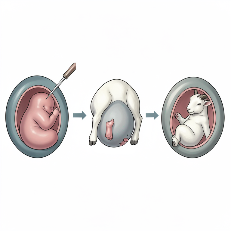
Management of Anasarcous Foetus
AI-generated conceptual illustration (Malabari goat). It explains the idea and does not show study data.
View source evidence ↗Figures & tables
From the original paper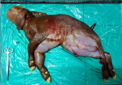 Figure 1
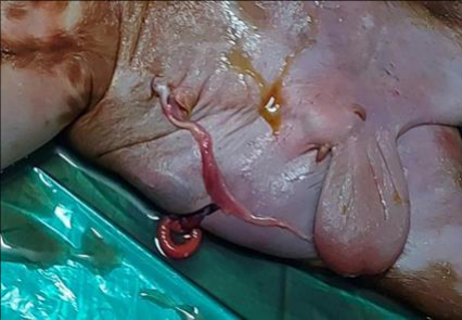 Figure 2
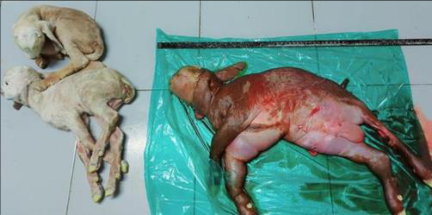 Figure 3
Introduction
Foetal anasarca is most frequently reported in cattle, but there have been few occurrences reported in goats (Selvaraju et al., 2018) [15] . This can occur to a single foetus or twins. A recessive autosomal trait causes foetal anasarca which is characterized by oedema leads to foetal hydrops in cattle. Foetal anasarca in Australian Dexter, Belted Galloway, Maine-Anjou, Short-horn cattle, and Spanish sheep has been documented (Roberts, 1986) [13] . Generally, surgical intervention is resorted to relieve an anasarcous foetus with ascites as per-vaginal delivery is often difficult due to absolute foetal oversize. Per-vaginal delivery of an anasarcous foetus in one of the triplet kids is rare occurrence in goats. Present article reports foetal anasarca with ascites and its successful management in a Malabari doe.
History and clinical signs
A full term four-year-old female Malabari goat having a parity of three was presented with the anamnesis of animal's inability to deliver a second foetus after delivering one live female kid. On physical examination, the rectal temperature was around 102.50 o F, pale conjunctival mucous membrane, oedematous vulva and foetal hind limbs were hanging out of the vulva. On vaginal examination, foetus was in posterior longitudinal presentation, lumbo-sacral in position with extended hind limbs. Foetus was found to be larger in size, with a distended abdomen that fluctuate with pressure, indicating foetal ascites, and lodged in the pelvic inlet. Foetal reflexes were absent, indicating a dead foetus.
Treatment and Discussion
Animal was restrained by giving epidural anaesthesia with 3 mL of 2 per-cent lignocaine Hcl at first inter-coccygeal space. To reduce the foetal size, subcutaneous fluid was drained as well as abdominocentesis was carried out, to remove intra-abdominal fluid (Figure 2). After draining out the fluids, foetal size was reduced, and lubrication was done with carboxy methyl cellulose solution. On gentle traction, a dead male anasarcous foetus was relieved pervaginally (Figure 1). Further, vaginal examination revealed presence of another live foetus which was tracted out per-vaginally (Figure 3).
The animal was administered with Inj. Meloxicam (1 mg/kg I/M), Inj. 5% DNS, Inj. Ringers lactate and Cleanex bolus I/U and antibiotic Inj. Ceftriazone (10 mg/kg intravenously), was followed up for five days. Goat, popularly known as poor man's cow, has a greater economic importance for farmer's livelihood. Dystocia in far m animals is one of the major factors that results in economic loss either due to loss of dam or foetus (Kumar et al., 2020) [7] . Dinesh et al. (2020) [4] reported that developmental abnormalities can occur in domestic animals which may result in structural abnormality of foetus leading to monstrosities. Foetal anasarca is one of the developmental abnormalities which occurs in caprine. Anasarca is a generalized oedematous condition of body and less reported in case of small ruminants (Prabaharan et al., 2016; Sarangom et al., 2020) [11,14] . Craig (2000) [3] reported that foetal anasarca or dropsy of the tissue beneath the skin of the foetus were seen in calf, kids and foals. Though the cause of foetal anasarca is unknown, it is thought to be caused by circulatory disturbances of liquid exchange and may be of placental origin (Roberts, 1986) [13] . Foetal ascites may occur due to obstruction or lack in lymphatics, which restricts the removal of peritoneal fluid and causes accumulation of fluid in subcutaneous space and this may be due to existence of the autosomal recessive allele which affects the embryological development of normal lymph nodes (Chandrashekaran et al., 2015). Foetal anasarca will be carried to the term, but there is lack of progress in second stage of parturition due to an increase in foetal volume from excessive fluid in the subcutaneous (especially in head and limbs), umbilical and inguinal ring as well as hydrocoele (Noakes, 2018) [9] . Foetal anasarca in goats were also reported previously (Laiju et [8,5,2,11,1,7] . Jayachandra et al. (2013) [5] reported that a rarely mild hydrops of the amnion or allantois and oedema of the placenta may accompany foetal anasarca. Most anasarcous foetuses are expelled dead. Usually, pregnancy is maintained full term in case of foetal anasarca alone. In some circumstances, embryotomy may be resorted to drain excess fluid (Roberts, 1986) [13] . Most often anasarcous foetus are presented posteriorly and prolong the stage of the labour (Noakes, 2018) [9] , in the present case also the anasarcous foetus was in posterior presentation. Forced extraction was used to remove large anasarcous foetuses, while fetotomy techniques such as limb amputation and evisceration were used to remove overly large foetuses (Periyannan, 2021 and Prasath et al., 2023) [10,12] . The delivery of a large anasarcous foetus usually requires surgical intervention (Kumar et al., 2005) [6] . The anti-natal diagnosis of most of the commonly occurring foetal complications of gestation is partly possible with ultrasonogram, and such pregnancies should be carefully monitored and terminated (Laiju et al., 2012) [8] .
Conclusion
A rare instance of foetal anasarca with ascites and successful per-vaginal removal of the anasarcous foetus in a Malabari doe has been documented, as well as the delivery of two live foetuses on per-vaginum concurrently. Abdominocentesis and embryotomy can be directed for the management of congenital dropsical conditions such as anasarca as an alternative to caesarean section (invasive), which may result in economic loss for farmers raising small ruminants such as Malabari goats.
Acknowledgement
AcknowledgementThe authors are thankful to the faculty of Department of Animal Reproduction, Gynaecology and Obstetrics, Kerala Veterinary and Animal Sciences University, College of Veterinary and Animal Sciences, Pookode, Kerala for providing necessary facilities and guidance throughout the study of clinical cases.
References
15 in the original paper- Baruti M, Bhuyan M, Bhuyan D, Singh B, Deka R. Dystocia due to foetal anasarca in an Assam hill goat. Int J Chem Stud. 2018;6(1):948-949.
- Chandrasekaran D, Selvakumar S, Kumar RS, Pothiappan P, Das AK, Balasubramanian S. Pervaginal delivery of Anasarcous foetus in a Tellicherry doe. Indian J Anim Reprod. 2015;36(1):60-61.
- Craig JF. Fleming's Veterinary Obstetrics. Greenworld Publishers, 2000, p. 271-273.
- Dinesh M, Kalaiselvan E, Manikandan R. A rare case of Holocardius acephalus monster with foetal anasarca in a non-descriptive goat kid. Pharma Innov J. 2020;9(11):14.
- Jayachandra HK, Kulkarni H, Magadum S, Badami S. Dystocia due to foetal anasarca with achondroplasia in a goat: a case report. Int J Food Agric Sci. 2013;3(3):116118.
- Kumar S, Bhatt P, Prasad JK, Rawat AK, Maurya SN, Kumar S. Dystocia due to foetal anasarca in a crossbred cow. Indian J Anim Reprod. 2005;26:177-178.
- Kumar G, Verma A, Singh R, Pratap B. Dystocia due to foetal anasarca in goat: A case report. J Entomol Zool Stud. 2020;8(4):241-242.
- Laiju MP, Mohan MR, Bastin PF. Foetal anasarca twins with hydroallantois in Malabari does. J Indian Vet Assoc Kerala. 2012;10(1):52-53.
- Noakes DE, Parkinson TJ, England GC, editors. Arthur's Veterinary Reproduction and Obstetrics. Elsevier Health Sciences; 2018.
- Periyannan M, Selvaraju M, Senthilkumar K, Subhash A, Gawhane MP, Gopikrishnan D. Rare incidence of dystocia due to foetal anasarca with ascites in a Jersey crossbred heifer. Pharma Innov J. 2021;10(10):560-562.
- Prabaharan V, Sivakumar A, Jayaganthan P, Raja S, Vijayarajan A, Kumar SS. Dystocia due to foetal anasarca and ascites with live foetus in a doe. Int J Sci Environ Technol. 2016;5(4):2586-2589.
- Prasath RA, Ajay Eswarr M, Sathriyan G, Mohammed Zubair S, Aruneshwaran EL. Successful management of dystocia due to fetal Anasarca in a Mecheri Ewe: A case report. Int J Vet Sci Anim Hus. 2023;8(5):218-219.
- Roberts SJ. Veterinary Obstetrics and Genital Diseases. 3rd ed. CBS Publishers and Distributors, 1986.
- Sarangom SB, Mathai VM, Harshan HM, Prasad CP, Muraleedharan K, Sundaran S. Dystocia due to foetal anasarca and foetal ascites with concurrent live foetus in a multiparous Malabari doe. J Anim Health Prod. 2020;8(4):168-170.
- Selvaraju M, Varudharajan V, Prakash S, Ravikumar K, Senthilkumar K. Bull Dog Monster Dystocia with Schistocephalus fissilabrus, Fetal Ascites, and Anasarca in a Doe. Indian Vet J. 2018;95(11):57-58.