
Research article · 2024 · Full text
International Journal of Biology SciencesAntimicrobial agents and techniques used in modern microbiology
Volume 6(2) · Pages 173–177 · DOI: 10.33545/26649926.2024.v6.i2c.251 ↗
Research areasProbiotics and Fermented Foods
Abstract
Author-writtenOver the past 20 years, there has been an unprecedented increase in the number of new antibiotics developed through the purposeful use of antimicrobial agents. This growth has been attributed to extensive research efforts that have taken advantage of the expanding body of knowledge describing the interactions between antibiotics and their targets in bacterial cells. The development of additional classes of antimicrobial drugs has frequently benefited from knowledge gathered from one class. When it came to beta-lactams, data on the correlations between structure and activity obtained from cephalosporins and penicillins was quickly applied to cephamycins, monobactams, penems, and carbapenems to find drugs with a significantly higher potency that were broad-spectrum. For patients to receive the best possible care, a pathogen's resistance to antibiotics must be quickly identified, and the proper antimicrobial treatment must then be administered. Traditional techniques for detecting bacterial resistance, like automated instruments, broth microdilution, and disc diffusion, are mainly standardized and are frequently employed. However, the results cannot be retrieved before 48 hours after the sample is received, which could result in the misuse or prolonged usage of broad-spectrum antibiotics. Therefore, there is a need to create and implement new, quicker, standardized, sensitive, focused, and reliable procedures with consistent outcomes into everyday microbiological laboratory practice. Antimicrobial Susceptibility Testing (AST) of bacterial pathogens is a crucial procedure in clinical microbiology laboratories to ascertain susceptibility to antimicrobial drugs and identify potential drug resistance. This study focuses on the tools and techniques that can be used in clinical microbiology labs to assess the effectiveness of antibiotics, both now and in the near future.
Summary
Source-checkedPlain-language reading of the full paper, prepared with AI and checked against the text. Each point links to the authors' own words.
Why it matters
- Rapid identification of a pathogen's antibiotic resistance is essential for providing patients with the most effective care. View evidence ↗
- Standardized traditional resistance detection methods often take at least 48 hours to provide results, which can lead to the inappropriate or extended use of broad-spectrum antibiotics. View evidence ↗
The question
- The aim of this study is to review the current and future tools and techniques used in clinical microbiology laboratories to evaluate antibiotic effectiveness. View evidence ↗
What they did
- The authors conducted a thorough analysis of in vitro tests used to evaluate the antibacterial properties of both synthetic and natural substances. View evidence ↗
What they found
- Plant-derived polyphenolics, such as flavonoids and tannins, exhibit antibacterial activity by disrupting cell membranes and inhibiting essential enzymes. View evidence ↗
- Defensins, the largest family of antimicrobial peptides found in mammals, birds, and insects, cause cell death by breaking through microbial barriers. View evidence ↗
- Lysozymes found in human secretions like tears and saliva cause bacterial cell lysis by breaking down the cell walls. View evidence ↗
- Silver nanoparticles act as antimicrobial agents by damaging intracellular components and disrupting bacterial cell membranes. View evidence ↗
- Zinc oxide nanoparticles lower cell viability by producing Reactive Oxygen Species (ROS) that cause membrane leakage of DNA, proteins, and sugars. View evidence ↗
- Agar diffusion assays evaluate drug efficacy by measuring the zone of inhibition, which is the area where microbial growth is suppressed. View evidence ↗
- The Minimum Inhibitory Concentration (MIC) is the lowest amount of an agent that prevents visible bacterial growth, determined using either agar or broth dilution methods. View evidence ↗
- Bacterial synergy can be achieved by combining drugs that target different metabolic phases or by using beta-lactamase inhibitors with vulnerable antibiotics. View evidence ↗
- Combining bacteriostatic and bactericidal agents, such as penicillin and chlortetracycline, can result in antagonistic effects. View evidence ↗
What it means
- Due to the rise of Antimicrobial Resistance (AMR), the authors conclude that there is a critical need for the development of new antimicrobials and the prudent use of existing ones. View evidence ↗
- The authors recommend a comprehensive strategy to manage resistance that includes vaccination, improved hygiene, and antimicrobial stewardship. View evidence ↗
Visual summary
1 visualsIllustrations explain the paper; they do not show study data.
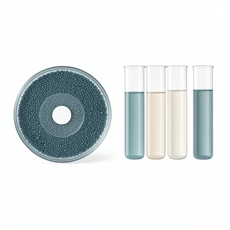
Antimicrobial Testing Methods
AI-generated conceptual illustration (Agar plates with bacterial lawns and glass test tubes containing liquid broth). It explains the idea and does not show study data.
View source evidence ↗Introduction
Every bacterium is targeted by a particular mechanism and distinct inhibitory activity of an antimicrobial drug. As a result, the effectiveness of antimicrobial drugs against a target bacteria determines which ones to use. Since every antimicrobial agent has a unique set of actions that impact germs in a distinct way, there is no one "magic bullet" antimicrobial agent that will be effective against every pathogenic and rotting microorganism [1] . This is because different bacteria have different physiologies and distinct antimicrobial mechanisms. Microorganisms can be simply categorized (aerobes or anaerobes), their optimal growth temperature (thermophilic, mesophilic, or psychrotrophic), their cell-wall composition (gram-positive and gram-negative), their growth stage (spores or vegetative cells), or their resistance to acid and osmosis. This classification can be very useful in the selection of particular antimicrobial agents. Antimicrobial substances either eradicate or stop the growth of microorganisms [2] . Antimicrobial agents have a history that predates the first century. Egyptians employed lard, honey, and lint as a wound treatment circa 1550 BC. Later, moldy bread was used to cure infected wounds in Egypt, Greece, Egypt, Serbia, and other ancient civilizations. Scientific discoveries have revealed that honey has significant hydrogen peroxide content, whereas moldy bread contains unprocessed antibiotics that are bacterially lethal [3] . Our ancestors treated microbial diseases with inorganic antibacterial compounds like copper and silver even before the development of contemporary healthcare institutions. Natural goods are abundant sources of biologically active molecules that have the potential to fight infections. These products are produced from plants, animals, and microbes. Furthermore, laboratory-created synthetic antimicrobial agents have shown a great deal of promise in the fight against microbial diseases. Appropriate antimicrobial screening and evaluation techniques are crucial to maximizing the potential of these drugs. By offering vital information on the efficacy and mechanisms of action of novel antimicrobial drugs, in vitro antimicrobial assays are essential to their discovery and development. Gaining a deeper understanding of these techniques would aid researchers in more effectively identifying possible antibiotic candidates and assist address the mounting threat posed by AMR. Even though broth dilution techniques and disk diffusion are commonplace technologies, they might not be as reproducible or quick [4] . Conversely, more recent techniques like as impedance testing, bioluminescence, and flow cytometry provide more throughput and sensitivity but may be more expensive and difficult to use in areas with limited resources. Therefore, it is crucial to conduct a thorough analysis of the in vitro tests that are used to evaluate the antibacterial properties of both natural and manufactured substances [5] .
Sources of antimicrobials agents
Antimicrobial activity has been reported from a wide range of natural sources, including plants, animals, and microorganisms, as well as synthetic chemicals, nanoparticles, and other materials.
Plant sources
Plants naturally produce a wide variety of bioactive chemicals as part of their defense mechanism. Numerous of these substances, also referred to as phytochemicals, are quite successful in thwarting microbial infections. The phytochemicals are interesting candidates for the development of antimicrobial drugs because they use a variety of mechanisms to prevent the growth and survival of microorganisms, including fungus, viruses, and bacteria. Polyphenolics, which include flavonoids, tannins, quinones, coumarins, and others, have demonstrated noteworthy antibacterial capabilities through the disruption of microbial cell membranes, inhibition of important enzymes, and interference with crucial cellular processes, among other mechanisms. Terpenoids are yet another significant class of bioactives derived from plant extracts that show strong antibacterial activity through rupturing microbial membranes, blocking the synthesis of proteins, or interfering with vital metabolic processes [6] . Certain terpenoids, like essential oils, have demonstrated remarkable efficacy against various bacteria, making them ideal candidates for antimicrobial uses. By rupturing microbial cell membranes, saponins glycosides that are present in a wide variety of plant species also demonstrate antibacterial activities. Furthermore, alkaloids, a different class of bioactive molecules originating from plants, have also shown impressive antibacterial qualities. Historically, medications based on alkaloids, including quinine and berberine, have been used to treat infectious disorders. Furthermore, plants generate specific amps known as antimicrobial peptides, like defensins, which target the membranes of microorganisms to further enhance their antibacterial effect. Another line of protection against infections is provided by lectins, which are proteins that bind to carbohydrates and have antibacterial qualities produced by certain plants. These various plant extractderived bioactive chemicals demonstrate their extraordinary potential as natural antibacterial agent sources [7] .
Animal sources
Antimicrobial peptides and proteins that are part of the animal kingdom's natural defense against pathogenic microbes are an invaluable resource. Similar to plants, a variety of animal taxa, such as insects, amphibians, reptiles, birds, and mammals, also generate amps that have broadspectrum activity against bacteria, fungi, and certain viruses. These amps can target and damage the cell membranes of microorganisms [8] . For example, amphibians, such as frogs and toads, secrete amps into their skin, which serve as a potent protection against germs. Numerous animals, including birds, insects, and mammals, also manufacture defensins, which are the largest family of amps. Defensins break through microbial barriers to cause cell death and stop infections in a number of anatomical locations, including the digestive and respiratory systems. Other antimicrobial compounds, such as lysozymes, lactoferrin, and similar proteins, are also derived from animal sources. The breakdown of bacterial cell walls by lysozymes, which are present in secretions such as milk, saliva, and tears, results in bacterial cell lysis. Lactoferrin is an iron-binding protein found in milk and body fluids that inhibits the growth of microorganisms by retaining vital minerals such as iron. Fish and crustaceans generate amps that have been shown to be active against bacteria, fungus, and viruses, such as piscidins and crustins [9] .
Microbial sources
The antimicrobial action of microbial products is remarkably diverse, since bacteria generate strong chemicals to survive in their surroundings and compete for resources. Of these, antibiotics are the most well-known and researched natural antibacterial substances. Antibiotics are substances produced by some bacteria and fungi that have the amazing capacity to stop other germs from growing. [10] . Penicillin, which was developed from the penicillium fungus, is a well-known example. It signaled a significant breakthrough in contemporary medicine and opened the door for a number of vital medicinal drugs to treat bacterial infections. Bacteriocins are a broad class of antimicrobial peptides that are also produced by microorganisms. By preventing spoilage and harmful bacteria in fermented foods, these tiny cationic peptides contribute to food preservation by their remarkable efficacy against closely related bacteria. Other bioactive chemicals such cyclic lipopeptides, lectins, and bacteriocin-like inhibitory substance (blis) are also part of the arsenal of microbial antimicrobial agents. These molecules exhibit strong activity against a variety of microbial infections. Antimicrobial chemicals are also produced by certain protists and archaea. For instance, some species of archaea are known to produce antimicrobial peptides called archaeocins that are effective against bacteria and other archaea and are believed to be involved in inter-microbial competition. Protist species that are aggressive against bacteria also produce antimicrobial peptides and proteins, such as amoebae [11] .
Synthetic antimicrobials
Chemically created and carefully engineered to have strong antibacterial qualities, synthetic antimicrobial compounds offer defense against the growing threat of bacterial resistance to traditional antibiotics. Synthetic antimicrobial substances with broad-spectrum antibacterial and antifungal action include triclosan. It frequently breaks down bacterial cell membranes in personal hygiene products like toothpaste and soaps. Another synthetic antibiotic called linezolid prevents gram-positive bacteria like streptococcus pneumoniae and staphylococcus aureus from synthesizing proteins. Quinolones are a class of synthetic antimicrobial drugs that block enzymes like topoisomerase IV and DNA gyrase, preventing bacterial DNA replication. Examples of these chemicals include ciprofloxacin and levofloxacin. Furthermore, artificial peptides that imitate the naturally occurring antimicrobial peptides in plants and animals have been created. One such synthetic peptide, pexiganan, breaks down bacterial cell membranes to show antimicrobial efficacy against both gram-positive and gram-negative bacteria. Other synthetic antimicrobial agents include metronidazole, which is used to treat infections caused by anaerobic bacteria and parasites by interfering with their DNA synthesis, and daptomycin, a semisynthetic lipopeptide antibiotic that breaks down bacterial cell membranes and is used to treat skin and bloodstream infections caused by gram-positive bacteria [12] .
Antimicrobial nanoparticles
Nanoparticles, which have a size range of 1 to 100 nm, have special physicochemical characteristics because of their small size and vast surface area. They be made artificially or spontaneously by plants, animals, and microbes. Strong antibacterial action of nanoparticles is demonstrated against viruses, fungi, and bacteria [13] . They are extremely efficient antibacterial agents because their tiny size improves their contact with microorganisms. One type of antimicrobial nanoparticle is silver nanoparticle, which damages intracellular components and breaks down bacterial cell membranes. Reactive Oxygen Species (ROS) produced by zinc oxide nanoparticles increase membrane lipid peroxidation, which results in membrane leakage of reducing sugars, DNA, and proteins and lowers cell viability. The combination of titanium dioxide nanoparticles, which have photocatalytic qualities and produce antimicrobial reactive oxygen species, and calcium oxide nanoparticles, which produce free radicals that damage bacterial cell membranes and the arrangement of polyunsaturated phospholipids, causes ROS to be produced.
Copper nanoparticles also interact with proteins and DNA. [14] .
Methods to evaluate antimicrobial activity Agar diffusion based screening of antimicrobial activity
Antibacterial research frequently uses economical, agar diffusion-based assays, such as disk diffusion, well diffusion, agar plug, and agar spot tests, to assess the antibacterial activity of test substances. These techniques work by preventing the growth of the test microorganism that has been inoculated on the agar surface through the diffusion of antimicrobial chemicals from paper discs, wells, or plugs into the nearby agar medium. The relative efficacy of the test drug against the particular microbe under examination can be evaluated by researchers by measuring the resulting zone of inhibition, which reflects the area where microbial growth is blocked or suppressed by the agent. An important tool in antimicrobial research is the agar diffusion experiment, which yields qualitative information about how well drugs work against particular microbes [15] .
Cross streak method
An distinctive variant of agar diffusion-based assays is the cross-streak method. This approach involves streaking the test microbe and the indicator microbe(s) across each other on the agar surface, as opposed to the previously reported assays where the antimicrobial agent is deposited on the agar medium. The development of the other microorganisms at the intersection points is inhibited if the test microorganism generates an antibiotic agent, which diffuses into the agar media. This approach is used not only to evaluate antimicrobial activity but also to investigate intermicroorganism antagonistic relationships [16] .
Co-culture assay
Similar to the cross-streak approach, the co-culture assay entails the simultaneous development of two microorganisms in a common environment, allowing for direct contact between them. The test microbe's inhibitory action is then verified by measuring and comparing the indicator organism's growth to a control. This technique is frequently used to evaluate the probiotic lactic acid bacteria's antibacterial activity against infections [17] .
Poisoned food technique
Since ancient times, the poisoned food methodology has been used as an antimicrobial screening tool, with a primary focus on assessing antifungal activity. The poisoned food method includes directly mixing the test substance with the growth medium into which the test microorganism is subsequently introduced, as opposed to agar diffusion assays, in which the test substance diffuses from a paper disk into agar. The test organism's growth inhibition is then evaluated. This method has been used extensively to study plant extracts' antibacterial properties. Even if the poisoned food method is being used today, more delicate approaches are rapidly taking its place. However, it is still a useful method for determining the antibacterial activity of substances, particularly for assessing the efficacy of natural products, extracts, and synthesized compounds against microbes [18] .
Agar dilution and broth dilution methods
Two very comparable approaches for figuring out the MIC of antibacterial drugs are the broth dilution method and the agar dilution method. The MIC is the lowest amount of an antimicrobial agent that prevents a specific bacterium from growing visibly. Agar dilution uses agar plates, while broth dilution uses liquid broth tubes. This is the primary methodological distinction between the two dilution techniques. In both techniques, the medium is first supplemented with varying quantities of the antimicrobial agent, and then a fixed number of microbial cells are added. By tracking the microorganism's growth or lack thereof the MIC can be ascertained. Both the broth and agar dilution techniques are crucial for helping determine which antimicrobial medication is best and for tracking how an antibiotic's susceptibility varies over time [19] .
Antimicrobial gradient diffusion test
By integrating the essential concepts of antimicrobial agent dilution and diffusion, the antimicrobial gradient diffusion test provides an alternate method for assessing antimicrobial activity. The gradient diffusion test is intended to identify the mic, in contrast to agar diffusion assays, which are mostly used as screening instruments. By diluting of the antimicrobial agent on a specific test strip, a concentration gradient covering a range of concentrations is produced using this method. After the test organism is injected into agar plates, the test strips are placed on top of the agar. The antimicrobial ingredient then starts to slowly seep into the agar from the strip, creating a measurable zone of inhibition. Developed in the 1980s, this technique has grown in favor as a substitute for the agar dilution method when determining the Minimum Inhibitory Concentration (MIC) of antibiotics against bacteria. It is noteworthy that the test strips utilized in the gradient diffusion test are mostly accessible through commercial channels and are commonly referred to as electronic test strips. Rather than being used as tools for a broad screening of antimicrobial activity or assessing the effectiveness of new antimicrobial drugs, these strips are mainly utilized for the accurate evaluation of antibiotic susceptibility among microorganisms. In clinical microbiology labs, the e-test approach plays a crucial role in directing treatment choices and assisting in the wise selection of suitable antibiotics to treat bacterial infections [20] .
Mechanism to reduce bacterial resistance
The development of antimicrobial resistance will be significantly slowed down by the careful selection of new drugs. Adhering to good hygiene standards can prevent the spread of plasmids, hinder the growth of drug-resistant bacteria in hospitals, and postpone the emergence of these organisms in the general public. It needs to be on the lookout for signs of antibiotic resistance in the community and hospital. Combining anti-cell wall medicines with aminoglycosides, using β-lactamase inhibitors with antibiotics that are vulnerable to β-lactamase, and combining medications that target different phases in bacterial metabolic or synthetic pathways are some significant instances of bacterial synergy. The combination of carbenicillin and gentamicin to treat p aeruginosa infections and penicillin and streptomycin to treat Enterococcus faecalis and Endocarditis are two examples of synergy that has proven clinically significant. In order to inhibit S. aureus, K. pneumoniae, H. influenzae, and anaerobic organisms like bacteroides species all of which are resistant to amoxicillin or ampicillin when they contain β-lactamases the β-lactamase inhibitor clavulanic acid or sulbactam has recently been combined with amino penicillins. Sulfamethoxazole and trimethoprim work together to kill several bacteria by targeting two different aspects of the folic acid cycle. Lastly, there is synergy between combinations of two penicillins that impact gramnegative bacteria at separate stages of cell wall formation. This is valid for the combination of penicillins or cephalosporins, which bind to pbp lb or 3, and mecillinam (amdinocillin), which binds to pbp 2 of bacteria [21] . Combining bacteriostatic and bactericidal agents can lead to antagonistic effects. The combination of penicillin and chlortetracycline for the treatment of pneumococcal meningitis is a typical example. Since tetracyclines, penicillins, and cephalosporins are used to treat mixed infections like pelvic inflammatory disease caused by gonorrhoeae and chlamydia, this impact has not been addressed from a molecular perspective. While antagonism can be demonstrated in test tubes and certain β-lactam antibiotics can produce β-lactamases that inactivate other β-lactams, it is unclear how these findings relate to infections in humans [22] .
Conclusion
Antimicrobial methods and substances are essential in contemporary microbiology for managing and stopping the spread of infectious illnesses. These substances, which include antiseptics, antifungals, antibiotics, and antivirals, are made expressly to target and prevent the growth of different diseases, so safeguarding human health. The effectiveness of these therapies has been greatly increased by developments in antimicrobial procedures, including the creation of new medications, resistance testing, and the application of innovative delivery methods. Molecular diagnostics, antibiotic susceptibility testing, and bioinformatics have improved the speed and accuracy of pathogen identification, enabling the administration of appropriate and efficient therapies. Ongoing research and development of novel antimicrobials is also required, as the prevalence of Antimicrobial Resistance (AMR) rises. This presents a serious problem and calls for the cautious and prudent use of these medicines. A complete plan to combat infectious diseases and manage resistance must also include enhanced hygiene habits, vaccination campaigns, and the integration of antimicrobial stewardship initiatives. In summary, although antimicrobial agents and contemporary methodologies have revolutionized the discipline of microbiology and substantially diminished the incidence of infectious illnesses, persistent attentiveness, ingenuity, and conscientious application are necessary to maintain their efficacy and safeguard worldwide health.
References
20 in the original paper- Leanse LG, Dos Anjos C, Mushtaq S, Dai T. Antimicrobial blue light: A 'Magic Bullet' for the 21st century and beyond? Adv Drug Deliv Rev. 2022;180:114057.
- Kümmerer K. Antibiotics in the aquatic environment-a review-part I. Chemosphere. 2009;75(4):417-434.
- Mandal MD, Mandal S. Honey: its medicinal property and antibacterial activity. Asian Pac J Trop Biomed. 2011;1(2):154-160.
- Arthur M, Courvalin P. Genetics and mechanisms of glycopeptide resistance in enterococci. Antimicrob Agents Chemother. 1993;37(8):1563-1571.
- Murray BE. New aspects of antimicrobial resistance and the resulting therapeutic dilemmas. J Infect Dis. 1991;163(6):1184-1194.
- Resistance, Limited Antibiotic Pipeline, and Societal Implications. Pharmacotherapy. 2017;37(1):71-84.
- Jackson N, Czaplewski L, Piddock LJ. Discovery and development of new antibacterial drugs: learning from experience? J Antimicrob Chemother. 2018;73(6):14521459.
- Norrby SR, Bergan T, Holm SE, et al. Evaluation of new beta-lactam antibiotics. Rev Infect Dis. 1986;8(Suppl 3):S235-S370.
- Schaberg DR. Resistant gram-positive organisms. Ann Emerg Med. 1994;24(3):462-464.
- Hossain TJ. Methods for screening and evaluation of antimicrobial activity: A review of protocols, International Journal of Biology Sciences https://www.biologyjournal.net advantages, and limitations. Eur J Microbiol Immunol. 2024;14(2):97-115.
- Miethke M, Pieroni M, Weber T, Brönstrup M, Hammann P, Halby L, et al. Towards the sustainable discovery and development of new antibiotics. Nat Rev Chem. 2021;5(10):726-749.
- Tommasi R, Brown DG, Walkup GK, Manchester JI, Miller AA. ESKAPEing the labyrinth of antibacterial discovery. Nat Rev Drug Discov. 2015;14(8):529-542.
- Gale EF, Cundliffe E, Reynolds PE, Richmond MH, Waring MJ. The molecular basis of antibiotic action. London: John Wiley & Sons; 1981.
- Hage DS, Anguizola JA, Bi C, Li R, Matsuda R, Papastavros E, et al. Pharmaceutical and biomedical applications of affinity chromatography: recent trends and developments. J Pharm Biomed Anal. 2012;69:93105.
- Franken H, Mathieson T, Childs D, Sweetman GM, Werner T, Tögel I, et al. Thermal proteome profiling for unbiased identification of direct and indirect drug targets using multiplexed quantitative mass spectrometry. Nat Protoc. 2015;10(10):1567-1593.
- Li X, Zolli-Juran M, Cechetto JD, Daigle DM, Wright GD, Brown ED et al. Multicopy suppressors for novel antibacterial compounds reveal targets and drug efflux susceptibility. Chem Biol. 2004;11(10):1423-1430.
- Liu A, Tran L, Becket E, Lee K, Chinn L, Park E, et al. Antibiotic sensitivity profiles determined with an Escherichia coli gene knockout collection: generating an antibiotic bar code. Antimicrob Agents Chemother. 2010;54(4):1393-1403.
- Lamsa A, Lopez-Garrido J, Quach D, Riley EP, Pogliano J, Pogliano K et al. Rapid Inhibition Profiling in Bacillus subtilis to Identify the Mechanism of Action of New Antimicrobials. ACS Chem Biol. 2016;11(8):2222-2231.
- Ribeiro da Cunha B, Fonseca LP, Calado CRC. Metabolic Fingerprinting with Fourier-Transform Infrared (FTIR) Spectroscopy: Towards a HighThroughput Screening Assay for Antibiotic Discovery and Mechanism-of-Action Elucidation. Metabolites. 2020;10(4):145.
- Sinha Roy R, Yang P, Kodali S, Xiong Y, Kim RM, Griffin PR, et al. Direct interaction of a vancomycin derivative with bacterial enzymes involved in cell wall biosynthesis. Chem Biol. 2001;8(11):1095-1106.