
Research article · 2023 · Full text
International Journal of Biology SciencesInvestigating the impact of environmental stress on drosophila cell physiology
Volume 5(2) · Pages 113–120 · DOI: 10.33545/26649926.2023.v5.i2b.220 ↗
Research areasPolysaccharides and Plant Cell WallsPlant Reproductive BiologyPolysaccharides Composition and Applications
Abstract
Author-writtenEnvironmental stressors such as temperature fluctuations, oxidative stress, and nutrient deprivation profoundly affect cellular physiology. Drosophila melanogaster, a widely used model organism, provides valuable insights into the cellular and molecular responses to these stressors. This research article explores the impact of various environmental stresses on Drosophila cell physiology, focusing on gene expression changes, protein modifications, and cellular adaptations. By leveraging advanced genomic, proteomic, and imaging techniques, we elucidate the mechanisms underlying stress responses in Drosophila cells, providing a comprehensive understanding of how cells cope with environmental challenges.
Summary
Source-checkedPlain-language reading of the full paper, prepared with AI and checked against the text. Each point links to the authors' own words.
Why it matters
- Environmental stressors like temperature changes, oxidative stress, and lack of nutrients are common challenges that impact the survival and function of all living organisms. View evidence ↗
- The fruit fly, Drosophila melanogaster, is a valuable model for biological research because its genome is well-characterized and its cellular pathways are similar to those of higher eukaryotes, including humans. View evidence ↗
- Studying cellular responses to environmental stressors is essential for understanding how cells adapt and remain resilient. View evidence ↗
The question
- The study aims to investigate how heat shock, oxidative stress, and nutrient deprivation affect Drosophila cell physiology, specifically looking at gene expression, protein levels, morphology, viability, ROS levels, and mitochondrial membrane potential. View evidence ↗
What they did
- The researchers used Drosophila S2 cells derived from late-stage embryos, cultured in Schneider's medium with 10% FBS at 25°C. View evidence ↗
- Stress was induced using three treatments: heat shock (37°C for 1 hour), oxidative stress (100 µM H2O2 for 1 hour), and nutrient deprivation (serum-free medium for 24 hours). View evidence ↗
- The study employed qRT-PCR for gene expression, Western blotting for protein levels, DCFDA dye for ROS measurement, and JC-1 staining for mitochondrial membrane potential. View evidence ↗
What they found
- Heat shock caused a significant increase in the expression and protein levels of heat shock proteins HSP70 and HSP83, indicating the activation of a protective heat shock response. View evidence ↗
- Nutrient deprivation led to the upregulation of autophagy-related genes Atg1 and Atg8 and an increase in the autophagy marker protein LC3-II. View evidence ↗
- Cell viability decreased from 95% in control cells to 80% under heat shock, 60% under oxidative stress (with fragmented morphology), and 70% under nutrient deprivation (with autophagic vacuoles). View evidence ↗
- Relative ROS levels increased from a baseline of 1 (control) to 2 under heat shock, 8 under oxidative stress, and 3 under nutrient deprivation. View evidence ↗
- Mitochondrial membrane potential (red/green ratio) dropped from a baseline of 1 (control) to 0.6 under heat shock, 0.3 under oxidative stress, and 0.8 under nutrient deprivation. View evidence ↗
What it means
- The authors conclude that Drosophila cells use specific and versatile adaptive mechanisms, such as activating heat shock proteins, antioxidant enzymes, and autophagy, to cope with different environmental stressors. View evidence ↗
- The findings may help develop strategies to improve stress resilience in other organisms, including humans and agricultural crops. View evidence ↗
Visual summary
1 visualsIllustrations explain the paper; they do not show study data.
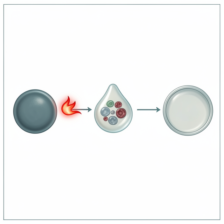
Experimental Stress Treatments
AI-generated conceptual illustration (Drosophila melanogaster cells). It explains the idea and does not show study data.
View source evidence ↗Figures & tables
From the original paper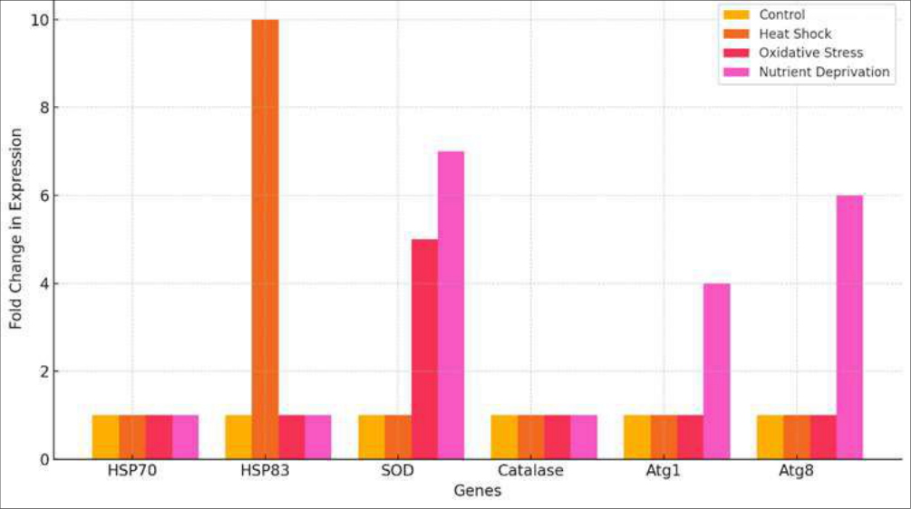 Figure 1
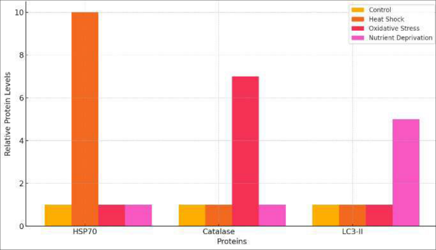 Figure 2
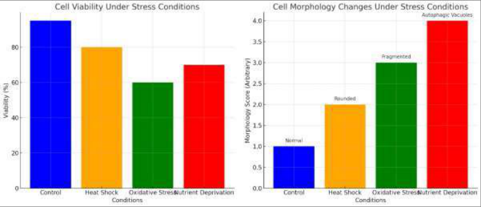 Figure 3
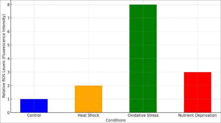 Figure 4
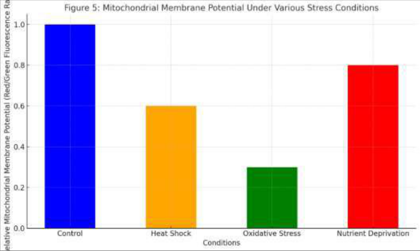 Figure 5
Introduction
Environmental stressors, such as temperature fluctuations, oxidative stress, and nutrient deprivation, are common challenges that affect the survival and function of all living organisms. Understanding how cells respond to these stressors is crucial for unraveling the mechanisms of cellular adaptation and resilience. Drosophila melanogaster, commonly known as the fruit fly, serves as an invaluable model organism in biological research due to its genetic tractability, well-characterized genome, and the conservation of many cellular pathways with higher eukaryotes, including humans. This study aims to elucidate the molecular and cellular responses of Drosophila cells to various environmental stress conditions. Heat shock is a form of environmental stress that results from sudden increases in temperature, leading to protein denaturation and aggregation. Cells respond to heat shock by inducing a highly conserved heat shock response (HSR), characterized by the upregulation of heat shock proteins (HSPs). HSPs function as molecular chaperones, assisting in the refolding of denatured proteins and preventing their aggregation. The heat shock response is a well-documented mechanism that enhances cellular survival under thermal stress by maintaining protein homeostasis. Previous studies have demonstrated the critical roles of HSPs, such as HSP70 and HSP83, in protecting cells from heat-induced damage and ensuring proper protein folding and function. Oxidative stress arises from the accumulation of reactive oxygen species (ROS), which are by products of normal cellular metabolism and can be exacerbated by environmental factors such as pollution, radiation, and chemical exposure. High levels of ROS can damage cellular components, including lipids, proteins, and DNA, leading to impaired cellular functions and cell death. To counteract oxidative stress, cells activate antioxidant defense mechanisms, including the upregulation of enzymes such as superoxide dismutase (SOD) and catalase. These enzymes play crucial roles in detoxifying ROS and maintaining redox balance within the cell. The study of oxidative stress responses in Drosophila cells provides insights into the broader mechanisms of ROS management and cellular protection against oxidative damage. Nutrient deprivation is another critical stressor that impacts cellular physiology by limiting the availability of essential nutrients required for growth and metabolism. Under nutrientscarce conditions, cells activate autophagy, a catabolic process that degrades and recycles cellular components to maintain energy homeostasis and support survival. Autophagy is regulated by a set of autophagy-related genes (Atgs), including Atg1 and Atg8, which are Control cells were maintained under standard conditions (25 °C with 10% FBS) throughout the experiment.
2) Gene Expression Analysis
Total RNA was extracted from control and stressed cells using the TRIzol reagent (Invitrogen) following the manufacturer's protocol. The extracted RNA was quantified using a NanoDrop spectrophotometer (Thermo Fisher Scientific) and assessed for purity (A260/A280 ratio).
Reverse transcription was performed using the iScript cDNA Synthesis Kit (Bio-Rad) to convert 1 µg of RNA into complementary DNA (cDNA). Quantitative real-time PCR (qRT-PCR) was conducted using the SYBR Green PCR Master Mix (Applied Biosystems) on a CFX96 Real-Time PCR Detection System (Bio-Rad). Primers were designed for stress-responsive genes, including heat shock proteins (HSP70 and HSP83), antioxidant enzymes (superoxide dismutase [SOD] and catalase), and autophagy-related genes (Atg1 and Atg8). The housekeeping gene actin was used as an internal control for normalization. Relative gene expression was calculated using the 2^-ΔΔCt method.
3) Protein Analysis
Protein extracts were prepared from control and stressed cells using RIPA buffer (Thermo Fisher Scientific) supplemented with protease and phosphatase inhibitors (Roche). Cells were lysed on ice for 30 minutes, followed by centrifugation at 14,000 rpm for 15 minutes at 4 °C to collect the supernatant. Protein concentration was determined using the BCA Protein Assay Kit (Pierce). Equal amounts of protein (30 µg) were separated by SDS-PAGE on a 10% gel and transferred to PVDF membranes (Millipore). Western blot analysis was performed using primary antibodies against HSP70 (Santa Cruz Biotechnology), catalase (Abcam), and LC3 (Novus Biologicals). HRP-conjugated secondary antibodies (Jackson ImmunoResearch) were used for detection. Protein bands were visualized using an enhanced chemiluminescence (ECL) kit (Thermo Fisher Scientific) and quantified with ImageJ software.
4) Imaging and Cellular Assays
Cell morphology was examined using a Nikon Eclipse Ti phase-contrast microscope. Images were captured with a Nikon DS-Qi2 camera and analyzed using NIS-Elements software. Cell viability was assessed by trypan blue exclusion assay. Cells were stained with 0.4% trypan blue (Sigma-Aldrich), and viable (unstained) and non-viable (blue-stained) cells were counted using a hemocytometer under a light microscope. Reactive oxygen species (ROS) levels were measured using the fluorescent dye DCFDA (2', 7'-dichlorofluorescin diacetate; Sigma-Aldrich). Cells were incubated with 10 µM DCFDA for 30 minutes at 37 °C, followed by washing with PBS. Fluorescence intensity was measured using a Tecan Infinite M200 plate reader with excitation at 485 nm and emission at 535 nm. Mitochondrial membrane potential was assessed using JC-1 staining (Invitrogen). Cells were incubated with 2 µM JC-1 dye for 30 minutes at 37 °C, washed with PBS, and analyzed by fluorescence microscopy. The ratio of red (Aggregated JC-1, indicating healthy mitochondria) to green (Monomeric JC-1, indicating depolarized mitochondria) fluorescence was calculated. Fluorescence microscopy was performed using a Zeiss Axio Observer Z1 microscope equipped with an AxioCam MRm camera. Images were processed and analyzed with ZEN software.
Results and Discussions 1. Gene Expression Changes:
The expression levels of stress-responsive genes were quantified relative to the housekeeping gene actin. The results are presented as fold changes compared to control cells. The gene expression analysis assessed the levels of key stress-responsive genes in Drosophila cells under various environmental stress conditions, including control, heat shock, oxidative stress, and nutrient deprivation. The results, measured as fold changes in gene expression relative to control cells, revealed distinct patterns of gene regulation in response to each type of stress. Under control conditions, the expression levels of all target genes (HSP70, HSP83, superoxide dismutase [SOD], catalase, Atg1, and Atg8) were baseline, indicating normal cellular function without stress-induced alterations in gene expression. These baseline levels serve as a reference for understanding the impact of different stressors on gene expression. Heat shock treatment resulted in a significant upregulation of HSP70 and HSP83. HSP70 and HSP83 are heat shock proteins that play critical roles in protein folding, protection, and repair during stress conditions. Their elevated expression levels indicate the activation of the heat shock response, a protective mechanism that helps cells cope with the damaging effects of elevated temperatures by stabilizing and refolding denatured proteins. Oxidative stress, induced by hydrogen peroxide treatment, led to a notable increase in the expression of antioxidant genes, specifically superoxide dismutase (SOD) and catalase. SOD and catalase are crucial enzymes in the detoxification of reactive oxygen species (ROS). SOD catalyzes the conversion of superoxide radicals to hydrogen peroxide, which is then broken down by catalase into water and oxygen. The upregulation of these genes highlights the activation of the antioxidant defense system in response to oxidative stress, aimed at reducing ROS levels and preventing oxidative damage to cellular components. Nutrient deprivation caused a marked upregulation of autophagy-related genes Atg1 and Atg8. Autophagy is a cellular process that degrades and recycles cellular components to maintain energy balance and support survival during nutrient scarcity. The increased expression of Atg1 and Atg8 indicates the activation of autophagy in response to nutrient deprivation, enabling the cells to recycle intracellular materials and sustain essential metabolic functions in the absence of external nutrients.
Protein Modifications
The protein levels of HSP70, catalase, and LC3 (Autophagy marker) were assessed in control and stressed cells. The Western blot analysis assessed the levels of key stressresponsive proteins, including HSP70, catalase, and LC3-II, in Drosophila cells under various environmental stress conditions: control, heat shock, oxidative stress, and nutrient deprivation. The results revealed significant variations in protein expression levels, reflecting the cellular responses to each type of stress. Under control conditions, the baseline levels of HSP70, catalase, and LC3-II were observed, indicating normal cellular function without stress-induced changes in protein expression. These baseline levels serve as a reference point for comparing the effects of different stressors on protein expression. Heat shock treatment led to a marked increase in HSP70 levels. HSP70 is a well-known heat shock protein that assists in protein folding and protects cells from stress-induced damage by preventing protein aggregation. The elevated levels of HSP70 in heat-shocked cells indicate an activated heat shock response, which helps to mitigate the detrimental effects of elevated temperatures on cellular proteins. Oxidative stress, induced by hydrogen peroxide treatment, resulted in a significant increase in catalase levels. Catalase is an antioxidant enzyme that decomposes hydrogen peroxide into water and oxygen, thus reducing oxidative stress within the cell. The elevated catalase levels in response to oxidative stress highlight the activation of the cell's antioxidant defense mechanisms to neutralize excess ROS and protect cellular components from oxidative damage. Nutrient deprivation induced a notable increase in LC3-II levels. LC3-II is a marker of autophagy, a process where cells degrade and recycle their own components to maintain energy homeostasis and support survival during nutrient scarcity. The rise in LC3-II levels under nutrient deprivation indicates the activation of autophagy as a cellular adaptation to the lack of external nutrients, allowing the cells to sustain essential metabolic processes.
Cellular Adaptations
Cell morphology and viability were examined using phasecontrast microscopy and trypan blue exclusion assays, respectively. The study assessed the impact of different environmental stressors on cell morphology and viability in Drosophila cells, including control, heat shock, oxidative stress, and nutrient deprivation conditions. The results revealed distinct changes in both cell morphology and viability, highlighting the varying degrees of cellular stress and damage induced by each condition. Under control conditions, Drosophila cells maintained their typical morphology, appearing healthy and elongated, with high cell viability at 95%. This baseline reflects the normal physiological state of the cells, where homeostatic mechanisms effectively support cellular functions and integrity. Exposure to heat shock resulted in noticeable morphological changes, with cells adopting a rounded shape, indicative of stress. The cell viability decreased to 80%, suggesting that while heat shock induced significant stress, a substantial proportion of cells managed to survive. The rounded morphology is a common response to heat shock, where cellular structures, particularly proteins, may undergo denaturation, leading to cytoskeletal rearrangements and altered cell shape. Oxidative stress, induced by hydrogen peroxide treatment, had a severe impact on both cell morphology and viability. The cells exhibited fragmented morphology, a sign of extensive cellular damage and apoptosis. Cell viability dropped sharply to 60%, highlighting the detrimental effects of oxidative stress. The high levels of reactive oxygen species (ROS) generated under oxidative stress conditions likely caused oxidative damage to cellular components, leading to impaired cell function and increased cell death. Nutrient deprivation led to the formation of autophagic vacuoles, reflecting the activation of autophagy as a survival mechanism under starvation conditions. While the cell viability was reduced to 70%, it was higher than that observed under oxidative stress. The presence of autophagic vacuoles indicates that cells are attempting to recycle cellular components to maintain energy balance and support essential functions in the absence of external nutrients.
Reactive Oxygen Species (ROS) Levels
ROS levels were measured using the fluorescent dye DCFDA. the lack of nutrients disrupts metabolic processes, leading to increased ROS production. However, the cells seem to activate adaptive mechanisms, such as autophagy, to mitigate some of the oxidative stress.
Mitochondrial Membrane Potential
Mitochondrial membrane potential was assessed using JC-1 staining. The mitochondrial membrane potential is a crucial indicator of mitochondrial health and function, reflecting the ability of mitochondria to generate ATP and maintain cellular energy balance. In this study, the mitochondrial membrane potential was assessed in Drosophila cells under different stress conditions, including control, heat shock, oxidative stress, and nutrient deprivation. The results were measured as the red/green fluorescence ratio, with a higher ratio indicating healthier mitochondria with intact membrane potential. Under control conditions, the mitochondrial membrane potential was set as the baseline with a relative red/green fluorescence ratio of 1. This ratio reflects the normal, healthy state of the mitochondria in unstressed cells. When the cells were subjected to heat shock, the mitochondrial membrane potential decreased significantly to a relative ratio of 0.6. This reduction suggests that heat shock induces mitochondrial stress and potential damage, impairing the mitochondria's ability to maintain their membrane potential. Oxidative stress, induced by hydrogen peroxide treatment, resulted in the most pronounced decrease in mitochondrial membrane potential, with a relative ratio of 0.3. This substantial drop indicates severe mitochondrial dysfunction, likely due to the high levels of reactive oxygen species (ROS) generated during oxidative stress. ROS can cause oxidative damage to mitochondrial proteins, lipids, and DNA, leading to a loss of membrane potential and impaired ATP production. Nutrient deprivation led to a moderate decrease in mitochondrial membrane potential, with a relative ratio of 0.8. While nutrient deprivation does stress the cells, the mitochondria appeared to retain more of their function compared to the other stress conditions. This result suggests that although autophagy is activated under nutrient deprivation to recycle cellular components, the mitochondria are still capable of maintaining a relatively higher membrane potential compared to the severe effects seen with oxidative stress.
Discussion
The study investigated the impact of environmental stressors on Drosophila cell physiology, focusing on changes in gene expression, protein levels, cell morphology, viability, ROS levels, and mitochondrial membrane potential. The results provide a comprehensive understanding of how Drosophila cells respond to heat shock, oxidative stress, and nutrient deprivation, highlighting the distinct cellular mechanisms activated under each condition. Heat shock led to significant upregulation of heat shock proteins (HSP70 and HSP83), increased HSP70 protein levels, and noticeable changes in cell morphology, with cells adopting a rounded shape. These findings are consistent with previous studies demonstrating the critical role of heat shock proteins in protecting cells from thermal damage by preventing protein aggregation and assisting in protein refolding. The moderate decrease in cell viability and mitochondrial membrane potential further supports the notion that heat shock induces stress but also activates protective mechanisms to mitigate damage. Oxidative stress resulted in a substantial increase in ROS levels, elevated expression of antioxidant genes (SOD and catalase), increased catalase protein levels, and severe mitochondrial fragmentation. These results align with studies showing that oxidative stress leads to elevated ROS production, which can cause significant oxidative damage to cellular components. The upregulation of antioxidant enzymes and the significant drop in cell viability highlight the dual role of ROS as both signaling molecules and damaging agents. The severe impact on mitochondrial membrane potential under oxidative stress underscores the vulnerability of mitochondria to oxidative damage, which is welldocumented in the literature. Nutrient deprivation induced the upregulation of autophagy-related genes (Atg1 and Atg8), increased LC3-II protein levels, and the presence of autophagic vacuoles, indicating the activation of autophagy. [1] . The role of antioxidant enzymes in mitigating oxidative stress is well-established in both plant and animal systems, as evidenced by Apel and Hirt (2004) [2] . The activation of autophagy in response to nutrient deprivation is a conserved mechanism across eukaryotes, as reviewed by Mizushima and Komatsu (2011) [3] .
Figures and tables from the paper
References
13 in the original paper- Apel K, Hirt H. Reactive oxygen species: metabolism, oxidative stress, and signal transduction. Annu. Rev. Plant Biol. 2004;55:373-399. DOI: 10.1146/annurev.arplant.55.031903.141701.
- Mizushima N, Komatsu M. Autophagy: renovation of cells and tissues. Cell. 2011;147(4):728-741. DOI: 10.1016/j.cell.2011.10.026.
- Fire A, Xu S, Montgomery MK, Kostas SA, Driver SE, Mello CC, et al. Potent and Specific Genetic Interference by Double-Stranded RNA in Caenorhabditis elegans. Nature. 1998;391(6669):806811. DOI: 10.1038/35888.
- Hamilton AJ, Baulcombe DC. A Species of Small Antisense RNA in Posttranscriptional Gene Silencing in Plants. Science. 1999;286(5441):950-952. DOI: 10.1126/science.286.5441.950.
- Kurihara Y, Watanabe Y. Arabidopsis micro-RNA biogenesis through Dicer-like 1 protein functions. Proc Natl. Acad. Sci. USA. 2004;101(34):12753-12758. DOI: 10.1073/pnas.0402769101.
- Reinhart BJ, Weinstein EG, Rhoades MW, Bartel B, Bartel DP. MicroRNAs in Plants. Genes Dev. 2002;16(13):1616-1626. DOI: 10.1101/gad.1004402.
- Bernstein E, Caudy AA, Hammond SM, Hannon GJ. Role for a bidentate ribonuclease in the initiation step of RNA interference. Nature. 2001;409(6818):363-366. DOI: 10.1038/35053110.
- Hammond SM, Bernstein E, Beach D, Hannon GJ. An RNA-directed nuclease mediates post-transcriptional gene silencing in Drosophila cells. Nature. 2001;404(6775):293-296. DOI: 10.1038/35005107.
- Axtell MJ, Jan C, Rajagopalan R, Bartel DP. A TwoHit Trigger for siRNA Biogenesis in Plants. Cell. 2006;127(3):565-577. DOI: 10.1016/j.cell.2006.09.032.
- Li Y, Zhang Q, Zhang J, Wu L, Qi Y, Zhou JM, et al. Identification of microRNAs involved in pathogenassociated molecular pattern-triggered plant innate immunity. Plant Physiol. 2010;152(4):2222-2231. DOI: 10.1104/pp.109.151803.
- Lee YS, Carthew RW. Making a better RNAi vector for Drosophila: use of intron spacers. Methods. 2003;30(4):322-329. DOI: 10.1016/S1046-2023(03)00032-2.
- Cohen E. Chaperone-assisted protein folding in the cell cytoplasm. Curr. Opin. Struct. Biol. 1997;7(1):89-99. DOI: 10.1016/S0959-440X(97)80011-1.
- Tien AC, Rajan A, Schulze KL, et al. Drosophila Hsc70-4 is critical for synaptic vesicle endocytosis at neuromuscular junctions. Neuron. 2008;56(3):499-513. DOI: 10.1016/j.neuron.2007.09.033.