
Research article · 2024 · Full text
International Journal of Veterinary Sciences and Animal HusbandryMicrobiological and histopathological study of E. Coli infection in African Grey Parrot (Psittacus erythracus)
Volume 9(3) · Pages 477–479 · DOI: 10.22271/veterinary.2024.v9.i3g.1459 ↗
Research areasIdentification and Quantification in FoodFish Ecology and Management StudiesFish Biology and Ecology Studies
Abstract
Author-writtenIn the field of avian medicine, enteric bacteria are regarded as important potential pathogens capable of causing either primary or opportunistic infections. This study is focused on evaluating the microbiological and histological aspects of E. coli infection in Psittacus erythracus. Within this research, a postmortem examination of an eleven-month-old grey parrot with a history of relocation was conducted. The heart blood smear and impression smear examination from the liver and lungs revealed clumps of numerous coccobacilli. Gross changes observed include pulmonary congestion with oedema, air sacculitis, pneumonia, severe hepatic and renal congestion, diffuse haemorrhages in the proventriculus, haemorrhagic enteritis and engorgement of meningeal blood vessels. Histopathological Severe interstitial haemorrhage with mild inflammatory infiltrates in the lungs. The heart exhibited Severe haemorrhage in between cardiac muscle fibers of epicardium with myocardial degeneration. Congestion, hemmorage, infiltration of mono-nuclear cells was also noted in liver and spleen. The kidneys exhibited Cell swellings with tubular epithelial degeneration. The case was identified as E. coli based on isolation studies.
Summary
Source-checkedPlain-language reading of the full paper, prepared with AI and checked against the text. Each point links to the authors' own words.
Why it matters
- Enteric bacteria are significant pathogens in avian medicine, as they can cause both primary and opportunistic infections. View evidence ↗
- Avian colibacillosis is caused by Avian Pathogenic E. coli (APEC), which is a Gram-negative, facultative anaerobic bacillus. View evidence ↗
- In captive parrots, E. coli is frequently linked to septicemic, respiratory, and digestive issues, manifesting in various forms such as hepatitis and pericarditis. View evidence ↗
The question
- The study aimed to evaluate the histological and microbiological characteristics of an E. coli infection in an African Grey Parrot (Psittacus erythracus). View evidence ↗
What they did
- The research was based on a postmortem examination of one eleven-month-old African Grey Parrot that had a history of relocation. View evidence ↗
- Field staining techniques were used to perform impression smear examinations on the lungs and liver. View evidence ↗
- Lesion swabs were cultured on MacConkey’s agar, Eosin Methylene Blue agar, and brain-heart Infusion agar at 37°C for 24 to 48 hours. View evidence ↗
- Organs were fixed in 10% neutral buffered formalin, embedded in paraffin, and 5-um-thick sections were stained with Haematoxylin-Eosin for microscopic analysis. View evidence ↗
What they found
- The isolated bacteria were confirmed as E. coli through pink colonies on MacConkey agar (indicating lactose fermentation) and a green metallic sheen on EMB agar. View evidence ↗
- Histopathology of the lungs showed vasculitis and severe interstitial hemorrhage accompanied by mild inflammatory infiltrates. View evidence ↗
- The heart exhibited severe congestion, myocardial degeneration, and hemorrhage between the cardiac muscle fibers of the epicardium. View evidence ↗
- The liver showed severe sinusoidal congestion with mononuclear cell infiltration and mild hepatic degeneration characterized by vacuolization of the cytoplasm. View evidence ↗
- The spleen showed severe congestion, mild hemorrhage, and diffuse infiltration of mononuclear cells. View evidence ↗
- The kidneys exhibited necrotic changes in the renal tubules, tubular epithelial degeneration, and severe cell swelling. View evidence ↗
What it means
- The authors concluded that the African Grey Parrot died from an E. coli infection, with the liver being the most severely affected organ. View evidence ↗
Visual summary
1 visualsIllustrations explain the paper; they do not show study data.
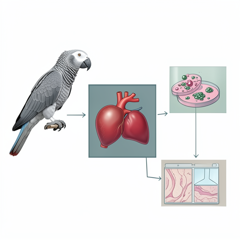
Diagnostic Workflow for Avian Colibacillosis
AI-generated conceptual illustration (African Grey Parrot (Psittacus erythracus)). It explains the idea and does not show study data.
View source evidence ↗Figures & tables
From the original paper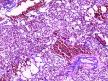 Figure 1
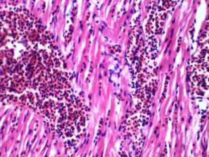 Figure 2
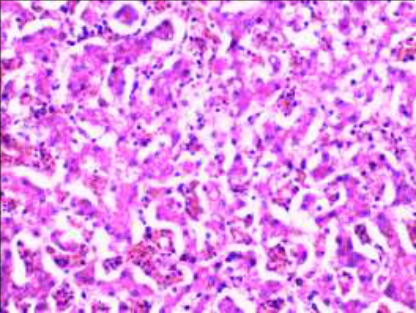 Figure 3
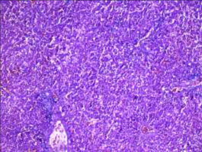 Figure 4
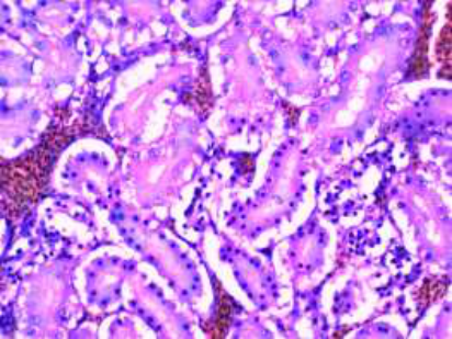 Figure 5
Introduction
Escherichia coli (E. Coli.) is a facultative anaerobic Gram-negative bacillus, Avian Pathogenic E. Coli (APEC) that causes avian colibacillosis. This disease may develop in different forms, such as: coligranuloma, cellulitis colisepticemia, airossacullitis, sinusitis, peritonitis, salpingitis, pericarditis, hepatitis, panopthalmitis and osteomyelitis; many species of E. coli species, which is commonly associated with digestive, respiratory, and septicemic problems in parrots kept in captivity (Ritchie et al., 1994) [8] . Whereas APEC is known to have caused the poultry industry to suffer large financial losses and to have caused septicemia and respiratory diseases. Nevertheless, certain E. Coli categorization investigations on wild birds (Steele et al., 2005 and Pedersen et al., 2006) [13,6] and captive psittacines have identified some degree of association between the disease and the particular pathotypes (Schremmer et al., 1999; Saidenberg 2009) [11,10] . There is ongoing debate regarding E. coli place in the normal microbiota of healthy pets who eat a regular diet consisting of grains, fruits, vegetables, and sprouts.
Materials and Methods
An eleven-month-old, recently dead African grey parrot. The bird was presented with a history of relocation and exhibited reduced appetite, greenish diarrhea and sudden death. Gross lesions observed include pulmonary congestion with oedema, air sacculitis, pneumonia, severe hepatic and renal congestion, diffuse haemorrhage on proventriculus, haemorrhagic enteritis and engorgement of meningeal blood vessels, post-mortem carried out by the Department of Veterinary Pathology, College of Veterinary and Animal Sciences, Mannuthy was the base study material. Impression smear examinations from the liver and lung were observed using the field staining technique. Swabs were collected from lesions and inoculated in nutrient broth, and then cultured on brain-heart Infusion agar (BHIA), Eosin Methylene Blue agar (EMB) and MacConkey's agar (MAC) were incubated at 37 o C for 24-48 hrs.
The colonies were identified by morphological, staining and biochemical techniques (Koneman et al., 1983; Quinn et al., 1994) [4,7] . Organs with gross lesions were removed from birds and fixed in 10% neutral buffered formalin. After dehydration in a graded ethanol series and clearing with xylene, the sample material was embedded in paraffin and 5-um-thick sections were stained with Haematoxylin-Eosin stain for observation under the light microscope (Suvarna 2019) [14] .
Results and Discussion
On impression smear examination of the liver and lung revealed clumps of numerous coccobacilli. In BHIA large, thick, greyish-white, moist, smooth colonies were isolated from the swab taken from the lungs and pericardium layer of the heart. On Gram staining organism appeared as Gramnegative rod-shaped bacteria. The isolated bacterium was confirmed to be E. Coli by culturing on selective and differential media. In MAC, the colonies appeared pink in colour suggesting lactose fermentation, which distinguishes E. coli and in EMB the colonies exhibited green metallic sheen. E. coli was also identified from multiple organs in a study conducted on red-spectacled Amazon (A. pretrei) parrots (Correa et al. 2013) [2] . Asymptomatic psittacine from a number of species, including Amazona spp. and Eupsittula spp. in captivity, have also been found to contain enteropathogenic E. Coli (Saidenberg et al., 2012) [9] . Avian colibacillosis aspects were investigated in a study conducted on a pet A. aestiva (Marietto-Goncalves et al. 2007) [5] , in which E. coli was isolated from several organs and the vacuoles observed including lesions, which consisted of infiltration of mononuclear nucleated cells in kidney and heart, congestion in kidneys, liver, spleen and lungs, which are consisted with the findings in this study. Colibacillosis frequently causes the following lesions: salpingitis with intense follicle necrosis and mixed infiltrate; cellulitis with intense heterophilic infiltrate, multinuclear giant cells and fibronecrotic plaques coagulative liver necrosis, airossaculitis, granulomas, acute lung haemorrhage and congestion (Crespo [3,1,12] . Histopathological examination revealed vasculitis and severe interstitial haemorrhage with mild inflammatory infiltrates in the lung (Fig. 1). In heart severe congestion, haemorrhage in between cardiac muscle fibres of epicardium with myocardial degeneration (Fig. 2). Liver shows a mild degree of hepatic degeneration with vacuolisation of hepatocytic cytoplasm (Fig. 3). Severe sinusoidal congestion with mononuclear cell infiltration. Spleen exhibits diffuse mononuclear cell infiltration with severe congestion and mild hemorrhage was observed (Fig. 4). The kidney showed severe swelling of cells with tubular epithelial degeneration and necrotic changes in the renal tubules (Fig. 5).
Conclusion
A pet, Psittacus erythracus succumbed to E. coli infection exhibiting mild to moderate lesions and liver was found to be the most affected organ. vasculitis and severe interstitial haemorrhage with mild inflammatory infiltrates found in the lungs during the histopathological examination. Severe congestion, and haemorrhage in between cardiac muscle fibres of epicardium with myocardial degeneration. The liver and spleen exhibited signs of congestion, haemorrhage, neutrophil infiltration, and vacoulation. Swelling of cells with tubular epithelial degeneration were noticed in the kidney.
Acknowledgement
AcknowledgementThe authors thank the Dean, College of Veterinary and Animal Sciences, Mannuthy for providing the necessary facilities to carry out this work.
References
10 in the original paper- Andrade CL, Ferreira GF, Franco RM, Nascimento ER, Tortelly R. Alteracoes patologicas identificacao da Escherichia coli como agente causal da celulite aviária em frangos de corte inspecionados em um matadouro em Sao Paulo. Brazilian Journal of Veterinary Medicine. 2006;13(3):139-143.
- Correa IMO, Flores F, Schneiders GH, Pereira LQ, Brito BG, Lovato M. Deteccao de fatores de virulência de Escherichia coli e análise de Salmonella spp. em psitacideos. Pesquisa Veterinaria Brasileira. 2013;33(2):241-246.
- Crespo R, Walker RL, Nordhausen R, Sawyer SJ, Manalc RB. Sal-pingitis in Pekin ducks associated with concurrent infection with Treta trichomonas spp. and Escherichia coli. Journal of Veterinary Diagnostic Investigation. 2001;13:240-245.
- Koneman EW, Stephen D, Dowell J, Sommers S. Color Atlas and Textbook of Diagnostic Microbiology Philadelphia: Lippincott, 1983.
- Goncalves MGA, Lima ET, Siqueira JL, Fiho ARL. Colisepticemia em papagaio-verdadeiro (Amazona aestiva). Revista Brasileira de Saúde e Produção Animal. 2007; 8(1):56-60.
- Pedersen K, Clark L, Andelt WF, Salman MD. Prevalence of Shiga toxin-producing Escherichia coli and Salmonella enterica in rock pigeons captured in Fort Collins, Colorado, Journal of Wildlife Diseases. 2006;42(1):46-55.
- Saidenberg AB, Teixeira RHF, Guedes NMR, Allgayer MC, Melville PN, Benites NR. Molecular detection of Enteropathogenic Eschericia coli in asymptomatic captive Psittacines. Pesquisa Veterinaria Brasileira. 2012;32(9):922-926.
- Schremmer C, Lohr JE, Wastlhuber U, Kosters J, Ravelshofer K, Steinruck H, et al. Enteropathogenic Escherichia coli in Psittaciformes. Avian Pathology. 1999;28:349-354.
- Seeley KE, Baitchman E, Bartlett S, Debroy C, Garner MM. Investigation and control of an attaching and effacing Escherichia coli outbreak in a colony of captive budgerigars (Melopsittacus undulatus). Journal of Zoo and Wildlife Medicine. 2014;45(4):875-882.
- Steele CM, Brown RN, Botzler RG. Prevalences of zoonotic bacteria among seabirds in rehabilitation centers along the Pacific Coast of California and Washington, USA, Journal of Wildlife Diseases. 2005;41(4):735-744.
Cited by 1
From OpenAlex, OpenCitations and reference lists read by Scinovex
- Prevalence and antimicrobial resistance profile of potentially zoonotic enterobacterales isolated from macaws in zoos of Northeastern Brazil ↗
Bruno Pessoa Lima, Adson Ribeiro Marques, N. M. Pascoal Filho, Cibelle Mara Pereira de Freitas, Lúcia Travassos da Rosa Costa, Lucas Silva Melo et al. · Brazilian Journal of Microbiology · 2025
❝ 1