
Research article · 2024 · Full text
International Journal of Advanced Biochemistry ResearchMolecular evidence of Elephant Endotheliotropic Herpes Virus (EEHV) from Maharashtra, India
Volume 8(3S) · Pages 672–679 · DOI: 10.33545/26174693.2024.v8.i3si.856 ↗
Research areasHerpesvirus Infections and TreatmentsPoxvirus research and outbreaksAnimal Disease Management and Epidemiology
Abstract
Author-writtenBackground: Elephant Endotheliotropic Herpes Virus (EEHV) is a significant disease of elephants and has been reported in captive and free-ranging elephants. The disease considerably impacts conservation efforts due to high mortality rates in juvenile elephants. The case presentation discusses the investigation of elephant calf mortality due to EEHV supplemented with molecular, histopathological, and phylogenetic investigations. Case-presentation: Arjun, a five-year-old Asian elephant (Elephas maximus), was found dead after a brief illness. A post-mortem examination was performed, and samples were collected for histopathology and molecular diagnosis. PCR and sequencing confirmed EEHV 1A. The sequence was similar to sequences of EEHV from Assam, India, and Switzerland. The amino acid alignment revealed four substitutions in the U38 protein, due to which the distinct virus was placed separately at the base of the phylogenetic tree. Conclusion: EEHV is a lethal infection in Asian elephants. Molecular techniques confirmed the classical gross and histopathological lesions. EEHV-1A, confirmed in this case, is considered virulent circulating field strain and sequencing efforts are a must to monitor the virus. Considering the dynamic nature of the virus, the herding nature of the species, its impact on juvenile calves, and the lifelong shedding of the virus from adult carriers, it is required to keenly investigate the prevalence and impact of the disease in free-range and captive elephants.
Summary
Source-checkedPlain-language reading of the full paper, prepared with AI and checked against the text. Each point links to the authors' own words.
Why it matters
- Elephant Endotheliotropic Herpes Virus (EEHV) is a major threat to elephant conservation because it causes high death rates among juvenile elephants. View evidence ↗
- Known as the 'calf killer,' EEHV primarily affects young elephants between 0 and 8 years of age. View evidence ↗
- While EEHV has been reported in Asian elephants in India, there is a lack of detailed investigations using molecular assays and phylogenetic analysis. View evidence ↗
The question
- The study aims to perform a post-mortem examination and provide molecular confirmation of EEHV in a specific case of an Asian elephant. View evidence ↗
What they did
- The subject was Arjun, a five-year-old Asian elephant from the Kamlapur Elephant Camp in Maharashtra, India. View evidence ↗
- DNA for molecular investigation was isolated from several organs, including the heart, lungs, liver, kidney, tongue, intestine, and lymph nodes. View evidence ↗
What they found
- Histopathology revealed sinusoidal congestion in the liver, erythrocyte extravasation in the heart, and hemorrhagic changes in the tongue, lungs, and spleen. View evidence ↗
- Comparison of the U38 protein revealed four specific amino acid substitutions (S28W, L47F, D124N, and R127I) out of 135 sequences. View evidence ↗
- PCR screening of other elephants in the herd returned negative results for all EEHV serotypes. View evidence ↗
What it means
- The authors conclude that due to the virus's dynamic nature and the species' herding behavior, there is an urgent need to investigate EEHV prevalence in both captive and free-ranging elephants. View evidence ↗
- The case highlights the vulnerability of Asian elephants to EEHV and the necessity of comprehensive monitoring and management strategies. View evidence ↗
Visual summary
1 visualsIllustrations explain the paper; charts are replotted from the paper's own table values.
Elephants / Ganesh and Basanti and more: Serum SGOT (mg/dl) vs Serum SGPT (mg/dl)
Replotted from Table 1 (page 5) of the original paper. Every bar is the paper's own table value.
View source evidence ↗Figures & tables
From the original paper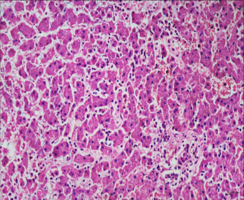 Figure 1
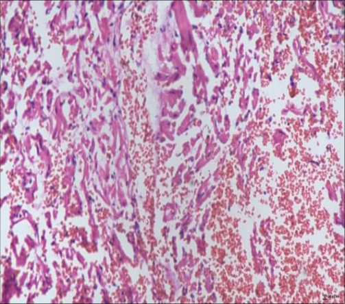 Figure 2
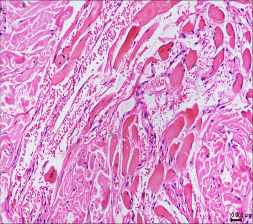 Figure 3
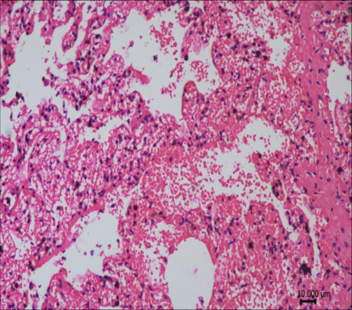 Figure 4
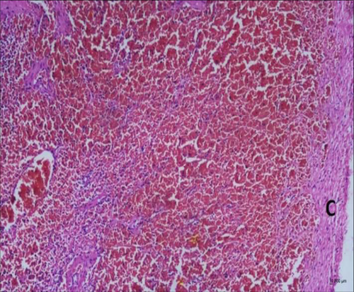 Figure 5
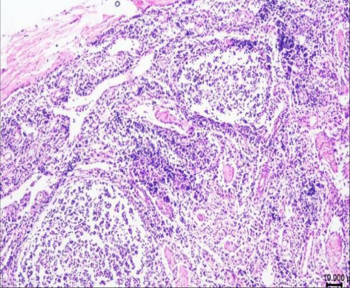 Figure 6
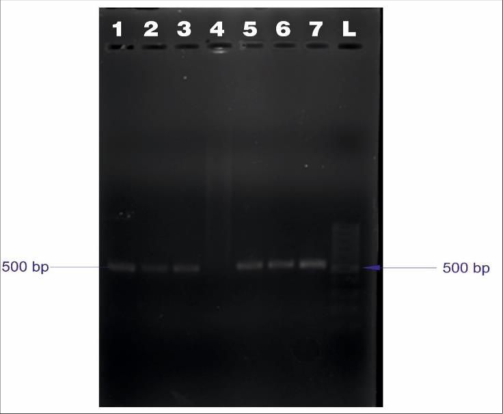 Figure 7
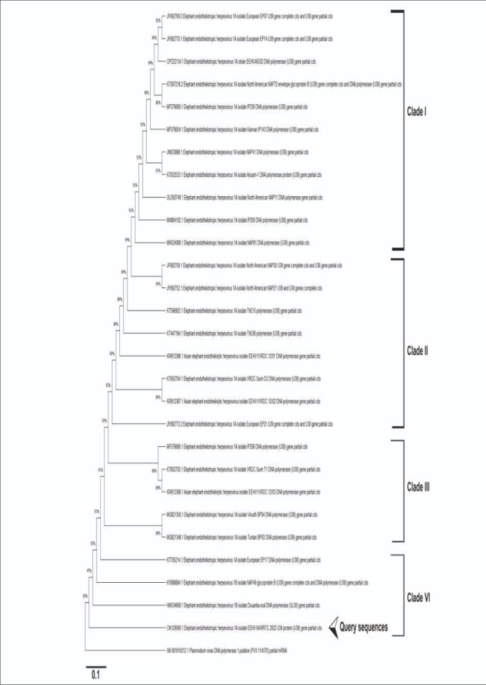 Figure 8
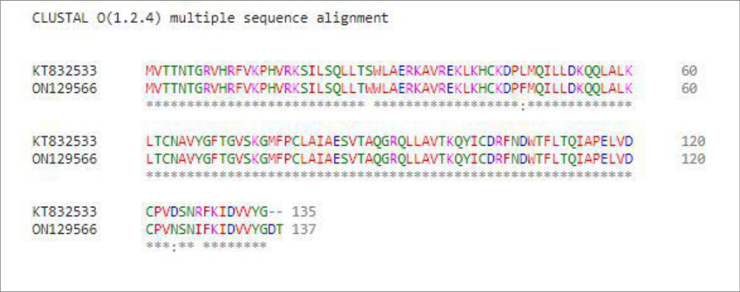 Figure 9
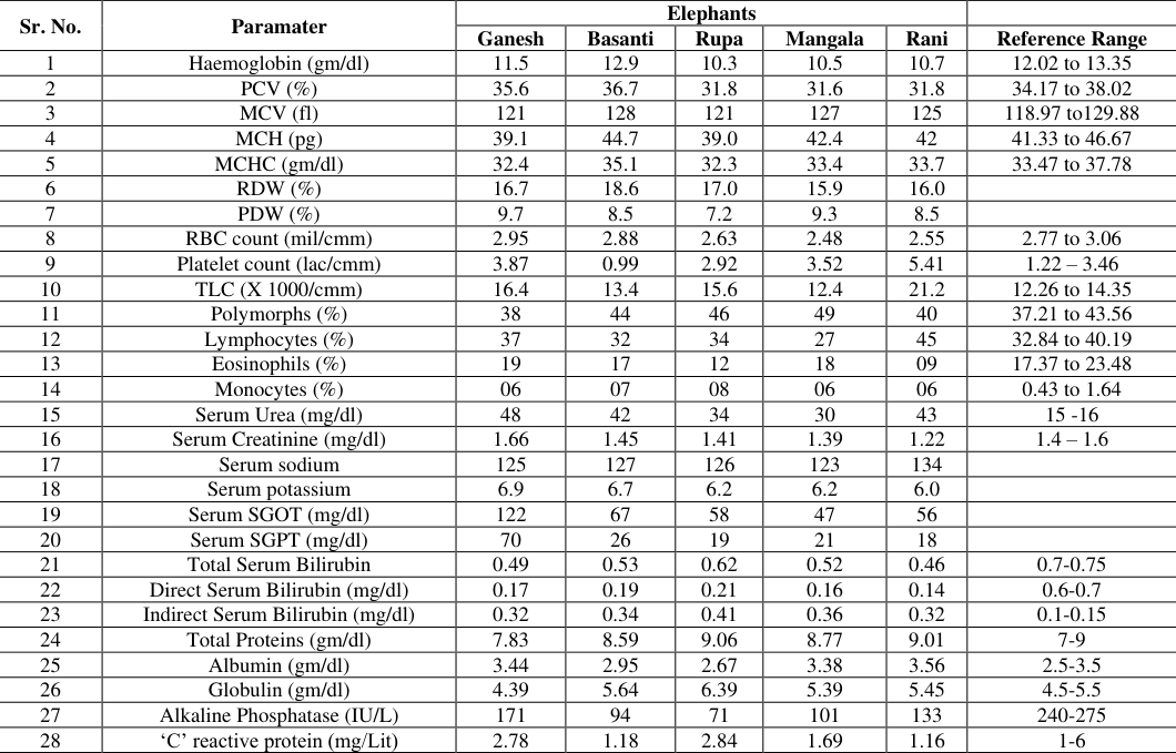 Table 1
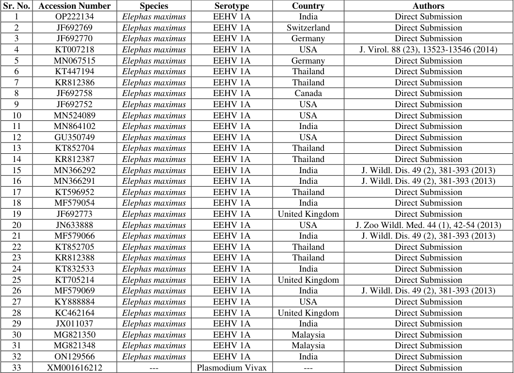 Table 2
Introduction
Elephant Endotheliotropic Herpes Virus (EEHV) is a significant disease of elephants and has been reported in captive and wild elephants worldwide. The disease appears to be lethal in Asian elephants, while in the African counterparts, the disease manifestations are mild [1] . A virus of the Proboscivirus genus, Betaherpesvirinae subfamily, Herpesviridae family and Herpesvirales order causes the disease. There are over 100 known herpes viruses, of which EEHV IA & EEHV IB have contributed to most fatalities in Asian elephants [2,3] . There are seven known serotypes of EEHV circulating in the Asian elephants. The disease is called 'calf killer' since young elephants aged 0 to 8 years are more susceptible to the infection, and many infected cases are fatal. The virus is shed in the secretions of the infected elephants, including the trunk secretions. The transmission of the virus is majorly due to secretions of the trunk. However, herds reporting deaths due to EEHV fail to exhibit the virus by molecular screening. There is no evidence of vertical transmission of the virus. The viremia in affected cases is inconsistent, so diagnosing the disease is challenging. Recent studies have pointed out the spike in viremia before the onset of clinical disease in juvenile elephants. Between 2006 and 2018, fifty-eight cases of EEHV were reported in Thailand; EEHV1 contributed to 76% of infections, followed by EEHV4, contributing to 20% of infections. The overall mortality rate was 69% [4,5] . There are treatment protocols in the clinical management of EEHV using antivirals like famciclovir and acyclovir with feeble success. The disease is prevalent in captive and wild elephants and does not show predilection based on sex. The disease has been reported in more than ten Southeast Asian countries where wild, captive and domestic elephant populations are predominantly based.
In many of the isolated herds in India having 5-10 elephants, only a few juveniles have survived to adulthood. Elephants are endangered in the Indian subcontinent due to illegal hunting, habitat loss, human-animal conflict, etc. EEHV can significantly impact the conservation of Asian elephants as the disease takes a heavy toll on juvenile elephants. A 1.2-5.6% molecular prevalence has been reported in Asian elephants from Thailand; however, the seroprevalence ranging from 42.3% to 43.9% has been determined [6] . A seroprevalence of 100% was reported in captive elephants from the USA [7,8] . Despite identifying the virus in 1971, very scarce information on the mode of transmission, incubation period, predisposing factors etc., is available. There have been a few reports of EEHV in Asian Elephants in India [9] . However, few detailed investigations have been undertaken based on molecular assays and phylogeny. The current case report deals with the post-mortem examination and molecular confirmation of EEHV in an Asian Elephant.
Case History
Arjun, a five-year-old Asian Elephant (Elephas maximus), was noticed with lethargy, fever, dullness and diarrhoea at the Kamlapur Elephant Camp, Gadchiroli, Maharashtra, India. The animal received supportive treatment in the form of fluid and antibiotics. The animal succumbed on day 3 of the treatment. The animal exhibited lacrimation & cyanosis of the tongue on the penultimate day. The animal was unable to stand and showed signs of dullness & weakness. To investigate the cause of death, a systematic post-mortem examination was undertaken at the request of the Deputy Conservator of Forests, Forest Department, Government of Maharashtra State. The gross pathological lesions noticed during the post-mortem examination included congestion of blood vessels of the liver, focal ulceration of the tongue, generalised haemorrhage, enteritis, myocardial hypertrophy and petechial haemorrhages; heart, liver, kidney, intestine, lungs etc. Samples were preserved in formalin and processed for histopathological examination. To obtain more insights on the cause of death, molecular investigations were carried out for which DNA was isolated from the lungs, heart, tongue, liver, lymph node, intestine, and kidney and preserved at -20°C till further use. To rule out the infection, samples of all animals in the herd were collected for EEHV screening. A Polymerase Chain Reaction (PCR) for EEHV 1,2,3,4,5,6 & 7 was carried out targeting the U38 protein of the DNA polymerase gene as described in the reference using Proflex Thermal Cycler (Mfg. Thermo Fischer, CA, USA) [10]. The amplicon was purified using QIAquick® PCR Purification Kit (Mfg. Qiagen, MD, USA) as per the manufacturer's instructions. The amplicon was sequenced using Genetic Analyser 3500 (Mfg. Thermo Fisher, CA, USA) using the Forward & Reverse primers. The obtained sequence was trimmed at ambiguous reads, and a consensus sequence was created using Mega X Software. The nBLAST tool of the NCBI (National Center for Biotechnology Information) was used to ascertain the identity of the virus. The sequence was submitted to NCBI using the BankIt tool for further retrieval. To understand the evolutionary relationship of the virus with the reported EEHV cases from India and abroad, a phylogenetic study was undertaken using Mega X Software [11] . A total of 32 sequences were retrieved from the NCBI database. Indian strains were given priority (Table 2). To maintain tree consistency, a neighbour-joining tree was constructed using Mega X software with a bootstrap value of 1000. FMDV was included as an outgroup.
| Sr. No. | Accession Number | Species | Serotype | Country | Authors |
|---|---|---|---|---|---|
| 1 | OP222134 | Elephas maximus | EEHV 1A | India | Direct Submission |
| 2 | JF692769 | Elephas maximus | EEHV 1A | Switzerland | Direct Submission |
| 3 | JF692770 | Elephas maximus | EEHV 1A | Germany | Direct Submission |
| 4 | KT007218 | Elephas maximus | EEHV 1A | USA | J. Virol. 88 (23), 13523-13546 (2014) |
| 5 | MN067515 | Elephas maximus | EEHV 1A | Germany | Direct Submission |
| 6 | KT447194 | Elephas maximus | EEHV 1A | Thailand | Direct Submission |
| 7 | KR812386 | Elephas maximus | EEHV 1A | Thailand | Direct Submission |
| 8 | JF692758 | Elephas maximus | EEHV 1A | Canada | Direct Submission |
| 9 | JF692752 | Elephas maximus | EEHV 1A | USA | Direct Submission |
| 10 | MN524089 | Elephas maximus | EEHV 1A | USA | Direct Submission |
| 11 | MN864102 | Elephas maximus | EEHV 1A | India | Direct Submission |
| 12 | GU350749 | Elephas maximus | EEHV 1A | USA | Direct Submission |
| 13 | KT852704 | Elephas maximus | EEHV 1A | Thailand | Direct Submission |
| 14 | KR812387 | Elephas maximus | EEHV 1A | Thailand | Direct Submission |
| 15 | MN366292 | Elephas maximus | EEHV 1A | India | J. Wildl. Dis. 49 (2), 381-393 (2013) |
| 16 | MN366291 | Elephas maximus | EEHV 1A | India | J. Wildl. Dis. 49 (2), 381-393 (2013) |
| 17 | KT596952 | Elephas maximus | EEHV 1A | Thailand | Direct Submission |
| 18 | MF579054 | Elephas maximus | EEHV 1A | India | Direct Submission |
| 19 | JF692773 | Elephas maximus | EEHV 1A | United Kingdom | Direct Submission |
| 20 | JN633888 | Elephas maximus | EEHV 1A | USA | J. Zoo Wildl. Med. 44 (1), 42-54 (2013) |
| 21 | MF579066 | Elephas maximus | EEHV 1A | India | J. Wildl. Dis. 49 (2), 381-393 (2013) |
| 22 | KT852705 | Elephas maximus | EEHV 1A | Thailand | Direct Submission |
| 23 | KR812388 | Elephas maximus | EEHV 1A | Thailand | Direct Submission |
| 24 | KT832533 | Elephas maximus | EEHV 1A | India | Direct Submission |
| 25 | KT705214 | Elephas maximus | EEHV 1A | United Kingdom | Direct Submission |
| 26 | MF579069 | Elephas maximus | EEHV 1A | India | J. Wildl. Dis. 49 (2), 381-393 (2013) |
| 27 | KY888884 | Elephas maximus | EEHV 1A | USA | Direct Submission |
| 28 | KC462164 | Elephas maximus | EEHV 1A | United Kingdom | Direct Submission |
| 29 | JX011037 | Elephas maximus | EEHV 1A | India | Direct Submission |
| 30 | MG821350 | Elephas maximus | EEHV 1A | Malaysia | Direct Submission |
| 31 | MG821348 | Elephas maximus | EEHV 1A | Malaysia | Direct Submission |
| 32 | ON129566 | Elephas maximus | EEHV 1A | India | Direct Submission |
| 33 | XM001616212 | --- | Plasmodium Vivax | --- | Direct Submission |
Table text extracted from the original PDF; superscript letters are shown inline. The original table image is below.
View original table image
Results
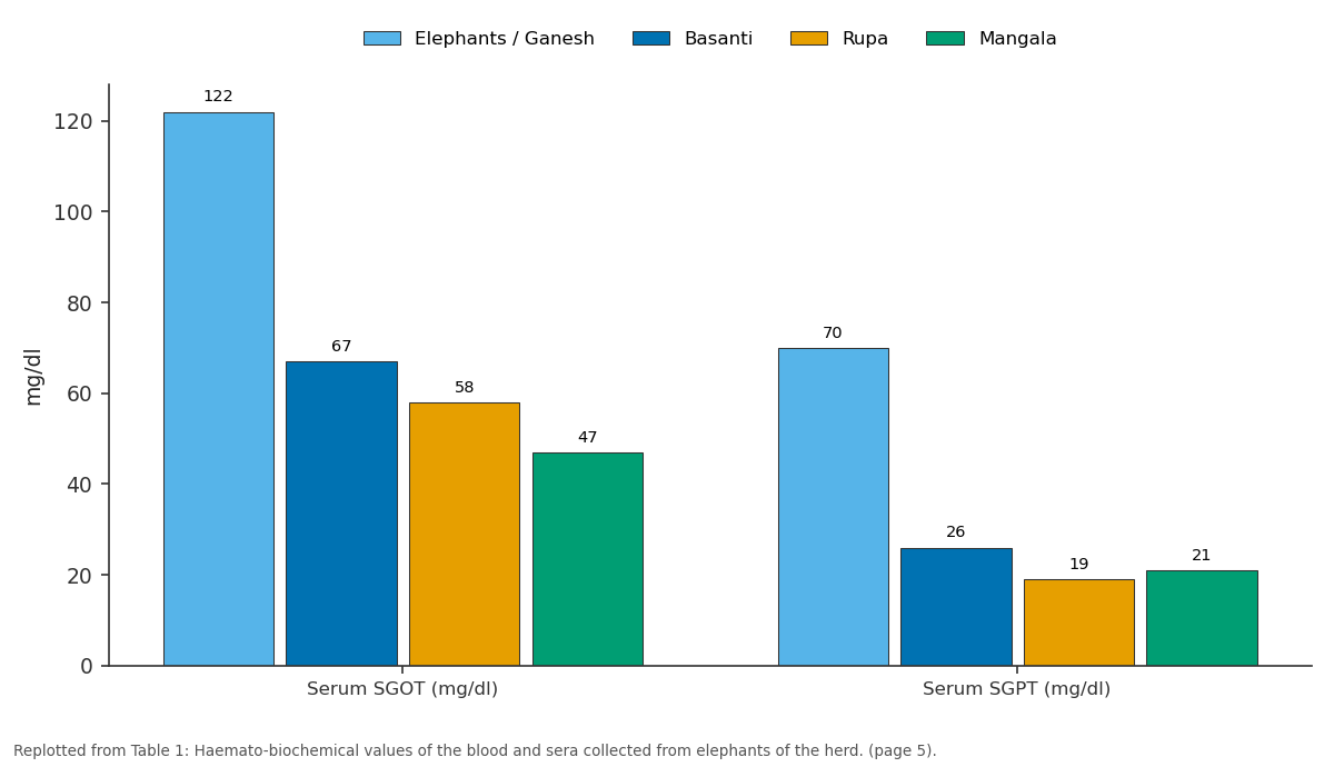
The Histopathological studies of the liver indicated sinusoidal congestion and degeneration of hepatocytes (Fig. 1); the heart indicated extravasation of erythrocytes in the myocardial tissue (Fig. 2); the tongue indicated Haemorrhagic changes in the skeletal muscle layer of the tongue accompanied by hyaline degeneration of skeletal muscles (Fig. 3), while, lungs and the spleen exhibited distinct haemorrhagic changes in the tissue (Fig. 4 &5). The Lymph node exhibited apparent depletion of lymphocytes in the lymphoid follicles (Fig. 6). The PCR provided an amplification of 500 bp, indicative of a positive result (Fig. 7). The sequence was found to be identical to the EEHV IA sequence reported in Asian elephants from India. (Accession No. OP222134). An accession number ON129566 was granted to the sequence by NCBI. After the confirmation of the death, it was essential to screen the rest of the herd for the infection; as a result, a thorough haemato-biochemical analysis was performed (Table 1). The platelet count was used as an early indicator of the infection. A very low platelet count was seen in the early haematology examination of elephant Basanti. Thus, screening was carried out to rule out the presence of EEHV in the foundation stock. The blood samples were screened with PCR and were found negative for all serotypes of EEHV. On nBlast analysis, the query sequence was found to be 98.92% identical to sequences of EEHV IA reported in Asian elephants from Assam, India (Accession No. OP222134) and Switzerland (Accession No. JF692769). The phylogenetic analysis differentiated the sequences into three distinct clades. Clade I consisted of EEHV IA sequences reported from Europe and North America and sequences from India. Clade II, on the other hand, was dominated by the EEHV sequences from Thailand and Malaysia. The query sequence was distinct and placed in Clade III with sequences from Europe and North America. Plasmodium vivax formed a distinct outgroup (Fig. 8).
| Sr. No. | Paramater | Elephants | — | — | — | — | — |
|---|---|---|---|---|---|---|---|
| Ganesh | Basanti | Rupa | Mangala | Rani | Reference Range | ||
| 1 | Haemoglobin (gm/dl) | 11.5 | 12.9 | 10.3 | 10.5 | 10.7 | 12.02 to 13.35 |
| 2 | PCV (%) | 35.6 | 36.7 | 31.8 | 31.6 | 31.8 | 34.17 to 38.02 |
| 3 | MCV (fl) | 121 | 128 | 121 | 127 | 125 | 118.97 to129.88 |
| 4 | MCH (pg) | 39.1 | 44.7 | 39.0 | 42.4 | 42 | 41.33 to 46.67 |
| 5 | MCHC (gm/dl) | 32.4 | 35.1 | 32.3 | 33.4 | 33.7 | 33.47 to 37.78 |
| 6 | RDW (%) | 16.7 | 18.6 | 17.0 | 15.9 | 16.0 | |
| 7 | PDW (%) | 9.7 | 8.5 | 7.2 | 9.3 | 8.5 | |
| 8 | RBC count (mil/cmm) | 2.95 | 2.88 | 2.63 | 2.48 | 2.55 | 2.77 to 3.06 |
| 9 | Platelet count (lac/cmm) | 3.87 | 0.99 | 2.92 | 3.52 | 5.41 | 1.22 – 3.46 |
| 10 | TLC (X 1000/cmm) | 16.4 | 13.4 | 15.6 | 12.4 | 21.2 | 12.26 to 14.35 |
| 11 | Polymorphs (%) | 38 | 44 | 46 | 49 | 40 | 37.21 to 43.56 |
| 12 | Lymphocytes (%) | 37 | 32 | 34 | 27 | 45 | 32.84 to 40.19 |
| 13 | Eosinophils (%) | 19 | 17 | 12 | 18 | 09 | 17.37 to 23.48 |
| 14 | Monocytes (%) | 06 | 07 | 08 | 06 | 06 | 0.43 to 1.64 |
| 15 | Serum Urea (mg/dl) | 48 | 42 | 34 | 30 | 43 | 15 -16 |
| 16 | Serum Creatinine (mg/dl) | 1.66 | 1.45 | 1.41 | 1.39 | 1.22 | 1.4 – 1.6 |
| 17 | Serum sodium | 125 | 127 | 126 | 123 | 134 | |
| 18 | Serum potassium | 6.9 | 6.7 | 6.2 | 6.2 | 6.0 | |
| 19 | Serum SGOT (mg/dl) | 122 | 67 | 58 | 47 | 56 | |
| 20 | Serum SGPT (mg/dl) | 70 | 26 | 19 | 21 | 18 | |
| 21 | Total Serum Bilirubin | 0.49 | 0.53 | 0.62 | 0.52 | 0.46 | 0.7-0.75 |
| 22 | Direct Serum Bilirubin (mg/dl) | 0.17 | 0.19 | 0.21 | 0.16 | 0.14 | 0.6-0.7 |
| 23 | Indirect Serum Bilirubin (mg/dl) | 0.32 | 0.34 | 0.41 | 0.36 | 0.32 | 0.1-0.15 |
| 24 | Total Proteins (gm/dl) | 7.83 | 8.59 | 9.06 | 8.77 | 9.01 | 7-9 |
| 25 | Albumin (gm/dl) | 3.44 | 2.95 | 2.67 | 3.38 | 3.56 | 2.5-3.5 |
| 26 | Globulin (gm/dl) | 4.39 | 5.64 | 6.39 | 5.39 | 5.45 | 4.5-5.5 |
| 27 | Alkaline Phosphatase (IU/L) | 171 | 94 | 71 | 101 | 133 | 240-275 |
| 28 | ‘C’ reactive protein (mg/Lit) | 2.78 | 1.18 | 2.84 | 1.69 | 1.16 | 1-6 |
Table text extracted from the original PDF; superscript letters are shown inline. The original table image is below.
View original table image
Discussion
India is one of the landscapes that harboured Asian elephants from time immemorial. The species is now endangered due to a lack of habitat, poaching for tusks and human-animal conflict. Though the country has a significant population of Asian elephants, only a few reports of EEHV have surfaced from the region. The population of elephants in India was 53,000 in 2012. There are two important populations of elephants in the country, first the northeastern frontier and the southern Deccan plateau population. The population of central India is limited to states of Madhya Pradesh, Chhattisgarh and Maharashtra. Though the population of wild elephants in the central Indian landscape is not significant, but the landscape is very important considering the migration and gene flow of the elephants. Till date, not more than five reports have emerged from India from 2000 [9,12,13,14,15] . The EEHV threatens the species as young calves suffer significant mortality due to the infection. The majority of the cases that have been reported from around the world are from domesticated and captive elephants. Very little information from free-ranging herds is available due to limited opportunities to sample from the wild stock. Though the case report is an isolated incidence of EEHV in an elephant calf from India, very little information on EEHV has been reported from the country. The central Indian landscape connects the eastern and the southern landscapes. Currently, very few herds of wild elephants are present in the landscape. There has been a history of EEHV in the herd in which the death was reported. The age, post-mortem lesions and clinical signs were suggestive of EEHV; hence, a detailed histopathological and molecular investigation was carried out. Blood samples were collected from all the animals in the herd and screened for EEHV.
A detailed study of histopathology, molecular methods & phylogenetic analysis was undertaken to generate epidemiologically significant data. It has been reported that elephant calves that are recently weaned are more prone to EEHV [16] . EEHV IA was previously reported in the country in 2013 (South India) [9] and 2017 (Assam) [13] , and the current case report provides further evidence of the circulating EEHV in India. The EEHV has an acute onset in young calves, and most of the gross post-mortem changes indicate haemorrhagic changes in the visceral organs. The changes are attributed to virus-induced increases in the permeability of blood vessels alone or in conjunction with the endothelial cell lysis [17] . The same was noticed in the case and was confirmed with histopathological studies. The PCR was performed to understand the tissue distribution of the virus in the visceral organs. The virus had even distribution in all the organs of the elephant except the lymph nodes. The tongue and heart had a good concentration of the virus and can be utilised as a tissue of preference for the detection of EEHV from post-mortem samples.
There are many EEHV serotypes that are reported in Asian elephants. An effort was made to screen all the serotypes of EEHV to accurately detect the circulating strain and rule out the possibility of a co-infection [18] . In the current study, the primers targeting EEHV 1, 2, 3, 4, 5, 6 & 7 were used, followed by identification based on sequencing. The reported sequence was similar to the reported sequence of EEHV 1A reported from Assam, India (Accession No. OP222134) and Switzerland (Accession No. JF692769). The sequence alignment and phylogenetic analysis were performed using the Mega X software, the KT832533 which is a previously reported sequence of EEHV from Tadoba-Andhari Tiger Reserve (Maharashtra, India) in 2013 was preferentially included. The sequence was aligned with the sequence to understand the changes in the two sequences at the U38 protein of the DNA polymerase gene. The amino acid alignment revealed four substitutions in the U38 protein; out of the 135 amino acid sequences compared, the following substitutions were noted S28W, L47F, D124N and R127I (Fig. 9). The sequence KT832533 has been reported from the same landscape of Maharashtra State of India, as the case under consideration; however, the sequences appear to be distinct from each other and share 98.91% identity and are placed in the Clade I of the phylogenetic tree. Hence the sequence formed a separate clade at the bottom of the tree. Summing up, Asiatic elephants have a cultural and aesthetic value in India; their ecological value in the ecosystem is colossal. EEHV is a significant disease of the Asiatic elephants; very little data on the status of the disease in the free range is available. Considering the dynamic nature of the virus, herding nature of the species, impact on juvenile calves and lifelong shedding of the virus from adult carriers, it is required to profoundly investigate the prevalence and impact of the disease in free range and captive elephants.
Conclusion
In conclusion, the comprehensive histopathological, molecular, and phylogenetic analyses conducted in response to a case of Elephant Endotheliotropic Herpesvirus (EEHV) infection in an Asian elephant calf provide valuable insights into the epidemiology and pathology of this significant disease. The observed histopathological changes, coupled with positive PCR results and molecular sequencing, confirmed the presence of EEHV IA, with a sequence closely resembling strains previously reported in both India and Switzerland. Furthermore, phylogenetic analysis highlighted distinct clades of EEHV IA strains, underscoring the need for continued surveillance and research on this virus's genetic diversity and distribution. This case underscores the vulnerability of Asian elephants to EEHV and emphasizes the importance of comprehensive monitoring and management strategies to safeguard both wild and captive populations of this culturally and ecologically significant species in India.
Acknowledgement
AcknowledgmentThe authors acknowledge the kind approval of the Principal Chief Conservator of Forest (Wildlife) & Chief Wildlife Warden, Maharashtra State, for carrying out the research work. Forest Development Corporation of Maharashtra Ltd., Nagpur and Office of the Divisional Manager, Gorewada Project, Nagpur, for the facilities provided for the research. Deputy Conservator of Forest, Sironcha & Director, Wildlife Research & Training Centre to facilitate the research work at WRTC, Gorewada, Nagpur.
Figures and tables from the paper
References
14 in the original paper- Hayward GS. Conservation: clarifying the risk from herpesvirus to captive Asian elephants. Vet Rec. 2012;170(8):202.
- Fickel J, Richman LK, Montali R, Schaftenaar W, Göritz F, Hildebrandt TB, Pitra C. A variant of the endotheliotropic herpesvirus in Asian elephants (Elephas maximus) in European zoos. Vet Microbiol. 2001;82(2):103-109.
- Latimer E, Zong JC, Heaggans SY, Richman LK, Hayward GS. Detection and evaluation of novel herpesviruses in routine and pathological samples from Asian and African elephants: identification of two new probosciviruses (EEHV5 and EEHV6) and two new gammaherpesviruses (EGHV3B and EGHV5). Vet Microbiol. 2011;147(1-2):28-41.
- Yun Y, Sripiboon S, Pringproa K, Chuammitri P, Punyapornwithaya V, Boonprasert K, et al. Clinical characteristics of Elephant Endotheliotropic Herpesvirus (EEHV) cases in Asian elephants (Elephas maximus) in Thailand during 2006-2019. Vet Q. 2021;41(1):268-279.
- Boonprasert K, Punyapornwithaya V, Tankaew P, Angkawanish T, Sriphiboon S, Titharam C, et al. Survival analysis of confirmed elephant endotheliotropic herpes virus cases in Thailand from 2006-2018. PLoS One. 2019;14(7):e0219288.
- Prompiram P, Wiriyarat W, Bhusri B, Paungpin W, Jairak W, Sripiboon S, et al. The occurrence of Elephant Endotheliotropic Herpesvirus infection in wild and captive Asian elephants in Thailand: Investigation based on viral DNA and host antibody. Vet World. 2021;14(2):545.
- Fuery A, Pursell T, Tan J, Peng R, Burbelo PD, Hayward GS, Ling PD. Lethal hemorrhagic disease and clinical illness associated with elephant endotheliotropic herpesvirus 1 are caused by primary infection: Implications for the detection of diagnostic proteins. J Virol. 2020;94(3):e01528-19.
- Hoornweg TE, Schaftenaar W, Maurer G, van den Doel PB, Molenaar FM, Chamouard-Galante A, et al. Elephant endotheliotropic herpesvirus is omnipresent in elephants in European zoos and an Asian elephant range country. Viruses. 2021;13(2):283.
- Zachariah A, Zong JC, Long SY, Latimer EM, Heaggans SY, Richman LK, et al. Fatal herpesvirus hemorrhagic disease in wild and orphan Asian elephants in southern India. J Wildl Dis. 2013;49(2):381-393.
- Bhusri B, Suksai P, Mongkolphan C, Tiyanun E, Ratanakorn P, Chaichoun K, et al. Detection of elephant endotheliotropic herpesvirus 4 in captive Asian elephants (Elephas maximus) in Thailand. Thai J Vet Med. 2017;47(1):97-102.
- Kumar S, Stecher G, Li M, Knyaz C, Tamura K. MEGA X: molecular evolutionary genetics analysis across computing platforms. Mol Biol Evol. 2018;35(6):1547.
- Stanton JJ, Nofs SA, Zachariah A, Kalaivannan N, Ling PD. Detection of Elephant Endotheliotropic Herpesvirus infection among healthy Asian elephants (Elephas maximus) in South India. J Wildl Dis. 2014;50(2):279-87.
- Mahato G, Sarma KK, Pathak DC, Barman NN, Gogoi P, Dutta M, et al. Endotheliotropic herpesvirus infection in Asian elephants (Elephas maximus) of Assam, India. Vet World. 2019;12(11):1790.
- Barman NN, Choudhury B, Kumar V, Koul M, Gogoi SM, Khatoon E, et al. Incidence of Elephant Endotheliotropic Herpesvirus in Asian elephants in India. Vet Microbiol. 2017;208:159-63.