
Research article · 2023 · Full text
International Journal of Veterinary Sciences and Animal HusbandryMolecular detection of Salmonella in broiler chicken
Volume 8(6) · Pages 208–210 · DOI: 10.22271/veterinary.2023.v8.i6d.838 ↗
Research areasSalmonella and Campylobacter epidemiologyIdentification and Quantification in Food
Abstract
Author-writtenSalmonella is one of the infectious diseases that can found in broiler chicken. Salmonella is also a potential pathogen that can cause disease in both animal and human. This study was designed as reference for detection and identification of Salmonella bacteria from broiler chicken feces using culture methods and combined with PCR analysis. 10 fecal samples were isolated from close house type farm at Tabanan district, Bali, Indonesia. The samples are transported using Stuart media, enriched using Selenite broth and cultured in Xylose-lysine-deoxycholate (XLD) agar. After the Salmonella colonies grow, the DNA are extracted, proceed to PCR analysis and sent for sequencing. The result showed that 60% of the samples are Salmonella enterica bacteria that confirmed by culture media and molecular method.
Summary
Source-checkedPlain-language reading of the full paper, prepared with AI and checked against the text. Each point links to the authors' own words.
Why it matters
- Salmonella is an infectious pathogen found in broiler chickens that can cause disease in both humans and animals. View evidence ↗
- Poultry and their products are considered a primary source of most human Salmonella infections. View evidence ↗
- In the United States, Salmonella enterica is responsible for roughly 1.4 million foodborne illness cases annually. View evidence ↗
The question
- The study aimed to establish a reference for detecting and identifying Salmonella in broiler chicken feces by combining culture methods with PCR analysis. View evidence ↗
What they did
- The researchers collected 10 random fecal samples from finisher-stage broiler chickens at a single close-house farm in Tabanan, Bali, Indonesia. View evidence ↗
- Fecal samples were transported to the laboratory using Stuart media. View evidence ↗
- Samples were incubated in Selenite media broth for 24 hours before being transferred to Xylose-lysine-deoxycholate (XLD) agar for identification. View evidence ↗
- The researchers used the MEGA 11 program and BLAST to identify Salmonella species from the sequencing results. View evidence ↗
What they found
- Out of 10 fecal swab samples, 6 (60%) produced black colonies on XLD agar, which is typical for Salmonella. View evidence ↗
- PCR analysis showed positive bands for all positive samples, and BLAST confirmed the presence of Salmonella enterica with 92.39% similarity. View evidence ↗
- Some XLD agar plates exhibited yellow colonies rather than the expected red colonies with black centers, suggesting potential lactose fermentation. View evidence ↗
- Most broiler chickens in the study did not show clinical signs of bacterial infection, which the authors attribute to the birds' age and immune systems. View evidence ↗
What it means
- The authors concluded that 60% of the samples were positive for Salmonella enterica, noting that molecular methods confirmed the results despite some inconsistencies with XLD agar. View evidence ↗
- The authors recommend using both microbiological culture and DNA molecular techniques together to detect Salmonella species. View evidence ↗
Visual summary
1 visualsIllustrations explain the paper; they do not show study data.
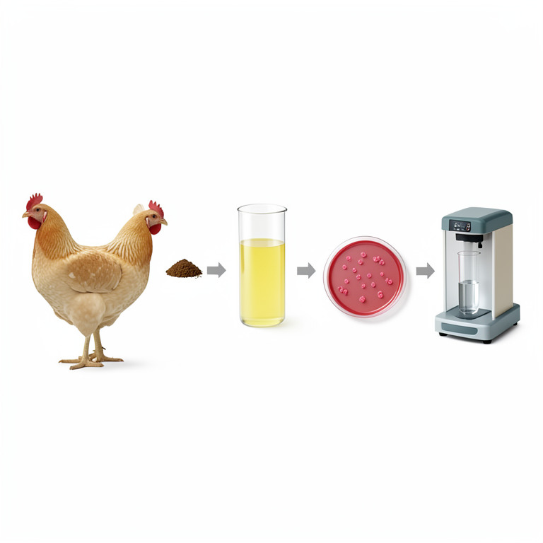
Salmonella Detection Workflow
AI-generated conceptual illustration (broiler chickens). It explains the idea and does not show study data.
View source evidence ↗Figures & tables
From the original paper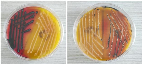 Figure 1
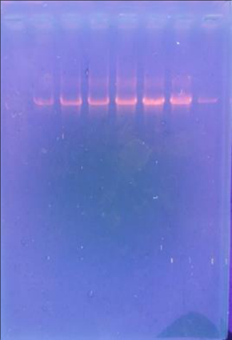 Figure 2
Introduction
Broiler chicken is a one of the livestock that is widely cultivated by breeders to meet the meat needs of consumers. Broiler chicken meat is the popular choice as food because it is cheap and easy to process. In order to provide the meat needs for the community and business purpose, many people start to open broiler chicken farm. The advantage of broiler chicken farming is the short production cycle, within 4-6 weeks broiler chickens can be harvested with a body weight of 1.5-1.56 kg (Yemima, 2014) [1] . However, there are many obstacles faced in producing healthy and quality chickens, one of the threats is infectious diseases and Salmonellosis are one of them. Salmonellosis are caused by Salmonella bacteria that included in the Enterobacteriaceae family of bacteria, there are 2 species in the genus, Salmonella enterica and Salmonella bongori. More than 2500 serotypes have been identified which are differentiated based on their antigenic composition (Bopp et al, 2003) [2] . Their size is about 2-5 µm long and 0.8-1.5 µm wide, straight rods, motile by peritrichous flagella, facultative anaerobic, and optimal growth temperature is 37 °C. D-Glucose and other carbohydrates are catabolized with acid and usually gas. They are oxidase negative, catalase positive, indole and Voges-Proskauer negative, methyl red and Simmons citrate positive. H 2 S is produced and urea is not hydrolyzed (Percival et al., 2004) [3] . Apart from being economically detrimental, salmonellosis will also have a negative impact on public health. Although there are many other pathogens, the transmission of Salmonella through food will cause the emergence of a disease (Winarsih et al., 2008) [4] . Salmonella enterica causes approximately 1.4 million cases of foodborne illness each year in the United States. Poultry and poultry products have been identified by several researchers as an important source of the majority of Salmonella infections in humans (Menconi et al., 2013) [5] . Animals that suffer from salmonellosis can become persistent carriers. Besides being found in feces, Salmonella can also be isolated from soil, water and waste contaminated with fecal material from salmonellosis animals (Ray, 2001) [6] . From many reason that Salmonella is a potential pathogen that can cause disease in both animal and human, early detection is important steps to take and with using PCR analysis it can increase the speed and efficiency of Salmonella detection (Singer et al, 2006) [7] . Most studies support the detection of Salmonella using PCR analysis, as its specificity and sensitivity are superior to those of culturing (Thomas et al, 2009) [8] . This study was designed for detection and identification of Salmonella bacteria from broiler chicken feces using culture methods and combined with PCR analysis.
Materials and Methods Sample
The samples are 10 random feces of finisher stage of broiler chicken in one close house type farm at Tabanan district, Bali, Indonesia. After the feces were taken, it transferred to Stuart media transport, and directly brought to the laboratory.
Culture Method
After the sample was arrived at laboratory, the sample was transferred to Selenite media broth and incubated for 24 hours. After 24 hours, Selenite media will be cloudy, and it is ready to proceed to the Xylose-lysine-deoxycholate (XLD) agar for further identification. XLD agar is the most commonly used highly selective medium for the recovery of enteric pathogens from fecal specimens (Taylor, 1965) [9] . Some Salmonella produces black dots in the middle of the colony as a result of the production of H 2 S gas (Afriyani et al., 2016) [10] . After the black colony of Salmonella grow, it will proceed to the molecular analysis.
Molecular Method
The colony were taken from the XLD agar and the DNA is isolated using GeneJET Genomic DNA Purification Kit and the PCR analysis for Salmonella bacteria are using (F:50 -ACAGTGCTCGTTTACGACCTGAAT-30, R:50 -AGACGGCTGGTACTGATT ATAAT-30) primer (Rozen & Skaletsky, 2000) [11] . After the DNA are obtain, the DNA will be sent for sequencing. The sequencing result will be proceeded to BLAST using MEGA 11 program to identify the Salmonella species. From 10 sample of fecal swab, 6 sample (60%) are producing black colony on XLD agar. The black colony identified as Salmonella bacteria according to Bell and Kyriakides (2002) [12] research that conclude the typical colonies of Salmonella on Xylose Lysine Deoxycholate (XLD) media are black in color, Zaraswati (2006) [13] also added statement that microbial colonies of Salmonella reduce thiosulphuric acid to sulfate so that the colonies appear black. But in this research, some of the XLD agar plates had yellow colonies instead of the expected red colonies with black centers. According to Leonard et al. (2015) [14] the yellow-pigmented colonies appear due to lactose fermentation by the microorganisms. Escherichia coli grow as yellow colonies on XLD while Salmonella is known as non-lactose fermenters and appear as pink with black center colonies. There is some controversy however, with using XLD for Salmonella detection as there are Salmonella serovars which have horizontally inherited the lactose fermentation gene from E. coli. Several studies have also reported that S. enterica serovar Typhimurium and other S. enterica serovars which grow as yellow colonies on XLD agar (McDonough et al, 2000) [15] . As this finding open many possibilities in the future for specific media agar that completely differentiate the Salmonella and E. coli. It is recommended that both microbiological culture methods and DNA molecular techniques are concurrently applied for the detection of Salmonella spp. even though culturing is more laborious and time consuming while molecular techniques are quick and more sensitive (Ahmed, 2014) [16] . The PCR result showed a good band on all the positive samples, and the BLAST result are confirmed that the sample is Salmonella enterica with 92.39% similarity. S. enterica is responsible for infections in humans and animals, with serovars Enteritidis and Typhimurium being the most reported (Andino & Hanning, 2015) [17] . Avian salmonellosis can be broadly divided into two main types based on infection biology. The majority of broad-host range S. enterica serovars are capable of infecting the chicken, usually leading to a period of colonization of lower gastrointestinal tract (Beal et al, 2004) [18] . Salmonella usually infects chickens via the fecal-oral route with spread from the intestinal tract primarily at the distal ileum and caeca of the bird (Barrow et al, 2012) [19] . Although infection with these serovars can lead to systemic disease in chicks or immunocompromised animals, in healthy immunocompetent animals of a week of age or more, infection leads to little or no signs of disease (Wigley, 2014) [20] . In this research most of the clinical sign of the broiler chicken are not showing any sign of bacterial infection, probably it connected with the chicken immune system and their ages.
Conclusion
From 10 fecal sample of broiler chicken, it is showed that 6 (60%) sample were positive with Salmonella enterica bacteria. Even there is some incompatibility identification using XLD media agar, but it was confirmed by molecular method.
Acknowledgement
AcknowledgementsThe author is thankful to the Udayana University Research Grant Project, all veterinary laboratory colleague, and students who participated in this research.
Figures and tables from the paper
References
17 in the original paper- Winarsih W, Priosoeryanto BP, Lay BW, Wibawan IWT, Kompiang IP. Gambaran mikroskopis hati ayam broiler yang diberi probiotik dan infeksi Salmonella subklinis. Jumal Patologi Veteriner Indonesia. 2008;1(1):33-40.
- Menconi A, Reginatto AR, Londero A, Pumford NR, Morgan M, Hargis BM, et al. Effect of Organic Acids on Salmonella Typhimurium Infection in Broiler Chickens. International Journal of Poultry Science. 2013;12(2):7275.
- Ray B. Fundamental Food Microbiology. 2 nd ed. CRC Press; 2001. Boca Raton, Florida.
- Singer RS, Cooke CL, Maddox CW, Isaacson RE, Wallace RL. Use of pooled samples for the detection of Salmonella in feces by polymerase chain reaction. Journal of Veterinary Diagnostic Investigation. 2006;18:319-325. DOI: 10.1177/104063870601800401.
- Thomas D, Rodrigo A, Hernandez M, Ferrus MA. Validation of real-time PCR and enzyme-linked fluorescent assay-based methods for detection of Salmonella spp. in chicken faces samples. Food Anal. Methods. 2009;2:180-189.
- Taylor WI, Schelhart D. Isolation of Shigellae. I. Xylose lysine agar: New media for isolation of enteric pathogens. Am J Clin Pathol. 1965;44:471-475.
- Afriyani A, Darmawi D, Fakhrurrazi F, Manaf ZH, Abrar M, Winaruddin W. Isolation of Salmonella sp. in Feces of Broiler Chicks at Ulee Kareng Market Banda Aceh. Jurnal Medika Veterinaria. 2016;10(1):74-76.
- Rozen S, Skaletsky H. Primer 3 on the WWW for general users and for biologist programmers. Methods Mol. Biol. 2000;132:365-386.
- Bell C, Kyriakides A. Salmonella: A Approach to the Organism and Its Control in Foods. Blackell Publishing Ltd; 2002. London.
- Zaraswati D. Mikrobiologi Farmasi: Cetakan Ke Tiga. Universitas Hasanuddin Press; 2006. Makasar.
- Leonard SR, Lacher DW, Lampel KA. Acquisition of the lac operon by Salmonella enterica. BMC Microbiol. 2015;15:173.
- McDonough PL, Shin SJ, Lein DH. Diagnostic and public health dilemma of lactose-fermenting Salmonella enterica serotype Typhimurium in cattle in the northeastern United States. J. Clin. Microbiol. 2000;38:1221-1226.
- Ahmed OB, Asghar A, Abd El-Rahim I, Al H. Detection of Salmonella in food samples by culture and polymerase chain reaction methods. J. Bacteriol. Parasitol. 2014;5:187. Available online: https://www.longdom.org/open-access/detection-ofSalmonella-in-food-samples-by-culture-and-polymerasechain-reaction-methods-2155-9597.1000187.pdf (accessed on 6 October)
- Andino A, Hanning I. Salmonella enterica: Survival, colonization, and virulence differences among serovars. Sci. World J. 2015;2015:520179.
- Beal RK, Powers C, Wigley P, Barrow PA, Smith AL. Temporal dynamics of the cellular, humoral and cytokine responses in chickens during primary and secondary infection with Salmonella enterica serovar Typhimurium. Avian Pathol. 2004;33:25-33. doi: 10.1080/03079450310001636282.
- Barrow PA, Jones MA, Smith AL, Wigley P. The long view: Salmonella -the last forty years. Avian Pathol. 2012;41:413-20. doi: 10.1080/03079457.2012.718071.
- Wigley P. Salmonella enterica in the Chicken: How it has Helped Our Understanding of Immunology in a NonBiomedical Model Species. Front Immunol. 2014 Oct 10;5:482. doi: 10.3389/fimmu.2014.00482. PMID: 25346731; PMCID: PMC4193332.