
Research article · 2023 · Full text
International Journal of Veterinary Sciences and Animal HusbandryAnalysis of lactic acid bacteria present in raw samples of milk in Wayanad district of Kerala
Volume 8(5S) · Pages 140–146 · DOI: 10.22271/veterinary.2023.v8.i5sc.727 ↗
Research areasProbiotics and Fermented FoodsMilk Quality and Mastitis in Dairy CowsOral microbiology and periodontitis research
Abstract
Author-writtenLactic acid bacteria are the bacteria present in milk and milk samples and they are group of catalase-negative, nonspore forming microorganisms. Upon gram staining Lactic acid bacteria (LAB) are gram positive and rod shaped. The genera Lactococcus and Lactobacillus are frequently linked to milk and dairy products. A total number of 50 fresh raw milk samples of 200 ml each was collected from various sources in Wayanad district of Kerala like ILFC Pookode, local households and nearby dairy chilling units, aseptically in sterile autoclavable bottles. Our study mainly focusses on the species level identification of Lactobacillus spp. and Lactococcus spp. We used two type of agar in our research work, for the isolation of Lactobacillus spp. de Man Rogosa and Sharpe (MRS) agar was used and M17 agar was used for the isolation of Lactococcus spp. 50 samples each were cultured on de Man Rogosa and Sharpe (MRS) agar and M17 agar. Colonies having typical characteristics were taken for preliminary identification by Gram staining and Biochemical tests. Purity of culture were maintained by obtaining pure culture. 16S rRNA gene sequencing was used for further identification of lactic acid bacteria (of Lactobacillus and Lactococcus) upto species level. The study was successful in species level identification of LAB from raw milk. We can conclude that raw sample milk contains abundance of lactic acid bacteria.
Summary
Source-checkedPlain-language reading of the full paper, prepared with AI and checked against the text. Each point links to the authors' own words.
Why it matters
- Lactic acid bacteria (LAB) and the substances they produce are important for preserving biological materials and improving microbial quality. View evidence ↗
- Raw milk serves as a natural source of LAB, which are used to create various dairy products. View evidence ↗
The question
- The primary goal of the study was to identify Lactobacillus and Lactococcus species present in raw milk. View evidence ↗
What they did
- The researchers collected 50 raw milk samples (200 ml each) from households, chilling units, and ILFC Pookode in the Wayanad district of Kerala. View evidence ↗
- Two types of agar were used for isolation: MRS agar for Lactobacillus species and M17 agar for Lactococcus species. View evidence ↗
- The study used PCR amplification and 16S rRNA gene sequencing to identify the LAB isolates at the species level. View evidence ↗
What they found
- All 50 isolates from MRS agar were Gram-positive, rod-shaped, and appeared as singles, pairs, or clusters. View evidence ↗
- All 50 isolates from M17 agar were Gram-positive and cocci-shaped, appearing as singles, pairs, or clusters. View evidence ↗
- All tested LAB isolates were negative for both catalase and oxidase activities. View evidence ↗
- PCR results were positive for 29 of the 50 samples from MRS agar and 27 of the 50 samples from M17 agar. View evidence ↗
- Sequencing of MRS isolates revealed high similarity to Lactobacillus plantarum, with similarity percentages ranging from 98.97% to 99.25%. View evidence ↗
- Sequencing of M17 isolates showed similarity to Lactococcus garvieae, with similarity percentages between 97.72% and 99.56%. View evidence ↗
What it means
- The authors conclude that raw milk contains a wide variety of lactic acid bacteria, which may provide health benefits. View evidence ↗
Figures & tables
From the original paper Table 1
 Table 2
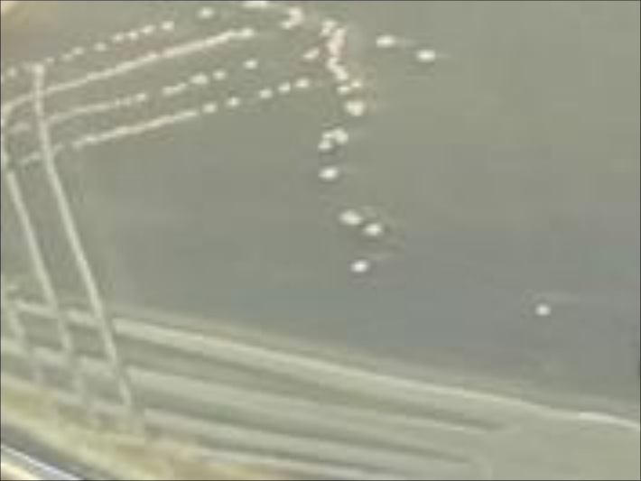 Figure 2
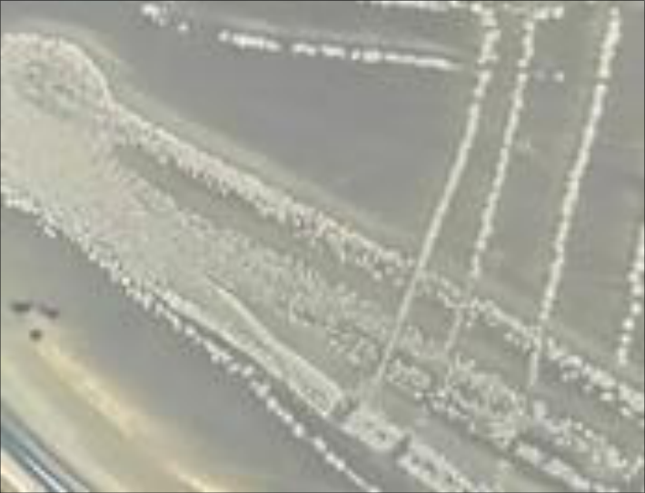 Figure 1
 Table 2
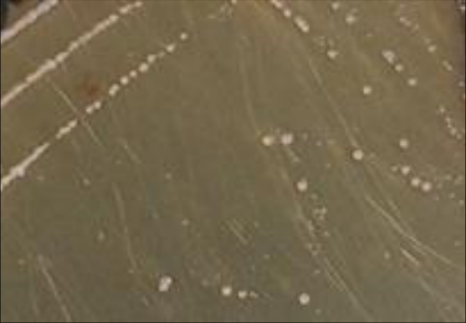 Figure 5
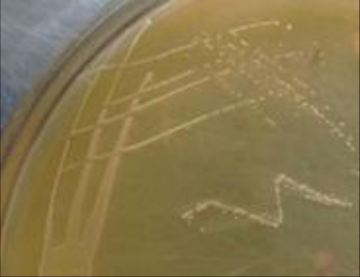 Figure 4
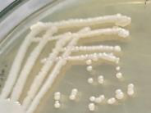 Figure 3
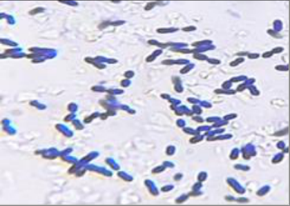 Figure 8
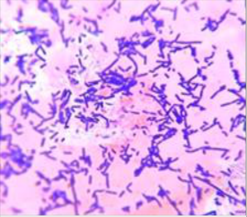 Figure 7
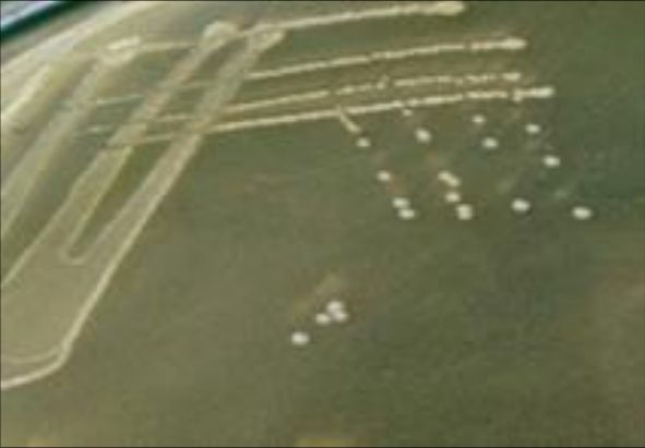 Figure 6
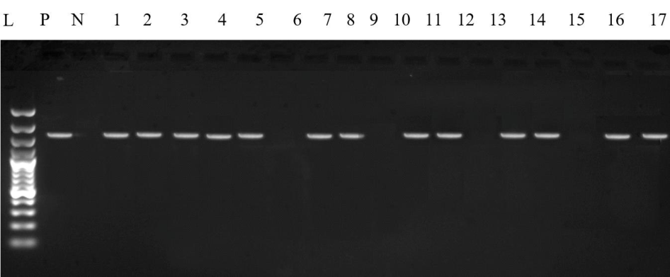 Figure 9
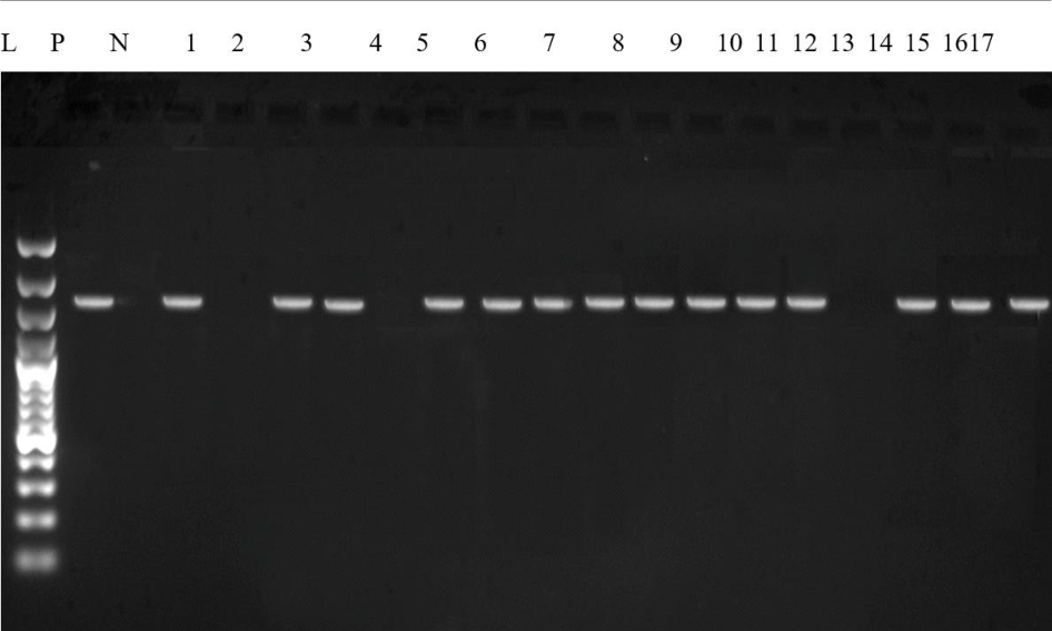 Figure 10
Introduction
Humans have been consuming animal milk for about 6, 000 years and it forms an important part of their daily eating habits. Humans consume various types of milk including cow, buffalo, goat, camel, yak, etc. [1] . Milk and milk products are a well-known source of lactobacilli. Lactic acid bacteria (LAB) belong to various genera of the Lactobacillaceae family. Lactic acid bacteria are generally represented as gram-positive, non-spore-forming, non-pigmented, catalase-negative, and oxidase-negative organisms [2] . Lactic acid bacteria includes generas Lactobacillus, Enterococcus, Lactococcus, Leuconostoc, Streptococcus, Tetragenococcus, Vagococcus, Weisella, Lactoshaera, Melisococcus, Oenococcus, Pediococcus, Microbacterium, Bifidobacterium [3] . LAB produce various substances such as Bacteriocin, Lactic Acid, Ethanol, Formic Acid, Acetone, Hydrogen Peroxide, Diacetyl, Exo-polysaccharides etc. [4] . Lactic acid bacteria and their metabolites have major role in improving microbial quality as well as in biological preservation. [5] . To initiate desired fermentation under controlled conditions, LAB starter cultures are purposefully added to the milk."Generally Recognized as Safe" group of lactic acid bacteria are of genera Lactococcus, Bifidobacterium and Lactobacillus [6] . These cultures affect the production, flavor, and texture of fermented foods so they are unavoidable in the dairy industry [7] . Native source of LAB is the raw milk and can be used for preparation of various milk products [8] . New useful strains of LAB can be discovered with knowledge of the composition and amount of LAB in raw milk that can be used as starting cultures for the development of a new, high-quality product as well as for new applications. In our research work, the 16S rRNA sequencing technique is used to discover the species of isolated LAB. By comparing the sequences of the molecular bases, especially the 16S rRNA gene, it is possible to determine the relationship between the bacteria.
Materials and Methods
Materials and Methods
Collection of sample
A total number of 50 fresh raw milk samples of 200 ml each was collected from various sources like ILFC Pookode, local households and nearby dairy chilling units, aseptically in sterile autoclavable bottles from Wayanad district of Kerala. Milk samples were serially numbered from 1-50 on the collection bottles.
Isolation of bacteria
Serial dilution method was done with Phosphate Buffered Saline for isolation of LAB [9,10] . One ml of the sample was mixed with 9 ml of phosphate buffered saline, and a dilution was prepared until 10 -7 . Then, 0.1 ml of the solution was transferred to a sterile petri dish containing MRS and M17 agar. The spread-plate method was followed. Plates were then incubated at optimum conditions for LAB growth. MRS agar plates were incubated anaerobically at 28-30 °C for 24-48 hours and M17 agar plates were kept at incubation temperature of 30-35 °C for 48 hours. All the agar plates were placed in inverted manner in incubator until the bacterial growth become evident. After incubation isolated colonies were restreaked on agar plates until pure cultures were isolated.
Characterization of Isolates
All the pure cultures isolated from dairy samples were studied for their morphological and staining reaction: Morphological examination was carried out by examining colony characteristics of all the isolates. Staining method followed was Gram staining (1984).
Biochemical characteristics
The biochemical tests performed are catalase test and oxidase test.
Storage of isolates
Thirty percentage (v/v) glycerol was used to store the selected LAB isolates at -20 °C.
16S rRNA sequencing of bacteri
PCR was used for amplification of DNA of selected LAB isolates and 16S rRNA gene sequencing was done for the identification of LAB (9). The procedure for 16S rRNA sequencing is as follows:
2.6.1 DNA Isolation DNA from samples were extracted using HiPurA Multi-Sample DNA Purification Kit. The extracted DNA were stored at -20 °C for future research.
Amplification of 16S rRNA gene
PCR was employed for amplification of 16 srRNA gene. Two universal 16S rRNA primers were used for PCR.
Electrophoresis of the amplified PCR product
A 10 μl of the PCR-amplified product was electrophoresed for 40 minutes at 100 volts in 1X TAE buffer on an agarose gel stained with 1.5 percent (w/v) ethidium bromide. The gel was then visualized and documented using a gel documentation system. As a molecular weight marker, a DNA ladder (100 bp+) was employed.
Sequencing of the PCR Products
PCR product of 6 random LAB isolates from both MRS and M17 agar was outsourced to Protein Design Private Limited in Bengaluru, Karnataka, for sequencing. Samples.
Sequence alignment of the 16S rRNA gene
The 16S rRNA gene sequences of the isolates were compared using the BLAST analysis (http://blast.ncbi.nlm.nih.gov/Blast.cgi). After then, these sequences were matched to those of homologous sequences.
Results and Discussion
The present study was conducted to analyse the species of the Lactic acid bacteria (LAB) present in raw sample of milk in Wayanad district of Kerala using 16 srRNA sequencing as tool.
For this study 50 fresh raw milk samples were collected from various sources like ILFC Pookode, local households and nearby dairy chilling units. LAB isolates were obtained by culturing in MRS and M17 agar. Identification and biochemical tests of LAB were performed. DNA amplification of selected isolates was done by 16SrRNA sequencing. The results are presented in the sections below:
Isolation and identification of lab
The serial dilution (10-dilution procedure) was used to isolate specific lactic acid bacteria from raw milk samples. One milliliter of the material was combined with nine milliliters of phosphate buffered saline to create a dilution that was then diluted to a final concentration of 10-7. A sterile petri dish containing MRS and M17 agar was next loaded with 0.1 ml of the solution and then spread-plate technique was used. After complete solidification, invert the prepared Petri dish and incubate in an incubator at the optimum conditions for each of the agar plates.
Until bacterial growth was visible, the MRS plates were then incubated anaerobically at 28-30 ºC for 24-48 hours. M17 agar plates were likewise maintained for 48 hours inverted at their incubation temperature of 30-35 ºC till bacterial growth occurred. Colonies were randomly selected, picked up, and purified by streaking on respective plates.
Colony characteristics of isolated LAB
Observations of colony morphology were done after getting a pure culture in a Petri dish.
Colony observations were put into groups based on size, pigmentation, edge formation and elevation. Colony observations were in accordance with the findings of researchers [11] . Colony observations were in accordance with the findings of researchers [11] .
Morphology and staining reaction
In MRS agar, out of 50 LAB isolates, all the isolates were rod shaped and occurred as singles, pairs, or clusters. All the LAB isolated using MRS Agar showed a positive gram staining result; hence, all the LAB isolated are Gram positive (Fig. 7 and Fig. 8). All the isolates had violet colour after gram staining. This was in accordance in observation by researchers [12] . All 50 isolates isolated from M17 agar were cocci shaped and occurred as singles, pairs, or clusters. All the isolates showed a positive gram staining result; hence, all LAB isolates are Gram positive these observations were similar to findings by researchers [13] . They isolated LAB from milk, curd and colostrum using MRS agar and observed rod shaped bacteria. Similar observations were made by researcher [14] that Lactococcus spp. are lactic acid bacteria with coccoid shapes.
Results on biochemical tests 3.4.1 Catalase test
All the LAB isolates were catalase negative. This matches with observations of researchers [15] . According to these researchers, lactic acid bacteria fail to produce the catalase enzyme, which converts hydrogen peroxide into water and oxygen and thus yields a negative catalase test result.
Oxidase test
All the LAB isolates were oxidase negative. Researchers [16] explained this phenomenon that in the oxidase test, tetramethyl p-phenylene diamene dihydrochloride, could not be oxidised by the bacterial cultures to produce the purplecoloured compound. Hence LAB were oxidase negative.
Conclusion
The study was conducted to analyse the species of the Lactic acid bacteria (LAB) present in raw sample of milk in Wayanad district of Kerala using 16 srRNA sequencing as molecular tool.
In raw milk, a vast variety of lactic acid bacteria have been found. Therefore, raw milk can offer enormous amounts of lactic acid bacteria and thus enhance health.
Figures and tables from the paper
| Set | Primer name | Sequence 5’-3’ | Target region | Product size | Reference |
|---|---|---|---|---|---|
| 1 | 16SrRNAFor | 5’- AGAGTTTGATCCTGGCTCAG-3’ | 16S rRNA | 1500 bp | (10) |
| 2 | 16SrRNA Rev | 5’-ACGGCTACCTTGTTACGACTT-3' | 16S rRNA | 1500 bp | (10) |
Table text extracted from the original PDF; superscript letters are shown inline. The original table image is below.
View original table image
| Observation | Type of colony | Type of colony | Type of colony |
|---|---|---|---|
| Size of colony | Small (Fig 4) | Medium (Fig 5) | Large (Fig 6) |
| Sample number | 2, 3, 4, 6, 7, 9, 12, 18, 23, 25, 27, 29, 32, 36, 37, 41, 43, 45, 46, 47, 48 | 28 | 1, 5, 8, 10, 11, 13, 14, 15, 16, 17, 18, 19, 26, 29, 30, 31, 33, 35, 38, 40, 42, 44, 49, 50 |
| Pigmentation | Nil | Nil | Nil |
| Edge formation | Circular | Circular | Circular |
| Elevation | Flat | Raised | Raised |
Table text extracted from the original PDF; superscript letters are shown inline. The original table image is below.
View original table image
References
12 in the original paper- Tapeshova SZ, Tokabasova AK, Atalikhova GB. Isolation and study of lactic acid bacteria cultures, yeast of natural cooking kumis starter for the goat's milk. Biological Science. 2013;12:68-71.
- Sarangdhar M, Dipak V, Sheela S. Isolation and Identification of lactobacilli from raw milk samples obtained from Aarey Milk Colony. International Journal of Scientific and Research Publications. 2015;5(4):1-5.
- Holzapfel WH, Haberer P, Geisen. Microorganisms in food and nutrition. American Journal of Clinical Microbiology. 2001;73:365s-373s.
- Rodriguez E, Arques JL, Rodriguez R, Nunez M, Medina M. Reuterin production by lactobacilli isolated from pig feces and evaluation of probiotic traits. Letters of Applied Microbiology. 2003;37:259-263.
- OSullivan L, Ross RP, Hill C. Potential of bacteriocinproducing lactic acid bacteria for improvements in food safety and quality. Biochimie. 2002;84:593-604.
- Tulini FL, Hymery N, Haertlé T, Le Blay G, De Martinis EC. Screening for antimicrobial and proteolytic activities of lactic acid bacteria isolated from cow, buffalo and goat milk and cheeses marketed in the southeast region of Brazil. Journal of Dairy Research. 2016 Feb;83(1):115124.
- Bin M, Bahieldin A, Alharbi MG, Al-Masaudi S, AlJaouni SK, Harakeh SM, et al. Isolation, Characterization Molecular and Probiotic Potential of Lactic Acid Bacteria in Saudi raw and fermented Milk. Compl Alt. Med. 2018;20:1-12.
- Malik V, Devi U, Yadav RNS, Mahanta J. Isolation, identification and antibacterial properties of L. plantarum from 'Khorisa', a traditional food product of Assam. International Journal of Pharmaceutical Res. and Health Care. 2014;4(4). ISSN No: 2249-5738.
- Rahmawati N, Syukri M, Darmawi D, Zachreini I, Yusuf M, Idroes R. February. Identification of lactic acid bacteria from etawa goat milk kopelma Darussalam Village, Banda Aceh. In IOP Conference Series: Earth and Environ. Sci. 2021;667(1):012022. IOP Publishing.
- Padmavathi T, Bhargavi R, Priyanka PR, Niranjan NR, Pavitra PV. Screening of potential probiotic lactic acid bacteria and production of amylase and its partial purification. Journal of Genetic Engineering and Biotechnology. 2018 Dec 1;16(2):357-62.
- Tadesse G, Ashenafi M, Ephraim E. Survival of E. coli O157: H7 Staphylococcus aureus, Shigella flexneri and Salmonella spp. in fermenting Borde', a traditional Ethiopian beverage. Food control. 2005;16(2):189-196.
- Thakur M, Deshpande HW, Bhate MA. Isolation and identification of lactic acid bacteria and their exploration in non-dairy probiotic drink. International Journal of Curr. Microbiol. Appl. Sci. 2017;6:1023-1030.