
Research article · 2023 · Full text
International Journal of Veterinary Sciences and Animal HusbandryIsolation and identification of streptococcus organisms in mastitis milk
Volume 8(4S) · Pages 28–31 · DOI: 10.22271/veterinary.2023.v8.i4sa.628 ↗
Research areasMilk Quality and Mastitis in Dairy CowsProbiotics and Fermented FoodsMicrobial infections and disease research
Abstract
Author-writtenThe study focused on isolating and identifying Streptococcus, a mastitis-causing organism, from raw milk samples obtained from different dairy farms. A total of 100 milk samples were collected, and microbiological testing was conducted to analyse the raw milk. Results showed that 27 samples (27%) tested positive for Streptococcus through cultural methods, while 33 samples (33%) tested positive using PCR assay. Further analysis of the samples revealed that out of 40 milk samples from organized dairy farms, 10 (25%) tested positive for Streptococcus through cultural methods. Among the 40 samples from unorganized dairy farms, 15 (37.5%) were positive for Streptococcus. For the 20 samples from individual farmers, 2 (10%) tested positive through cultural methods. Similarly, using PCR, 12 (30%) samples from organized dairy farms, 18 (45%) samples from unorganized dairy farms, and 3 (15%) samples from individual farmers tested positive for Streptococcus. Conducting microbiological testing is crucial in identifying the causative agents of mastitis and implementing appropriate control measures to eliminate the infection.
Summary
Source-checkedPlain-language reading of the full paper, prepared with AI and checked against the text. Each point links to the authors' own words.
Why it matters
- Mastitis is a major economic problem for dairy farmers because it significantly reduces milk production and income. View evidence ↗
- Estimates suggest that mastitis can cause a 70% reduction in overall farm production. View evidence ↗
- The disease is defined by mammary gland inflammation, which causes pathological tissue changes and increases somatic cells, specifically leukocytes, in the milk. View evidence ↗
The question
- The research aimed to isolate and identify Streptococcus, a known cause of mastitis, from raw milk samples collected from various dairy farms. View evidence ↗
What they did
- The researchers collected and analyzed a total of 100 raw milk samples using microbiological testing. View evidence ↗
- Samples were aseptically gathered from dairy farms in and around Hyderabad that were known for high mastitis incidence. View evidence ↗
- The Californian Mastitis Test (CMT) was used to detect subclinical mastitis using 20 ml half-udder milk samples collected from disinfected teats. View evidence ↗
- Streptococcus was isolated using blood agar and subsequently confirmed using a PCR technique for all phenotypically confirmed isolates. View evidence ↗
What they found
- Overall, 27% of samples were positive for Streptococcus via cultural methods, while 33% were positive via PCR assay. View evidence ↗
- The traditional cultural method was found to be 81.8% as efficient as the PCR assay in detecting the bacteria. View evidence ↗
- In organized dairy farms, 25% of samples were positive by culture and 30% were positive by PCR. View evidence ↗
- Individual farmers had the lowest positivity rates, with 10% positive by culture and 15% positive by PCR. View evidence ↗
- The 197 bp PCR product for Streptococcus remained stable at -20°C but degraded when stored at 4°C over time. View evidence ↗
What it means
- The authors concluded that Streptococcus incidence is most prevalent in unorganized dairy farms. View evidence ↗
- The authors concluded that PCR is more efficient than cultural methods for isolating the bacteria. View evidence ↗
Figures & tables
From the original paper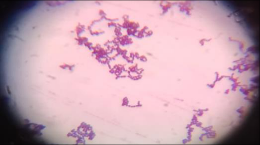 Figure 3
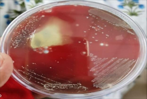 Figure 2
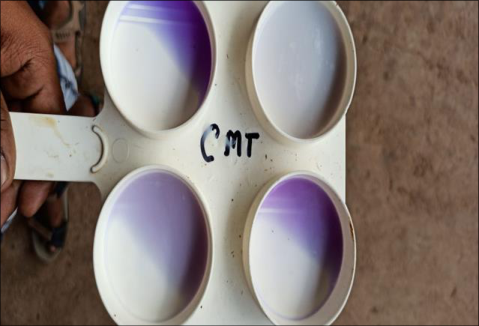 Figure 1
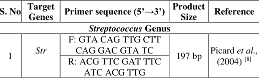 Table 1
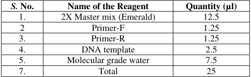 Table 2
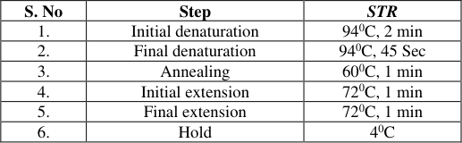 Table 3
 Table 4
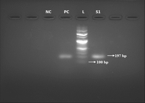 Figure 4
Introduction
Mastitis is a significant economic concern for dairy farmers, as it can lead to substantial losses in milk production and, consequently, impact the farmer's income. It has been estimated that mastitis alone causes a 70% reduction in farm production [1] . Mastitis is characterized by inflammation of the mammary gland [2] . This inflammatory condition leads to abnormalities in milk, such as increased somatic cells, particularly leukocytes, and pathological changes in the mammary tissue. Additionally, clinical signs of mastitis include heat, redness, swelling, hardness, and pain [3] . Mastitis exists in both clinical and subclinical forms, with clinical mastitis exhibiting detectable changes in mammary tissue, while subclinical mastitis is mainly recognized through the somatic cell count in milk [4] . The development of mastitis is influenced by multiple factors due to its complex nature. The udder of a cow provides an ideal environment for microbial growth, and when optimal conditions in terms of temperature, nutrition, and lack of external influences are present, harmful organisms can proliferate rapidly, leading to udder damage and the development of mastitis [5] .
Materials and Methods
Materials and Methods
Sample Collection
Milk samples were aseptically collected from various dairy farms in and around Hyderabad, known to have a high incidence of mastitis. A total of 100 milk samples were collected using sterile vials and stored at 4°C until further processing.
Californian Mastitis Test (CMT)
The CMT was performed to detect subclinical mastitis. Teats were disinfected using alcohol before collecting individual 20 ml half-udder milk samples under sterile conditions. These samples were kept at 4 °C until bacterial cultural examination within 24 hours. CMT scores ranged from (-) for negative results to (+ + +) for different degrees of positive reactions [6] . A commercial CMT reagent was used, and the CMT results were observed. (Fig 01).
Sample Processing
The milk samples were centrifuged at 2000 rpm for 15 minutes, and the supernatant was discarded. The remaining sediment was resuspended in a small volume (0.05 ml) of saline solution [7] . Diluted milk samples were then aseptically inoculated into nutrient broth, followed by incubation at 37 °C for 24 hours. After the incubation period, Gram staining and morphological analysis were performed. Streptococcus microorganisms were isolated using blood agar. All media were purchased from Hi Media, Mumbai and prepared in the laboratory following standard procedures.
Isolate Identification
The obtained pure cultures were subjected to primary tests, such as the catalase test. Catalase-negative cultures were streaked onto nutrient agar slants and preserved at 4 °C. Pure cultures from the slants were further tested using various biochemical tests following standard procedures. a) Hemolytic pattern: On five percent sheep blood agar plates, β-hemolysis (clear zone of clearance) and α-hemolysis (greenish discoloration around and beneath the colonies) were observed (Fig: 02).
PCR assay Extraction of Genomic DNA from isolated Streptococcus
Genomic DNA was extracted from the isolated Streptococcus strains. The pure isolates were cultured in 3 ml of Luria Bertani (LB) broth for 24 hours at 37 °C. Genomic extraction was performed using the Maricon DNA Bacterial plus kit, following these steps: 1. A highly turbid LB broth culture (1 ml) was transferred to a microcentrifuge tube and centrifuged at 10,000 rpm for 10 minutes to collect the bacterial cell pellet. 2. The supernatant was carefully removed to avoid disturbing the pellet. 3. Rapid lysis buffer (200μL) was added to the tube containing bacterial pellets, which were then resuspended by vortexing for 5 minutes. 4. The tube was heated at 100 °C for 5 minutes using a rotary thermal block set, followed by snap chilling for 10 minutes. 5. After centrifugation at 10,000 rpm for 10 minutes, the supernatant (150μl) was transferred to a new microcentrifuge tube. A portion of this supernatant was used for quality, purity, and concentration analysis and stored at -20 °C for up to three weeks. 6. The aliquot containing genomic DNA was used in PCR reactions for further genomic studies.
Estimation of Quality, Purity and Concentration of DNA
The quality, purity, and concentration of isolated DNA were determined using agarose gel electrophoresis and Nano Drop™ 2000/2000c spectrophotometers. Gel electrophoresis showed that the extracted genomic DNA did not exhibit any shearing, indicating its high quality. The DNA samples were then analysed by measuring their absorbance at 260 and 280 nm. All samples exhibited an A 260 /A 280 ratio higher than 1.8, confirming their suitability for PCR amplification.
Molecular Detection of Streptococcus Genus
To confirm the presence of Streptococcus spp., PCR technique was employed for all previously phenotypically confirmed isolates. Specific Str gene primers synthesized by Eurofins Genomics Pvt Ltd, Bangalore, India were used. Details regarding primer sequences, amplicon sizes, reaction mixture composition, and cycling conditions can be found in Table 01, 02, and 03 respectively. Following the completion of the PCR reaction, the amplified products were briefly stored at 4 °C before undergoing analysis using agarose gel electrophoresis. Lastly, out of the 20 milk samples obtained from individual farmers, 2 (10%) samples were positive for Streptococcus using the culture method, while the PCR method detected Streptococcus in 3 (15%) samples. The cultural method exhibited a 66.66% efficiency compared to the PCR assay.
Agarose Gel Electrophoresis for visualization of PCR Products
Agarose Gel Electrophoresis for visualization of PCR Products
Incidence of Streptococcus in milk samples
In this study, 100 milk samples were collected from various sources. Traditional cultural methods identified Streptococcus in 27% of the samples, while PCR assays detected the bacterium in 33% of the samples. The cultural method showed an 81.8% efficiency compared to PCR. Hegde et al.
(2013) [9] reported a higher incidence of 34.5% for Streptococcus in milk samples collected from different dairy farms using cultural methods, which was more than the 27% incidence observed in our study. Similarly, Elango et al.
(2010) [10] reported a higher incidence (35.08%) in Namakkal milk samples using cultural methods. Conversely, a lower incidence (18.1%) was reported by Preethirani et al. (2015) [11] using PCR assay on mastitis milk from Bangalore.
For the 40 milk samples obtained from organized dairy farms, Streptococcus was detected in 25% and 30% of the samples using cultural and PCR assays, respectively. The cultural method exhibited an 83.33% efficiency compared to PCR. Hegde et al. (2013) [9] reported a higher incidence (36.5%) of S. agalactiae in milk samples from organized dairy farms, which was more than the 25% incidence found in our study. In contrast, Preethirani et al. (2015) [11] reported a lower incidence (21.8%) in organized dairy farms from Bangalore using PCR assay. Mekibib et al. (2010) [15] reported a lower incidence (7.2%) of S. agalactiae in mastitis milk from organized dairy farms in Ethiopia compared to our cultural method (25%).
For the 40 milk samples obtained from unorganized dairy farms, Streptococcus was detected in 37.5% and 45% of the samples using cultural and PCR assays, respectively. The cultural method exhibited an 83.33% efficiency compared to PCR. Hegde et al. (2013) [9] reported a lower incidence (32.8%) of Streptococcus in milk samples from unorganized dairy farms, which was less than the 37.5% incidence found in our study. Similarly, Preethirani et al. (2015) [11] reported a lower incidence (16.6%) in unorganized dairy farms from Bangalore using PCR assay.
Comparing the milk samples from unorganized and organized dairy farms, our study found a slightly higher incidence (45%) of Streptococcus in unorganized farms compared to organized farms (30%). However, Hedge et al. (2013) [9] reported a higher incidence (36.5%) in organized dairy farms compared to the incidence (32.8%) in unorganized dairy farms.
For the 20 milk samples from individual farmers, 10% tested positive for Streptococcus using cultural methods, while 15% tested positive using PCR. The cultural method exhibited a 66.66% efficiency compared to PCR. The incidence in milk samples from individual farmers was lower compared to milk samples from both organized and unorganized dairy farms.
Standardization of PCR assay for Streptococcus
To standardize the PCR assay for Streptococcus, we optimized the annealing temperature, primer concentration, template volume, and cycling conditions. The specific PCR product of 197 bp (Fig: 04) for Streptococcus was found to be stable when stored at -20°C, as storing it at 4°C for a longer duration resulted in product degradation possibly due to the action of thermo-stable endogenous nucleases (Gibson and McKee, 1993) [16] .
Conclusion
This study found that the incidence of Streptococcus was highest in unorganized dairy farms compared to both organized dairy farms and individual farmers. Additionally, the PCR method demonstrated higher efficiency in isolating the bacteria compared to the cultural method.
Figures and tables from the paper
| S. No. | Name of the Reagent | Quantity (µl) |
|---|---|---|
| 1. | 2X Master mix (Emerald) | 12.5 |
| 2 | Primer-F | 1.25 |
| 3. | Primer-R | 1.25 |
| 4. | DNA template | 2.5 |
| 5. | Molecular grade water | 7.5 |
| 7. | Total | 25 |
Table text extracted from the original PDF; superscript letters are shown inline. The original table image is below.
View original table image
| S. No | Step | STR |
|---|---|---|
| 1. | Initial denaturation | 940C, 2 min |
| 2. | Final denaturation | 940C, 45 Sec |
| 3. | Annealing | 600C, 1 min |
| 4. | Initial extension | 720C, 1 min |
| 5. | Final extension | 720C, 1 min |
| 6. | Hold | 40C |
Table text extracted from the original PDF; superscript letters are shown inline. The original table image is below.
View original table image
| S. No | Source | No of samples | Cultural method | PCR assay | % of cultural method compared to PCR assay |
|---|---|---|---|---|---|
| 1 | Organized dairy farm | 40 | 10 (25%) | 12 (30%) | 83.33 |
| 2 | Unorganized dairy farm | 40 | 15 (37.5%) | 18 (45%) | 83.33 |
| 3 | Individual farmers | 20 | 2 (10%) | 3 (15%) | 66.66 |
| Total | 100 | 27 (27%) | 33 (33%) | 81.8 |
Table text extracted from the original PDF; superscript letters are shown inline. The original table image is below.
View original table image
References
14 in the original paper- Sumathi BR, Gomes AR, Krishnappa G. Antibiogram profile based dendrogram analysis of Escherichia coli serotypes isolated from bovine mastitis. Veterinary World. 2008;1(2):37.
- Suojala L, Pohjanvirta T, Simojoki H, Myllyniemi AL, Pitkala A, Pelkonen S, et al. Phylogeny, virulence factors and antimicrobial susceptibility of Escherichia coli isolated in clinical bovine mastitis. Veterinary Microbiology. 2011;147(3-4):383-388.
- Ranjan R, Gupta MK, Singh KK. Study of bovine mastitis in different climatic conditions in Jharkhand, India. Veterinary World. 2011;4(5):205-208.
- Al-Zainy ZOA, Al-Jeburii KOS. Prevalence of Grampositive bacteria in buffalo mastitis in Iraq. Int J Adv Res. 2015;3(4):216-225.
- Whyte D, Walmsley M, Liew A, Claycomb R, Mein G. Chemical and rheological aspects of gel formation in the California Mastitis Test. Journal of Dairy Research. 2005;72(1):115-121.
- Zecconi A, Piccinini R, Zepponi A, Ruffo G. Recovery of Staphylococcus aureus from centrifuged quarter milk samples. Journal of Dairy Science. 1997;80(11):30583063.
- Picard FJ, Ke D, Boudreau DK, Boissinot M, Huletsky A, Richard D. Use of tuf sequences for genus-specific PCR detection and phylogenetic analysis of 28 streptococcal species. Journal of Clinical Microbiology. 2004;42(8):3686-3695.
- Hegde R, Isloor S, Prabhu KN, Shome BR, Rathnamma D, Suryanarayana VVS et al. Incidence of subclinical mastitis and prevalence of major mastitis pathogens in organized farms and unorganized sectors. Indian Journal of Microbiology. 2013;53:315-320.
- Elango A, Doraisamy KA, Rajarajan G, Kumaresan G. Bacteriology of sub clinical mastitis and antibiogram of isolates recovered from cross bred cows. Indian Journal of Animal Research. 2010;44(4):280-284.
- Preethirani PL, Isloor S, Sundareshan S, Nuthanalakshmi V, Deepthikiran K, Sinha AY et al. Isolation, biochemical and molecular identification, and in-vitro antimicrobial resistance patterns of bacteria isolated from bubaline subclinical mastitis in South India. PLoS One. 2015;10(11): e0142717.
- Mallikarjunaswamy MC, Krishna Murthy GV. Antibiogram of bacterial pathogens isolated from bovine subclinical mastitis cases. Indian Veterinary Journal. 1997;74(10):885-886.
- Mohini kumari P, Janaki ram gupta B. Diagnosis and therapy of sub-clinical mastitis in post-parturient cows. Indian Veterinary Journal. 2002;79(1):89-89.
- Mekibib B, Furgasa M, Abunna F, Megersa B, Regassa A. Bovine mastitis: Prevalence, risk factors and major pathogens in dairy farms of Holeta Town, Central Ethiopia. Veterinary world. 2010;3(9):397-403.
- Gibson JR, McKee RA. PCR products generated from unpurified Salmonella DNA are degraded by thermos table nuclease activity. Letters in Applied Microbiology. 1993;16(2):59-61.