
Research article · 2022 · Full text
International Journal of Veterinary Sciences and Animal HusbandryIncidence of dermatophillosis among one humped camel (camelus dromedarius) provided to tamboul livestock market, Sudan
Volume 7(6) · Pages 29–32 · DOI: 10.22271/veterinary.2022.v7.i6a.452 ↗
Research areasNail Diseases and TreatmentsMycotoxins in Agriculture and FoodBee Products Chemical Analysis
Abstract
Author-writtenThe present study aimed to the determined incidence of Dermatophillosis among one humped camel (Camelus dromedarius) provided to Tamboul livestock market, Sudan. Three hundred camels of different ages and sex that came from different parts of Sudan were studied in different season’s assessment of some factors on the incidence of Dermatophillosis, and haematological and biochemical values for infected camels were done. The results showed that the incidence rate of Dermatophilus infection in camels was 3%, visually; loss of hair and accumulation of necrotic white materials around the neck, forelimb, thorax and sometimes all parts of the body was shown; the necrotic white materials were deep and hard attachment with skin. Microscopically the Dermatophilus congolensis under the microscope revealed cocci was used 10% KOH and Giemsa stain. According to age, Chi-square revealed a significantly higher incidence rate in the age group less than 6 years (88.9%) followed by 6-12 years (11.1%) years and not noted in ages more than 13 years. Therefore, a high significance variation in sex and all infected camels were noticed in male. While the season factor showed an insignificant effect on Dermatophilus infection. In addition, the results showed that Dermatophilosis was an insignificant effect on the mean of hematological and biochemical parameters (p>0.05). Dermatophillosis mixed with dermatophytosis was detected in most examined samples. Direct examination using 10% KOH is a reliable technique to determine chronic Dermatophillosis and the Strategic application of anti-biotic drugs mixed with anti-fungal drugs for the treatment of camel Dermatophillosis is strongly recommended.
Summary
Source-checkedPlain-language reading of the full paper, prepared with AI and checked against the text. Each point links to the authors' own words.
Why it matters
- Dermatophillosis is a skin disease caused by the bacterium Dermatophilus congolensis, which can affect humans as well as various wild and domestic animals. View evidence ↗
- The disease causes economic losses through decreased animal productivity, hide depreciation due to skin matting, and potential mortality in weak animals. View evidence ↗
- Infected animals may experience impaired reproductive performance, a loss of overall condition, and reduced milk production. View evidence ↗
The question
- The study aimed to determine the incidence of Dermatophillosis in one-humped camels (Camelus dromedarius) at the Tamboul livestock market in Sudan. View evidence ↗
What they did
- The researchers examined a sample size of 300 suspected camels of varying ages and sexes. View evidence ↗
- Diagnosis was performed using a direct examination technique where skin scrapings were mixed with 10% potassium hydroxide (KOH) for 0.5 to 1 hour. View evidence ↗
- Blood samples were collected from the jugular vein of camels across three seasons (winter, summer, and autumn), with 100 samples per season. View evidence ↗
What they found
- The incidence rate of Dermatophillosis among the 300 examined camels was 3% (9 camels). View evidence ↗
- Clinical signs in infected camels included hair loss and the accumulation of necrotic white materials on the thorax, forelimb, neck, or the entire body. View evidence ↗
- Age significantly influenced incidence, with the highest rate in camels under 6 years (88.9%), followed by those aged 6-12 years (11.1%), and no cases in camels over 13 years. View evidence ↗
- Gender showed a highly significant variation, as all infected camels in the study were male. View evidence ↗
- The season did not have a significant effect on the incidence of Dermatophilus congolensis infection (p > 0.05). View evidence ↗
- Most of the examined samples showed a mixed infection of dermatophillosis and dermatophytosis. View evidence ↗
What it means
- The authors concluded that the direct examination technique is reliable for the detection of chronic dermatophillosis. View evidence ↗
- The study recommends a treatment strategy for dermatophillosis that combines antibiotic and antifungal drugs. View evidence ↗
Visual summary
1 visualsIllustrations explain the paper; they do not show study data.
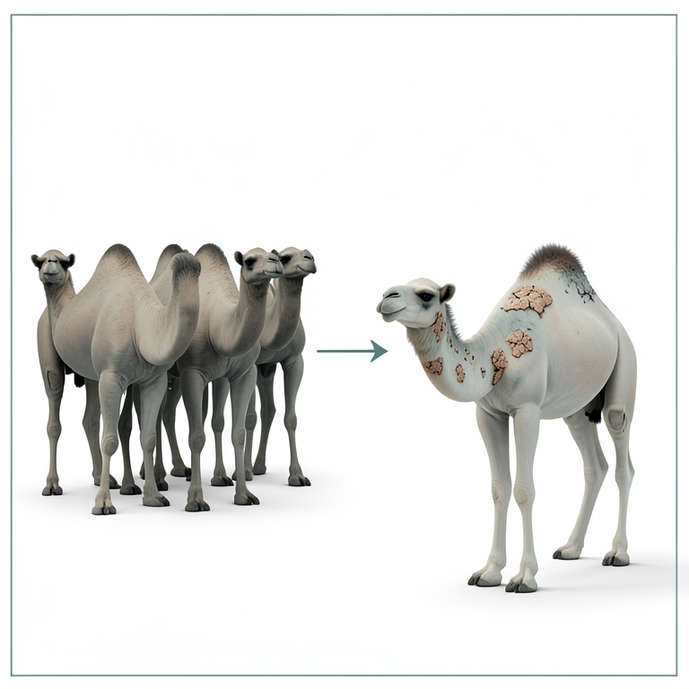
Incidence Distribution
AI-generated conceptual illustration (one-humped camels (Camelus dromedarius)). It explains the idea and does not show study data.
View source evidence ↗Figures & tables
From the original paper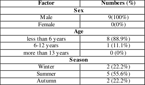 Table 1
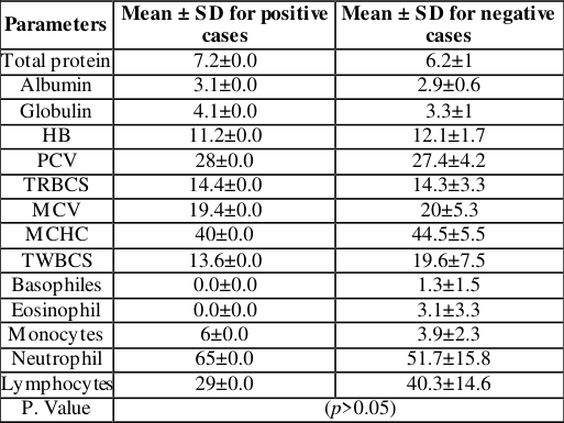 Table 2
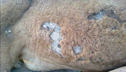 Figure 1
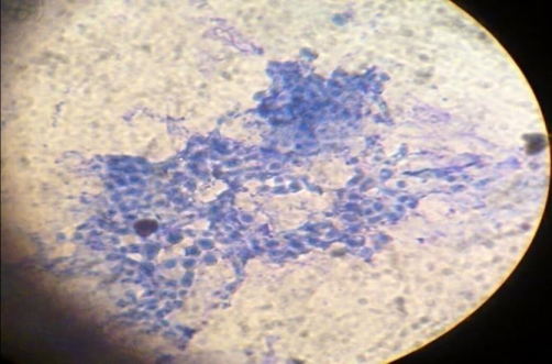 Figure 2
Introduction
Dermatophillosis is a skin disease caused by a bacterium called Dermatophilus congolensis; the disease affects many species of domestic and wild animals and occasionally humans [1] . This organism is found throughout the world, but is most prevalent in tropical and subtropical regions, and has a wide host range [2] . The disease is non-pruritic and is characterized by exudative, proliferative or hyperkeratosis dermatitis, accompanied by the production of crusts and folliculitis. Several factors are involved in the pathogenesis of Dermatophillosis; among them are mechanical injury to the skin, rainfall, tick infestation, concurrent diseases and stress that compromise the host immune system, humidity is a major predisposing factor in the spread and epidemiology of Dermatophillosis [3] . Dermatophillosis often lead to creates economic problems by severe skin matting resulting in hide depreciation and decrease animal productivity also mortality from severe cases in susceptible weak animals was detected [4,5] . In addition that the disease revealed a loss of condition impaired reproductive performance and decrease in milk production and a marked increase in the somatic cell counts in milk [6] . Camel Dermatophillosis has been reported in Kenya, Sudan and Saudi Arabia. In Sudan the outbreaks was in the Butana region, The fatality rates ranged from 0% in Saudi Arabia to 30% in Sudan, mixed infection involving Dermatophilus congolensis and Microsporum gypseum were founded [7] . Tick control in infected animals and environment is indicated to minimize the risk of Dermatophillosis [8] in study was carried out at Qas sim Region, Central of Saudi Arabia, to study dermatophytosis in a private farm of dromedary camels.
The prevalence of dermatophytosis in camels was 11.5% and significantly differed among different age groups (p ≤.0001 and odds ratio = 14.61), with higher prevalence among camels younger than three years (22.10%). Clinical signs of ringworm recorded in this study were non-pruriginous dry circumscribed discrete, crusty hairless lesion distributed over the head, neck, shoulder, limbs and flanks [9] . In man the disease has been characterized by pimples and multiple pustules (2-25) on the hands and forearms, containing a Serous or yellowish-white exudate. The lesions healed in 3 to 14 days, leaving a purplish-red scar [4] . Clinical diagnosis is confirmed by microscopic examination of Stained smears (Giemsa, methylene blue, or Wright's stain) made from exudates or scabs. [10] . the present study aimed to the determined incidence of Dermatophillosis among one humped camel (Camelus dromedarius) provided to Tamboul livestock market, Sudan
Materials and Methods
Materials and Methods
Study area
The study was conducted in Tamboul livestock market; it is one of the biggest markets of camels founded in Sudan, and located in East Gezira state and far about 150 km to the south of Khartoum; the capital of Sudan.
Study Design
A case-control study was applied, one hump camel suspected to be infected by Dermatophillosis was examined by 10% KOH direct examination, and assessment was done according to age, gender and season and description of the Dermatophilus congolensis visually and microscopically. Evaluated the effect of the disease on hematological and biochemical value, furthermore, used negative cases as a control.
Study population and samples size
Three hundred suspected camels (300) of different ages, sex, and season were examined. Samples size was calculated based on the formula N= Z 2 pq/d 2 [11] . And the prevalence of camel Dermatophillosis probability was taken according to Gitao et al., [12] that revealed 20% of camels in the Butana area were infected with Dermatophillosis.
Sample Collection
Skin scrapings of the lesion from three hundred suspected camels with clear skin lesions were collected with a blunt scalpel in sterile Petri dishes. All specimens were labelled and transferred to the laboratory for the diagnosis of Dermatophillosis using a direct examination technique.
Direct 10% KOH Examination technique
The collected skin scrapings were transferred to test tubes and mixed with a small amount of 10% potassium hydroxide solution and left to stand for 0.5-1 hours until the skin particles have partly disintegrated. The tubes were centrifuged at 3000 revolutions per minute. The supernatant fluid was discarded, and a drop of sediment was investigated under a microscope for the detection of the Dermatophilus congolensis [13] Clinical investigations Three hundred camels of different ages, sex and season (winter, summer and autumn) were examined visually for Dermatophillosis and clinical signs with lesions characterization for all suspected camels were detected.
Blood samples
Venous blood samples were obtained in three seasons; winter, summer and autumn (100 in each season). Blood samples were collected from the Jugular vein of suspected camels by Using a Vacationer EDTA tube, vacationer needle, syringe, needle, needle holder, and disinfectant for blood parameters. At the same time, 3 ml Blood without anticoagulant was used to obtain serum for biochemical tests; the blood was centrifuged at 3000 rpm for 15 minutes and the serum was kept in Eppendorf tubes and then stored at -20 ºC until used [14] .
Haematological and Biochemical parameters
Haematological parameters were measured according to Kemal, [14], and total protein and albumin were determined according to the manufacturer's instructions while globulin was measured according to the formula Globulin g/dl = (Total protein _Albumin).
Statistical Analysis
Data collected from this study were compiled using an appropriate statistical package SPSS version 16. Results were summarized as means ± standard deviation (S.D) and Levels of Significance were taken at (p≤ 0.05).
Results
The results of current study examined three hundred (300) skin scrape conducted in suspected camels provided to Tamboul livestock market 9(3%) out of 300 camels were infected with Dermatophillosis. Camels infected by Dermatophillosis were diagnosed clinically by loss of hair and accumulation of necrotic white materials around the neck, forelimb, thorax and sometimes all parts of the body; the necrotic white materials were deep and hard attachment with skin Figure (1). The Dermatophillus congolenses under a microscope revealed cocci after preparation by 10% KOH and stained by Giemsa stain Figure (2). According to age groups {(less than 6 years), (6-12) and (more than 13 years)} examined, the present study discovered high significant variation and higher incidence rate with camel Dermatophillosis was detected in age group less than 6 years (88.9%) followed by 6-12 years (11.1%) years and no cases in age more than 13 years. Also, chi-square detected high significance variation according to gender and all infected camels were noticed in males. The Incidence rate of Dermatophilus congolensis in camels according to season, the result was revealed no significant variations (p>0.05). In additions that, the present result also recorded there was no significant variations in the effect of camel Dermatophillosis on hematological and biochemical parameters shown in table 1&2.
| Factor | Numbers (%) |
|---|---|
| Sex | |
| Male | 9(100%) |
| Female | 0(0%) |
| Age | |
| less than 6 years | 8 (88.9%) |
| 6-12 years | 1 (11.1%) |
| more than 13 years | 0 (0%) |
| Season | |
| Winter | 2 (22.2%) |
| Summer | 5 (55.6%) |
| Autumn | 2 (22.2%) |
Table text extracted from the original PDF; superscript letters are shown inline. The original table image is below.
View original table image
Discussion
Dermatophilosis occurs most frequently in domestic animals, But is increasingly recognized in wildlife. D. congolensis was first described in domestic cattle in Zaire, Africa in 1910 [15] This present study recorded 3% of camels provided to Tamboul livestock markets from all over Sudan from 2015 to 2016 were infected with Dermatophillosis. The prevalence rate in this study was less than the results recorded by Gitao et al., [16] that revealed 20% of camels in the Butana area were infected with Dermatophillosis [17] . The prevalence of camel Dermatophillosis in Iran was 13.6%. This variation is due to the result of the wide use of ox tetracycline drugs among camel herds and also may be from different climates according to the duration of the study. Macroscopically and microscopically characterization for Dermatophilus congolensis in this study was in full agreement with other results discovered others researchers [16,17,18] . The study revealed higher significant different effect of sex on prevalence of Dermatophillosis; the incidence rate was more infected in males than females. This is Similar results were obtained by Nath (2010). While the current s tudy did not recorded significant different between season and incidence Dermatophillosis, its disagree with the results obtained by Gitano et al., [16] that recorded that Dermatophillosis was found to be more prevalent in the wet season (21.2%) compared to its prevalence in the dry season (14.5%). These different results return back to the environmental condition because season interference was found in a few last years in Sudan area. Other factor like prevailing of transmitting ticks and acacia tree could be behind this prevalence rates. The current study the effect of age detected Significant different (p<0.02) effect of age on Dermatophillosis, where founded high incidence rate in 1-5 age group was reported compare to other groups. Similar conclusion was reached previous studies [12, 16, 19] . Study reports the incidence of dermatophytosis in a circus dromedary aged about 2.5 years with emphasis on clinical signs, laboratory examination and treatment. Camel had skin problem persisting for a month [20] . In One-humped camel (Camelus dromedarius) in Al-Butana area, El-Gazira State, Sudan, among these; 18 camels (3.6%) were infected with mange, 50 camels (10%) were infected with ringworm and 4 camels (0.8%) were infected with dermatophilosis. There was significant difference in the prevalence of ringworm infection between male and female camels (p≤ 0.05), but no significant difference was observed in mange and dermatophillosis infection (p≤0.05 [21] . The infected animals were examined clinically and the outbreak was reported as mixed infection of camelpox and dermatophilosis. Hair matting particularly on the head, neck, and abdomen was the common skin lesion that observed on the infected animals. Lesions showed hairless brownish crusts with irregular sizes and pus exudation that left irregular border crusted lesions [22] Skin lesions were reported in 17 of 140 animals and were distributed throughout the animals' head, neck, back, and perineum. Lesions ranged in size from paintbrush matted hairs to wart-like lesions up to 2 cm in diameter. Skin scrapings were taken from living animals and stained directly with gram stain. There was severe multifocal exudative dermatitis with hyperkeratosis on histology. Neutrophils, mononuclear cells, and numerous branching filaments coccoid bodies infiltrated the dermis [23] . In the Brazilian study, cases occurred in all seasons, with 2 (18%) outbreaks in winter, 2 (18%) outbreaks in summer, 3 (28%) outbreaks in autumn, and 4 (36%) outbreaks in spring. The morbidity rate in cattle ranged from 2.5% to 80%, in horses from 66% to 100%, and in sheep from 9% to 10%. Outbreaks in sheep presented mortality ranging from 2% to 3% [24] .
Conclusions
The study concluded that chronic dermatophillosis was present in 3% of Sudanese camels. Dermatophilus infections and dermatophytosis were found in most of the samples tested. The direct examination technique was a reliable technique for detecting chronic dermatophillosis. The study recommended that for dermatophillosis, treatment strategic should be use anti-biotic drugs combined with anti-fungal.
Figures and tables from the paper
| Parameters | Mean ± SD for positive cases | Mean ± SD for negative cases |
|---|---|---|
| Total protein | 7.2±0.0 | 6.2±1 |
| Albumin | 3.1±0.0 | 2.9±0.6 |
| Globulin | 4.1±0.0 | 3.3±1 |
| HB | 11.2±0.0 | 12.1±1.7 |
| PCV | 28±0.0 | 27.4±4.2 |
| TRBCS | 14.4±0.0 | 14.3±3.3 |
| MCV | 19.4±0.0 | 20±5.3 |
| MCHC | 40±0.0 | 44.5±5.5 |
| TWBCS | 13.6±0.0 | 19.6±7.5 |
| Basophiles | 0.0±0.0 | 1.3±1.5 |
| Eosinophil | 0.0±0.0 | 3.1±3.3 |
| Monocytes | 6±0.0 | 3.9±2.3 |
| Neutrophil | 65±0.0 | 51.7±15.8 |
| Lymphocytes | 29±0.0 | 40.3±14.6 |
| P. Value | (p>0.05) |
Table text extracted from the original PDF; superscript letters are shown inline. The original table image is below.
View original table image
References
18 in the original paper- Ayalew Y, Assefa A, Mekonen N, Belete S, Ayisheshim AA. Review on camel dermatophilosis. Advances in Biological Research. 2015;9(5):363-372. DOI: 10.5829/idosi.abr.2015.9.5.95267
- Burd EM, Juzych LA, Rudrik JT, Habib F. Pustularder matitis caused by Dermatophilus congolensis. Journal of Clinical Microbiology. 2007;45:1655-1658.
- Yeruham I, Elad D, Perl S. Dermatophilosis in goats in the Judean foot hills. Revue de médecine Vétérinaire. 2003;154(12):785-788.
- Stewart GH. Dermatophilosis, a Skin Disease of Animals and Man. The Veterinary Record. 1997;99:534-536.
- Chatikobo P, Kusina NT, Hamudikuwanda H, Nyoni OA. Monitoring study on the prevalence of dermatophilosis and parafilariosis in cattle in a smallholder semi-arid farming area in Zimbabwe. Tropical Animal Health Production. 2004;36(3):207-215.
- Gitao CG, Agab H, Khalafalla AI. Outbreaks of Dermatophilus congolensis infection in camels (Camelus dromedarius) from the Butana region in Eastern Sudan. Rev. Sci. Tech. 1998;17(3):743-748.
- Osman SA. Camel dermatophilosis: clinical signs and treatment outcomes. Journal of Camel Practice and Research. 2014;21(2):199-204.
- Abdulaziz M, Almuzaini Salama A. Osman Elhassan MA. Saeed. An outbreak of dermatophytosis in camels (Camelus dromedaríus) at Qassim Region, Central of Saudi Arabia, Journal of Applied Animal Research. 2016;44(1):126-129. DOI: 10.1080/09712119.2015.1021806
- Acha PN, Szyfres B. Zoonoses and communicable diseases common to man and animals . 2003:580. Pan American Health Org.Thrusfield M. Sample size determination. Veterinary Epidemiology. 2007;3:185189.
- Thrusfield M. Sample size determination. Veterinary Epidemiology. 2007;3:185-189.
- Gitao CG, Agab H, Khalafalla AI. An outbreak of a mixed infection of Dermatophilus congolensis and Microsporum gypseum in camels (Camelus dromedarius) in Saudi Arabia. Rev. Sci. Tech. Off. Int. Epiz. 1998;17(93):749-755.
- Gitao CG, Agab H, Khalifalla AJ. Camel Dermatophilosis in Kenya, Sudan and Saudi Arabia. Proc. Third Ann. Meeting for Anim. Prod. Under Arid Conditions. 1998;2:93-107.
- Khodakaram-Tafti A, Khordadmehr M, Ardiyan M. Prevalence and pathology of dermatophilosis in camels (Camelus dromedaries) in Iran. Tropical animal health and production. 2012;44(1):145-148.
- Shaibu SJ, Kazeem HM, Abdullahi US, Fatihu MY, Yakubu B, Makinde AA, et al. Direct detection of Dermatophilus congolensis from skin scabs of dermatophilosis infected animals by polymerase chain reaction, WFL Publisher Science and Technology , Journal of Food, Agriculture & Environment. 2010;8 (3&4):577-579.
- Saravanan Kurinji S, Mani Saravanan Ma, Venkatesan Somu, Yogeshpriya Premalatha N. Dermatophytosis IN A NOMADIC CIRCUS Camel. Journal of Camel Practice and Research. 2020;27:367-370. 10.5958/22778934.2020.00053.3.
- Muna Tawfeeq Abd. Herd report: outbreak of mixed dermatophilosis and pox infection in camels (camelus dromedarius) in south Iraq. Adv. Anim. Vet. Sci. 2018;6(8):321-324.
- El-Neweshy M, Al Mawly J, Ali HA. First report of Dermatophilus congolensis infection in native Omani cattle. Journal of Agricultural and Marine Sciences [JAMS]. 2020;24,34-38. https://journals.squ.edu.om/index.php/jams/article/view/3 084
- Leonardo Schuler Faccini, Fabiano da Rosa Venancio, Taina dos Santos Alberti, Haide Valeska Scheid, Rosimeri Zamboni, Ana Lucia Schild. Dermatophilosis in ruminants and horses in the southern region of Rio Grande do Sulk between 2009-2019. Research, Society and Development. 2022;11(5)1-7. DOI: http://dx.doi.org/10.33448/ rsd-v11i5.28624
Cited by 1
From OpenAlex, OpenCitations and reference lists read by Scinovex
- Dermatophilosis in Young Omani Camels ↗
O. Mahgoub, M. H. Tageldin, Eugene Harper Johnson · Animal Science Cases · 2026
Abstract Skin diseases are a major health hazard in livestock, including camels, as they affect productivity, welfare, and food security in arid regions. The current study reports on an outbreak of a skin condition in young camels from Oman. Twelve healthy Omani camels were purchased from southern Oman (Dhofar) and shipped to Muscat for a trial on intensive camel feeding.…
❝ 0