
Research article · 2021 · Full text
International Journal of Veterinary Sciences and Animal HusbandryTreatment of transmissible venereal tumor without metastasis in mixed local Balinese dog by surgery and vincristine sulfate: A case report
Volume 6(3) · Pages 25–29 · DOI: 10.22271/veterinary.2021.v6.i3a.356 ↗
Research areasVeterinary Oncology ResearchVeterinary Orthopedics and NeurologyMicrobial infections and disease research
Abstract
Author-writtenA three-year-old mixed local Balinese female dog with 8.8 weight was examined with a mass and swelling in the vaginal area accompanied by blood discharge. Based on histopathological findings, the animal was diagnosed with a transmissible venereal tumor. After physical examination and radiographic evaluation, it can be concluded that there is no metastasis. Treatment is carried out by surgery to excise the tumor mass and followed by chemotherapy using vincristine sulfate at a dose of 0.025 mg/kg BW three times at intervals of seven days. Hematological examination before treatment showed lymphocytosis and microcytic hypochromic anemia, and after treatment showed microcytic hypochromic anemia without lymphocytosis. After surgery, the animals were given cefotaxime antibiotic intravenously for two days and continued with cefixime orally for five days. Mefenamic acid analgesia was given for three days orally. On the seventh day after surgery, the wound healed clinically. After two months of follow-up, there were no signs of tumor recurrence. Treatment with surgery and chemotherapy has been shown to be effective in treating TVT case without metastasis.
Summary
Source-checkedPlain-language reading of the full paper, prepared with AI and checked against the text. Each point links to the authors' own words.
Why it matters
- Transmissible venereal tumor (TVT) is a highly contagious malignant tumor in dogs that primarily affects the genital organs. View evidence ↗
- Unlike most neoplasms, TVT is not spontaneous but is transmitted through activities such as coitus, licking, or sniffing. View evidence ↗
- TVT is a recurring issue in Bali, with 55 cases reported between 2006 and 2010, frequently affecting roaming local dogs. View evidence ↗
The question
- The study aims to report the outcome of treating a female local Balinese dog with TVT using a combination of surgery and vincristine sulfate. View evidence ↗
What they did
- The subject was a three-year-old mixed local Balinese female dog weighing 8.8 kg with a vaginal mass and blood discharge present for four months. View evidence ↗
- The treatment protocol consisted of surgical excision of the tumor followed by three doses of vincristine sulfate (0.025 mg/kg BW) administered at seven-day intervals. View evidence ↗
- Post-operative care included intravenous cefotaxime (22 mg/kg BW) for two days, followed by oral cefixime (10 mg/kg BW) for five days. View evidence ↗
- Physical and radiographic examinations were used to confirm that the tumor had not metastasized. View evidence ↗
What they found
- Before surgery, the dog exhibited leukocytosis (WBC 23.4-26.5 x 10³/µL vs reference 6.0-17.0) and microcytic hypochromic anemia. View evidence ↗
- Histopathological analysis confirmed the diagnosis of TVT, identifying tumor cells as lymphoblasts with a homogenous polyhedral shape. View evidence ↗
- The surgical wound showed clinical healing by the seventh day following the procedure. View evidence ↗
- Follow-up examinations conducted over approximately two months showed no signs of tumor recurrence. View evidence ↗
- One week post-treatment, total leukocytes returned to normal (12.4 x 10³/µL), though hemoglobin and PCV remained low due to the bone marrow suppressive effects of vincristine sulfate. View evidence ↗
What it means
- The authors conclude that combining surgery with vincristine sulfate chemotherapy is highly successful for treating non-metastatic TVT without recurrence. View evidence ↗
- The authors recommend neutering or spaying street dogs to minimize the transmission of TVT. View evidence ↗
- The authors note that the patient's general health must be closely monitored during this treatment due to the potential side effects of chemotherapy. View evidence ↗
Visual summary
2 visualsIllustrations explain the paper; charts are replotted from the paper's own table values.
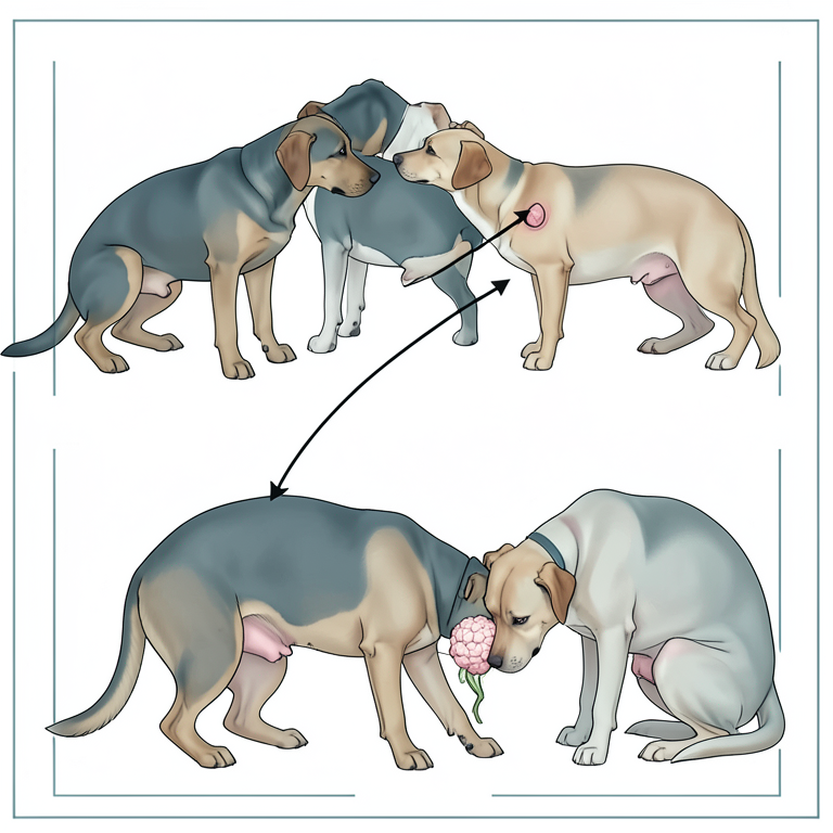
TVT Transmission Pathways
AI-generated conceptual illustration (Balinese dogs). It explains the idea and does not show study data.
View source evidence ↗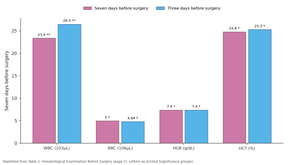
Seven days before surgery and Three days before surgery: WBC (103µL) vs RBC (106µL) vs HGB (g/dL) vs HCT (%)
Replotted from Table 2 (page 2) of the original paper. Every bar is the paper's own table value.
View source evidence ↗Figures & tables
From the original paper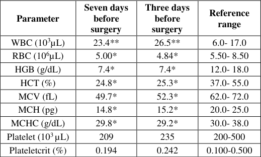 Table 2
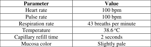 Table 1
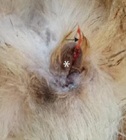 Figure 1
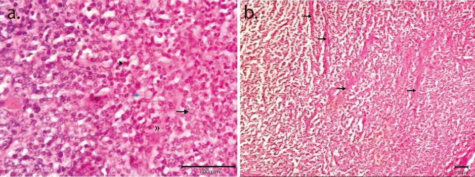 Figure 2
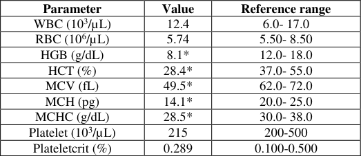 Table 3
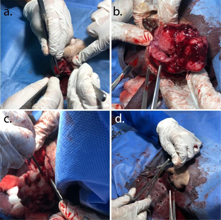 Figure 3
Introduction
Venereal sarcoma or generally known as transmissible venereal tumor (TVT) is a malignant tumor that affects the genital organ and is considered highly contagious in dogs. This disease is a serious problem around the world and occurs in the same frequency both male and female dogs who have uncontrolled sexual activity. This tumor has a unique pathogenesis compared to other neoplasms, because the growth is not spontaneous but is transmitted through coitus, (Foster, 2017) [7] , licking, and even sniffing (Behera et al., 2012; Foster, 2017; Stockmann et al., 2011) [3,7,16] . Dogs of all breeds and ages can be infected, but generally found in dogs with ages ranging from two to five years (Das and Das, 2000) [4] . This disease has also been reported in domestic dogs that have not been neutered or spayed (Strakova and Murchison, 2014) [15] . TVT cases in Bali are commonly found every year, Dharma et al. (2010) [5] reported that there were 55 cases in the period 2006-2010 which commonly affect roaming local dogs in the streets. The clinical sign of TVT is a cauliflower-like mass on the genital area or mucosal surface with blood discharge. TVT is classified into two groups based on the location of the tumor mass, genital and extragenital. In genital TVT, the tumor mass is located in posterior area of vagina, especially at vestibulovaginal junction in the female, and located in the caudal aspect of penis and preputium in the male. In extragenital TVT, the tumor mass is located in the outside area of the dog genital, such as in the oral area. This can occur because the dogs are licking or sniffing the genital area (Ganguly et al., 2016). TVTs cases that are still in the early stages, less than 1-year-old or without metastases, the success rate of treatment with surgery followed by chemotherapy with vincristine sulfate can reach 100%. However, if the case is longer and accompanied by metastases, the success rate of treatment is lower (Marcos et al., 2006) [10] . Treatment with surgery and chemotherapy preventing tumor cells from metastasis.
In this article, the authors report a case of transmissible venereal tumor in female local Balinese dogs treated with surgery and vincristine sulfate.
Case 2.1 Signalment and Anamnesis
A three-year-old mixed local Balinese female dog with a bodyweight of 8.8 kg was examined at the Department of Veterinary Surgery and Radiology, Animal Teaching Hospital, Udayana University with complaints of a mass in the vagina area and blood discharge since four months ago. Information from the owner stated that initially the mass was small and gradually grew bigger (Fig. 1). The mass began to appear three weeks after mating with stray dogs. The case dog is kept in the yard and sometimes plays around with other stray dogs. The food provided by the owner with homemade chicken liver twice a day. The dog's behavior is agile and has a good appetite. The dog has a history of rabies vaccinations and deworming. 1. The obvious clinical signs are swelling in the vaginal area accompanied by blood discharge and when palpated, the mass feels soft with irregular growth. Based on physical examination there is no sign of metastasis.
Physical Examination and Clinical Sign
Physical Examination and Clinical Sign
Hematological Examination
Seven days before surgery, the first hematological examination was carried out. Whole blood was collected in EDTA. The blood was then analyzed in hematological analyzer at Animal Teaching Hospital Faculty of Veterinary Medicine Udayana University. The hematological profile seven days before surgery shows the animal had microcytic hypochromic anemia and leukocytosis (Table 2).
| Parameter | Seven days before surgery | Three days before surgery | Reference range |
|---|---|---|---|
| WBC (103µL) | 23.4** | 26.5** | 6.0- 17.0 |
| RBC (106µL) | 5.00* | 4.84* | 5.50- 8.50 |
| HGB (g/dL) | 7.4* | 7.4* | 12.0- 18.0 |
| HCT (%) | 24.8* | 25.3* | 37.0- 55.0 |
| MCV (fL) | 49.7* | 52.3* | 62.0- 72.0 |
| MCH (pg) | 14.8* | 15.2* | 20.0- 25.0 |
| MCHC (g/dL) | 29.8* | 29.2* | 30.0- 38.0 |
| Platelet (103 µL) | 209 | 235 | 200-500 |
| Plateletcrit (%) | 0.194 | 0.242 | 0.100-0.500 |
Table text extracted from the original PDF; superscript letters are shown inline. The original table image is below.
View original table image
Radiographic Evaluation
Radiographic examination was performed to check for the potential for metastasis of TVT in several anatomical sites, but there were no signs of metastases. In this case it was confirmed that this TVT case was not accompanied by metastases.
Histopathological Examination
Histopathological Examination
Diagnosis and Prognosis
The final diagnosis is the dog has the transmissible venereal tumor. The prognosis is good because the tumor is still in an early stage and not accompanied with metastasis.
Treatment
Based on diagnosis and prognosis, the treatment is carried out by surgery to remove the tumor mass, then followed by chemotherapy. Microcytic hypochromic anemia at seven days before surgery is treated by administering Hematodin® (PT. Romindo Primavetcom, Jakarta, Indonesia) intramuscularly for seven days at a dose of 1 mL/5 kg BW to stimulate the hematopoietic system. Three days before surgery, a second hematological examination was performed, and the data is presented in Table 2. The animals had previously been fasted for 12 hours and abstained from drinking for three hours to prevent their vomiting and urination reflexed during surgery.
Prior to surgery, animals were premedicated using atropine sulfate (PT. Ethica Industri Pharmaceutical, Bantul, Indonesia) at a dose of 0.02 mg/kg BW subcutaneously, then induced with a combination of xylazine (Interchemie, Metalweeg, Netherlands) at a dose of 1.5 mg/kg BW and Ketamine (Ket-A-100, Agrovet Market, Lima, Peru) at a dose of 11 mg/kg BW intravenously. Surgery is performed with an incision in the vulva dorsally to reveal and find the mass of the tumor. The mass that has been found is then pulled forward and a total excision is started. The entire vagina is cleaned of the tumor mass and the normal tissue where the tumor is attached is excised (Figs. 3). The bleeding that occurs is treated with blood vessel ligation and administration of epinephrine (PT. Ethica Industri Pharmaceutical, Bantul, Indonesia) topically. Excision area then cleaned using 0.9% NaCl. The subcutaneous tissue was then sutured with a simple continuous pattern using chromic catgut 3.0 and the skin was sutured with a simple interrupted pattern with silk suture materials. Postoperative animal cases were carried out by cleaning the wound using povidone-iodine and administering antibiotics and analgesics. The antibiotic given was cefotaxime injection (PT. Dankos Farma, Jakarta, Indonesia) at a dose of 22 mg/kg BW intravenously three times a day for two days followed by administration of cefixime trihydrate antibiotic (PT. Dankos Farma, Jakarta, Indonesia) at a dose of 10 mg/kg BW per orally twice a day for five days. The analgesic given was 4% tolfenamic acid (Vetoquinol, Lure, France) at a dose of 4 mg/kg BW intramuscularly on the first day and followed by administration of 500 mg mefenamic acid (PT. Caprifarmindo Labs, Bandung, Indonesia) at a dose of 25 mg/kg BW once a day for two days. Chemotherapy was carried out three times with intervals of seven days. The first injection was carried out on first day after surgery, then the seventh day, and the last one is on the fourteenth day. Chemotherapy was carried out using vincristine sulfate (PT. Dankos Farma, Jakarta, Indonesia) at a dose of 0.025 mg/kg BW intravenously. One-week posttreatment, the hematological profile showed no abnormalities in total leukocytes, but microcytic hypochromic anemia was still present (Table 3). This case dog was then followed up for approximately two months, there were no signs of tumor recurrence. The owner then spays the dog intending to minimize the occurrence of recurring TVT.
| Parameter | Value | Reference range |
|---|---|---|
| WBC (103/µL) | 12.4 | 6.0- 17.0 |
| RBC (106/µL) | 5.74 | 5.50- 8.50 |
| HGB (g/dL) | 8.1* | 12.0- 18.0 |
| HCT (%) | 28.4* | 37.0- 55.0 |
| MCV (fL) | 49.5* | 62.0- 72.0 |
| MCH (pg) | 14.1* | 20.0- 25.0 |
| MCHC (g/dL) | 28.5* | 30.0- 38.0 |
| Platelet (103/µL) | 215 | 200-500 |
| Plateletcrit (%) | 0.289 | 0.100-0.500 |
Table text extracted from the original PDF; superscript letters are shown inline. The original table image is below.
View original table image
Discussion
Transmissible venereal tumor is an infectious disease in productive young dogs that most commonly occurs in dogs aged two to eight years and there is no breed predisposition and can infect both male and female dogs. According to Boscos et al. (2004), female dogs are more sensitive to this disease than male dogs because one male dog can infect several female dogs. Tumors grow rapidly on the vestibular and vulva in female dogs with a bright red color followed by hemorrhagic discharge. Surgical treatment accompanied by chemotherapy is considered effective for preventing the tumor from metastasis. Surgery is performed to completely remove the tumor mass and clean the surrounding tissue where the tumor is attached. However, surgical treatment is only performed when the tumor is accessible and not accompanied by metastasis. Surgery is effective if the TVT case is at stage I, the tumor nodule is small, where the tumor is easily accessible, and is non-invasive (Martins et al., 2005) [11] . TVT metastases are very rare, and when they do occur they usually affect the regional lymph nodes and surrounding cutaneous tissue (Faccini et al., 2019) [6] . Vincristine sulfate has been used widely as a single chemotherapy agent or combination therapy for the management of TVT cases. Therapy is generally carried out for two to seven weeks intravenously at a dose of 0.025 mL/kg BW depending on the severity of the TVT and the dog's resistance to chemotherapy (Das and Das, 2000) [4] . Vincristine sulfate is a chemical compound in the vinca alkaloid class derived from the Catharanthus roseus L. alkaloid plant (Ravina, 2011) [13] . Vincristine alkaloids have active substances that can inhibit cancer cells. This active substance can stop cancer cell division at the metaphase level, this causes the mitotic process stops at the metaphase stage so the cancer cells experience apoptosis (Avendano and Menendez, 2015) [2] . The results of the first hematological examination, performed a week before the operation showed the dogs had leukocytosis and microcytic hypochromic anemia. This anemia is caused by rupture of the tumor mass so that it sheds blood continuously. Das and Das (2000) [4] stated that the rupture of tumor masses was commonly found in TVT cases so the animal was prone to bleeding. Leukocytosis is common in dogs with TVT (Bahera et al., 2012; Kumar et al., 2018) [3,9] . This condition was then treated with Hematodin®. After the second hematological examination, three days before surgery, it showed an increase in hematocrit, mean corpuscular hemoglobin (MCH), and mean corpuscular hemoglobin concentration (MCHC) but not significant (Table 2). This is because the blood continues to flow in the dog's vagina. One-week post-treatment, the hematological profile showed no abnormalities in total leukocytes, but there is a decrease in Hb and PCV still occurs after treatment is due to the side effects of vincristine sulfate. Vincristine sulfate can suppress bone marrow activity so it directly affects erythropoiesis (Nak et al., 2005; Sandhu and Rampal, 2006) [12,14] . While the decrease of leukocytes one-week post-treatment is caused by suppression of progenitor cells in the bone marrow by vincristine sulfate (Nak et al., 2005) [12] . Study by Athar et al. (2001) [1] stated that the treatment of TVTs which begins with surgery only requires 2-3 times of chemotherapy with vincristine sulfate, healing will occur faster and there will be no recurrence. Meanwhile, in the case of transmissible venereal tumors that were only treated with a single vincristine sulfate without surgery, it required repeated chemotherapy up to 5-6 times to clear the tumor mass.
Conclusion
Surgical management of TVT case without metastasis followed by chemotherapy with vincristine sulfate has a considerable success rate without tumor recurrence.
Suggestion
Treatment of TVT cases with surgery followed by administration of vincristine sulfate needs to pay attention to the patient's general health condition because of the side effects. as an important note, surgery is only performed in cases of TVT without metastasis. Transmission of TVT in street dogs needs to be minimized by neutering or spaying, so the dog owners who release their dogs do not become infected.
Acknowledgement
AcknowledgementsWe would like to acknowledge to Dr. I Nyoman Dibia, DVM, M.P. (Pathologist from Animal Disease Investigation Center Bali) for the histopathologic diagnosis of the tumor biopsy sample.
Figures and tables from the paper
| Parameter | Value |
|---|---|
| Heart rate | 100 bpm |
| Pulse rate | 100 bpm |
| Respiration rate | 43 breaths per minute |
| Temperature | 38.6 oC |
| Capillary refill time | 2 seconds |
| Mucosa color | Slightly pale |
Table text extracted from the original PDF; superscript letters are shown inline. The original table image is below.
View original table image
References
16 in the original paper- Athar M, Suhail A, Muhammad G, Shakoor A, Azim F. Clinico-therapeutic studies on canine transmissible venereal tumour. Pakistan Veterinary Journal 2001;21(1):39-43
- Avendano C, Menendez JC. Anticancer Drugs Targeting Tubulin and Microtubules. In: Medicinal Chemistry of Anticancer Drugs, 2 nd Edition, Elsevier Science, Amsterdam, Netherland 2015, 359-390.
- Behera SK, Kurade NP, Shongsir WM, Durga PD, Krishna KM, Ranjan KM. Clinico-pathological findings in a case of canine cutaneous metastatic transmissible venereal tumour. Veterinarski arhiv 2012;82(4):401-410.
- Das U, Das AK. Review of canine transmissible venereal sarcoma. Veterinary Research Communication 2000;24:545-556
- Dharma DMN, Wirata IK, Supartika IKE. Canine Transmissible Venereal Tumor yang Didiagnosa di Balai Besar Veteriner Denpasar, 2006-2010. Animal Disease Investigation Center Denpasar, Ministry of Agriculture, Indonesia 2010. http://bbvdps.ditjenpkh.pertanian.go.id/wpcontent/uploads/2015/11/PENYEBARANTRANSMISSIBLE-VENEREAL-TUMOR.pdf (accessed 15 April 2021)
- Faccini LS, Legramanti WM, de Castro LT, Coelho ACB, Teixeria MC, Shild AL et al. Multiple Metastases of a Transmissible Venereal Tumor in a Dog. Acta Scientiae Veterinariae 2019; 47(1):444.
- Foster RA. Female Reproductive System and Mammae. In: Zachary JF (ed.) Pathologic Basis of Veterinary Disease, Sixth Edition, Elsevier, St. Louis, MO, USA 2017, 1147-1193.
- Ganguly B, Das U, Das AK. Canine transmissible venereal tumour: A review. Veterinary and Comparative Oncology 2013;14:1-12.
- Kumar VVVA, Kumari KN, Kumar KS, Kumar V, Lakshman M. Effect of Vincristine chemotherapy in TVT affected dogs. Journal of Pharmaceutical Innovation. 2018;7:163-166.
- Marcos R, Santos M, Marrinhas C, Rocha E. Cutaneous transmissible venereal tumor without genital involvement in a prepubertal female dog. Veterinary Clinical Pathology 2006;35:106-109.
- Martins MIM, de Souza FF, Gobello C. The Canine Transmissible Venereal Tumor: Etiology, Pathology, Diagnosis, and Treatment. In: Cocannon W, England G, Verstegen J, Forsberg (Eds.) Recent Advances in Small Animal Reproduction, International Veterinary Information Service, Ithaca, NY 2005, 161-167.
- Nak D, Nak Y, Cangul IT, Tuna B. A Clinicopathological Study on The Effect of Vincristine on Transmissible Venereal Tumour in Dogs, Journal of the Veterinary Medicine 2005;52:366-370.
- Ravina E. The evolution of drug discovery: from traditional medicines to modern drugs. Wiley, Weinheim, Germany 2011, 157-159.
- Sandhu HS, Rampal S. Essentials of Veterinary Pharmacology and Therapeutics, First Edition. Kalyani Publisher, New Delhi, India 2006, 1359-1381.
- Strakova A, Murchison EP. The Changing Global Distribution and Prevalence of Canine Transmissible Venereal Tumour. BMC Veterinary Research 2014;10:110.
- Stockmann D, Ferrari HF, Andrade AL, Lopes R, Cardoso, Tereza C, et al. Canine Transmissible Venereal Tumors: Aspects Related to Programmed Cell Death. Brazilian Journal of Veterinary Pathology 2011;4:67-75.
Cited by 7
From OpenAlex, OpenCitations and reference lists read by Scinovex
- Significance of hepatic enzymes: A review
Amit Kumar, Naveen Kumar, Anamika Anamika, Satya Satya · International Journal of Advanced Biochemistry Research · 2023
The assay of serum enzymes is very useful for the differential diagnosis and monitoring of various hepatobiliary disorders. Aspartate aminotransferases are found in the liver, cardiac muscle, skeletal muscle, kidneys, brain, pancreas, lungs, leukocytes, and erythrocytes in decreasing order of concentration. Alanine aminotransferases are found in the liver, muscle (cardiac and skeletal), kidneys, and erythrocytes and these are specific indicators of hepatocellular injury in dogs and cats not in large animals and pigs due to low activity in liver tissue. Alkaline phosphatases are markers of chol
- Treatment of canine transmissible venereal tumors in a male dog through surgery and chemotherapy ↗
Made Gede Adi Surya Saputra, I Wayan Wirata, I Gusti Ngurah Sudisma · ARSHI Veterinary Letters · 2025
Canine transmissible venereal tumors (CTVT) is a malignant tumor that affects the genital organs. It is highly contagious in dogs and is a significant global concern. This case report presents an effective and efficient treatment for CTVT in male dogs, offering valuable insights into veterinary care in small-animal oncology and surgery. A 2-year-old neutered male dog weighing 18,65 kg was…
❝ 1PDF - Surgical Procedure of a Transmissible Venereal Tumor in the Nostril of a Domestic Dog ↗
Hojat Hejazy, Seyed Amin Razavi, Faezeh Salari-Kakhk, Mohammad Shayeghi Alumnus · Small Animal Advances · 2025
Introduction: Canine transmissible venereal tumor (CTVT) is a contagious, round-cell neoplasm of mesenchymal origin that primarily affects the mucosal surfaces of the external genitalia in both male and female dogs. This neoplasm is naturally transmissible and sexually spread, particularly among reproductively active stray dogs. The present study investigated the clinical and histological signs, as well as surgical treatment, of CTVY…
❝ 1PDF - Vincristine sulfate in canine TVT: Confirming efficacy and expanding safety evidence through haematological and biochemical profiling
Tyson ., Amit Sharma, Ankit Ahuja, Vasu Sharma, Pravesh Kumar, Akshay Sharma · International Journal of Advanced Biochemistry Research · 2026
Canine transmissible venereal tumour (CTVT) is one of the most common neoplasm in dogs, with vincristine sulfate established as the first-line chemotherapeutic agent. Thirty-eight dogs with cytologically confirmed CTVT received vincristine sulfate at weekly intervals for 4–6 weeks. Characteristic clinical signs included serosanguinous genital discharge (86.84%), genital deformation (52.63%), and systemic manifestations such as anorexia (47.36%). Complete remission was achieved in 81.6% of cases after four doses and in an additional 15.8% after six doses, yielding an overall success rate of 97.
- Laporan Kasus: Keberhasilan Penanganan Transmissible Venereal Tumor pada Anjing Kacang Betina dengan Eksisi dan Kemoterapi ↗
Stephanie Ariella Gunawan, I Gusti Agung Gde Putra Pemayun, Anak Agung Gde Jayawardhita · jurnal veteriner · 2023
Transmissible Venereal Tumor (TVT)adalah tumor umum pada anjing yang ditularkan melalui perkawinan, jilatan, atau mengendus lesi tumor anjing penderita. Tujuan penulisan artikel ini adalah untuk mengungkapkan keberhasilan penanganan TVT pada anjing kacang melalui metode pembedahan dan kemoterapi. Anjing lokal/kacang betina berumur enam tahun dengan bobot badan 8,7 kg datang dengan keluhan adanya massa pada vagina disertai tetesan darah. Hasil pemeriksaan…
❝ 0PDF - Efficacy of oral fluralaner in the treatment of canine generalized demodicosis ↗
Peddineni Sree Krishna Sai, N Devadevi, Kavir Rajkumar, A. Abiramy Prabavathy, P Vijayalakshmi, Vijayakumar Vijayalakshmi · Journal of Research in Chemistry · 2022
The aim of the study was to assess the effectiveness of the fluralaner in the treatment of canine generalized demodicosis. Dogs presented with skin lesions were subjected to detailed clinical examination, skin scraping subjected for microscopic examination. Dogs turned positive for Demodexspecies were treated with single oral dose of fluralaner @ 30 mg/ kg body weight. After four weeks of…
❝ 0PDF - Coexisting upper tract urothelial carcinoma and renal tuberculosis: A rare presentation ↗
Muni Bhavani Itha, Yarlagadda Krishna Bharathi, Satyanarayana Veeragandham · Journal of Case Reports and Scientific Images · 2021
Tuberculosis (TB) is a global public health problem and is a leading cause of death, especially in underdeveloped and developing countries. In the majority of instances, renal tuberculosis is caused by the secondary hematogenous transmission of mycobacterium from pulmonary lesions to the renal cortex either during the initial TB infection or reactivation/endogenous reinfection of an old tuberculous focus. Upper tract