
Research article · 2023 · Full text
International Journal of Advanced Biochemistry ResearchSignificance of hepatic enzymes: A review
Volume 7(1) · Pages 95–100 · DOI: 10.33545/26174693.2023.v7.i1b.170 ↗
Research areasLiver Disease Diagnosis and TreatmentPancreatitis Pathology and TreatmentBiochemical and Molecular Research
Abstract
Author-writtenThe assay of serum enzymes is very useful for the differential diagnosis and monitoring of various hepatobiliary disorders. Aspartate aminotransferases are found in the liver, cardiac muscle, skeletal muscle, kidneys, brain, pancreas, lungs, leukocytes, and erythrocytes in decreasing order of concentration. Alanine aminotransferases are found in the liver, muscle (cardiac and skeletal), kidneys, and erythrocytes and these are specific indicators of hepatocellular injury in dogs and cats not in large animals and pigs due to low activity in liver tissue. Alkaline phosphatases are markers of cholestasis, present in most tissues but are in particularly high concentration in the osteoblasts of bone and the cells of the hepatobiliary tract, intestinal wall, renal tubules, and placenta. Gamma-glutamyl transferase occurs mainly in the cells of the liver, kidneys, pancreas, and prostate. Alcoholism and hepatocellular damage due to infectious hepatitis are causes of elevated plasma GGT activity. Sorbitol dehydrogenase is predominantly found in the liver and kidney and measurement of serum SDH activity has been shown to be valuable in assessing hepatocellular injury in various domestic species. Elevated serum arginase levels have been observed in naturally occurring liver diseases of horses, cattle, sheep, goats, and dogs. Ornithine carbamoyltransferase (OCT) is primarily found in the liver and is regarded as a liver-specific enzyme for detecting hepatocellular necrosis in domestic species. This review aims to highlight the importance of hepatic enzymes for diagnosis purposes.
Summary
Source-checkedPlain-language reading of the full paper, prepared with AI and checked against the text. Each point links to the authors' own words.
Why it matters
- The liver is essential for metabolic regulation, toxin elimination, protein production, and the digestion of fats and fat-soluble vitamins. View evidence ↗
- Liver failure symptoms may not manifest until a substantial portion of the organ's function, approximately 70% or more, is lost due to its high reserve capacity. View evidence ↗
- Diagnosing liver disease requires a multi-faceted approach combining clinical history, physical exams, biochemical tests, imaging, and biopsies. View evidence ↗
The question
- The objective of this review is to emphasize the significance of hepatic enzymes in the diagnosis of liver and hepatobiliary disorders. View evidence ↗
What they did
- The review examines the clinical use of measuring serum enzyme activity to assess diseases of the liver and bile ducts. View evidence ↗
- Liver injury is typically categorized by enzyme levels relative to the upper limit of normal (ULN), where 3-to 5-fold increases indicate significant injury and over 10-fold increases suggest moderate to marked injury. View evidence ↗
What they found
- In dogs, the activity of Alanine Aminotransferase (ALT) in the liver is roughly 10,000 times higher than in the blood plasma, making it a useful marker for liver damage. View evidence ↗
- Serum ALT is not routinely used for horses, cattle, sheep, and swine because these species have relatively lower hepatic ALT activity. View evidence ↗
- Aspartate Aminotransferase (AST) is a less specific indicator of liver disease than ALT because AST is also found in the heart, kidneys, and skeletal muscle. View evidence ↗
- Ornithine Carbamoyltransferase (OCT) is highly liver-specific in cattle and pigs, as almost all of its activity is located in the liver. View evidence ↗
- The highest increases in serum alkaline phosphatase (ALP) occur in animals suffering from biliary cirrhosis, cholangitis, or extrahepatic bile duct obstruction. View evidence ↗
- In neonatal calves, Gamma-glutamyltransferase (GGT) activity can be used to indicate successful passive immune transfer due to its relationship with immune globulin levels. View evidence ↗
- Elevated serum arginase levels are observed in naturally occurring liver diseases across several species, including dogs, goats, sheep, cattle, and horses. View evidence ↗
- Glutamate Dehydrogenase (GD) is a useful enzyme for evaluating hepatic necrosis specifically in cattle, goats, and sheep. View evidence ↗
What it means
- The authors conclude that while single enzymes provide value, combining multiple enzyme analyses with other biochemical markers improves the sensitivity and specificity of liver disease diagnosis. View evidence ↗
Visual summary
1 visualsIllustrations explain the paper; they do not show study data.
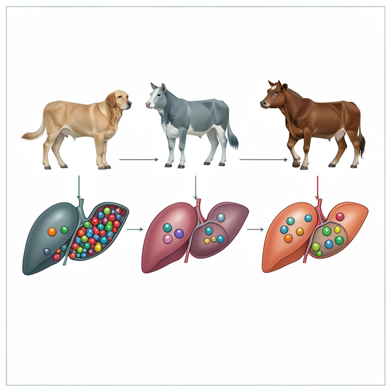
Comparative Enzyme Distribution
AI-generated conceptual illustration (Dog, cat, horse, and cow livers). It explains the idea and does not show study data.
View source evidence ↗Introduction
The liver plays a crucial role in various metabolic processes, such as regulating blood glucose levels, eliminating toxins and hydrophobic substances from the body, producing most of the proteins found in the blood plasma, and aiding digestion by producing and storing bile acids necessary for absorbing fats and fat-soluble vitamins. Any disruptions in these metabolic, excretory, synthetic, and digestive functions of the liver can lead to noticeable clinical symptoms of liver disease. It's worth noting that the liver has a significant reserve capacity, and symptoms of liver failure may not appear until around 70% or more of its functional ability is lost. Importantly, even when a substantial portion of liver cells is damaged, the liver has the remarkable ability to regenerate, allowing for potential recovery from acute injuries. To diagnose liver disease, a comprehensive approach involving the assessment of clinical history, physical examination, biochemical tests, hepatic imaging, and histopathological examination of liver biopsies is necessary. In clinical patients displaying symptoms and a history suggestive of liver disease, laboratory tests are conducted to confirm the diagnosis. These tests serve several purposes, including evaluating the extent of liver damage, predicting the prognosis, identifying treatable complications resulting from liver insufficiency (such as ascites and encephalopathy), and monitoring the patient's clinical progress [1] . The assessment of hepatocellular injury, which involves the loss of cellular integrity and the release of intracellular components, is commonly performed by measuring specific enzymes that leak out of damaged liver cells. These enzymes include alanine aminotransferase (ALT), aspartate aminotransferase (AST), lactate dehydrogenase (LD), sorbitol dehydrogenase (SDH), and glutamate dehydrogenase (GLD). Although less frequently used, arginase, isocitrate dehydrogenase, and fructose bisphosphatase can also be employed. Typically, significant liver injury is indicated by a 3-to 5-fold increase in blood levels of hepatic enzymes above the upper limit of normal (ULN), while increases exceeding 10-fold suggest moderate to marked injury. The effectiveness of these biomarkers relies on several factors. Firstly, they should be specific to the liver, allowing for an accurate assessment of liver damage.
Additionally, these biomarkers need to persist in the bloodstream, providing a measurable signal. Their persistence is influenced by their concentration within liver tissues, their location within subcellular compartments (with enzymes released more readily from the cytosol than mitochondria), and the rate at which their activity is cleared from the blood, known as their half-life. Hepatobiliary injury is evaluated by monitoring the presence of specific enzymes, known as cholestatic or induction enzymes, in the blood. The most commonly utilized enzymes for this purpose are alkaline phosphatase (ALP) and γglutamyltransferase (GGT), although occasionally 5'nucleotidase is employed. These enzymes are bound to cell membranes and their levels increase in the blood due to reflux into plasma caused by increased synthesis, canalicular injury, cholestasis, or solubilization by bile salts. Increased synthesis of these enzymes occurs in response to biliary epithelial hyperplasia, which is a proliferative and reparative reaction to liver injury. However, this limits their specificity for distinguishing hepatobiliary injury from hepatocellular injury. ALP has additional limitations as a blood biomarker for liver injury, as it also increases during bone growth, colostrum ingestion in neonates, liver synthesis induction by glucocorticoids and anticonvulsants (in dogs), and certain neoplasms, such as mammary tumors [1] .
Hepatic Enzymes
Liver disease is typically identified through the detection of increased levels of liver-specific enzymes in the bloodstream. While these enzymes are commonly referred to as "liver function tests," it is important to note that they don't directly measure the overall function of the liver.
Instead, they indicate changes in the integrity of the cell membrane of hepatocytes (liver cells), the death of hepatocytes or biliary epithelium, impaired bile production or flow (cholestasis), or the activation of enzyme synthesis processes [2] . The measurement of serum enzymes is commonly used in clinical practice to assess liver and bile duct diseases. These enzymes exhibit high levels of activity specifically within the liver. When there is liver cell damage or cholestasis (impaired bile flow), these enzymes are released into the bloodstream, leading to increased serum activity levels, which can be used as diagnostic markers. The duration of elevated serum enzyme activity depends on various factors such as molecular size, intracellular location, clearance rate from the bloodstream, rate of enzyme inactivation, and, in some cases (e.g., alkaline phosphatase [AP] and glutamyl transpeptidase [GGT]), the rate of enzyme production in the liver. Certain enzymes, including alanine aminotransferase (ALT), aspartate aminotransferase (AST), ornithine carbamoyltransferase (OCT), glutamic dehydrogenase (GD), sorbitol dehydrogenase (SDH), and arginase, show increased serum activity when there is liver cell necrosis. Elevated levels of alkaline phosphatase (AP), GGT and 5' nucleotidase (5'-ND) in the serum typically indicate either intrahepatic or extrahepatic cholestasis. Due to its strategic position between the splanchnic and systemic circulation, the liver is susceptible to exposure to numerous substances such as toxins, drugs and their byproducts, bacterial toxins, and infectious agents. These factors can impact the levels of liver enzymes in the bloodstream. When evaluating abnormal liver enzyme levels, it is important to consider the specific type of enzyme alteration (whether it is related to hepatocellular damage or cholestasis), the extent of the increase in serum enzyme activity, the rate at which the activity levels change (whether it increases or decreases), and whether there are fluctuations in enzyme activity over time or a consistent pattern of change. Taking these factors into account is crucial for a comprehensive clinical assessment of liver enzyme aberrations [1] .
Serum Alanine and Aspartate Aminotransferases
The levels of aminotransferases, specifically AST and ALT, in the bloodstream are measured to detect liver cell damage. These enzymes play a crucial role in transferring the αamino nitrogen from aspartate or alanine to α-ketoglutaric acid, resulting in the production of glutamate. AST and ALT are involved in important processes like gluconeogenesis (formation of glucose) and the synthesis of urea. In the liver, ALT facilitates the transfer of α-amino nitrogen from alanine to α-ketoglutarate, producing pyruvate, which can be used in gluconeogenesis. In muscle tissues, ALT converts pyruvate into alanine, which transports non-ionized nitrogen from the muscles to the liver as part of the glucose-alanine cycle for further processing. ALT activity is significantly higher in the liver compared to other tissues. In dogs, hepatic ALT activity is approximately 10,000 times higher than its concentration in the blood plasma. Similarly, cats, humans, and certain experimental rodent species also exhibit high hepatic ALT activity, making serum ALT measurement a routine practice for assessing liver cell damage. However, in horses, cattle, sheep, and swine, hepatic ALT activity is relatively lower, and as a result, serum ALT is not routinely measured in these species. The liver of all domestic species exhibits high activity levels of AST, and measuring its concentration in the bloodstream is a routine practice for evaluating liver cell damage. However, it's important to note that AST activity is also elevated in the kidney, heart, and skeletal muscle. As a result, increases in serum AST levels are considered less specific indicators of liver disease compared to elevations in serum ALT levels. The distribution of ALT and AST within the hepatocyte (liver cell) differs. In dogs, the majority of hepatic ALT and AST activity is found in the cytosol, while a significant portion of AST (20%) and a smaller portion of ALT are present in the mitochondria [3] . Additionally, the distribution of these transaminases within the acinar zones of the liver also varies. ALT exhibits the highest activity in Zone 1 hepatocytes, while AST has the highest activity in Zone 3 hepatocytes. The relative levels of ALT or AST in the bloodstream may reflect the specific acinar zone where liver injury has occurred [4] . The most significant increases in serum ALT levels are observed in cases of hepatocellular inflammation and necrosis. In such conditions, a progressive decrease in ALT activity may indicate recovery and a reduction of 50% or more in serum ALT activity over several days is considered a positive prognostic indicator. However, it is important to note that some animals with severe liver disease may have normal serum ALT activity. In these cases, a decline in serum ALT activity could indicate a significant reduction in viable hepatocytes or a decrease in transaminase synthesis (e.g., due to toxins like microcystins or aflatoxin). In all domestic species, AST activity is high in the liver, and an increase in serum AST activity is typically seen in both acute and chronic liver injuries. However, since AST activity is also elevated in other tissues like muscle, kidney, pancreas, and red blood cells, damage to these tissues can also result in elevated serum AST levels. Determining the specific source of increased serum AST activity is not a straightforward process, and there is no direct method for it. However, additional laboratory tests can be useful. For example, in cases of skeletal muscle disease (including trauma or degenerative conditions), measuring serum creatine kinase (CK) levels may provide valuable information. In muscle diseases, both AST and CK are expected to be elevated. In acute muscle injuries, CK levels may increase before AST reaches its maximum elevation, and CK levels typically decrease before AST activity fully declines. In cases where both myopathy and liver disease are present in dogs and cats, measuring serum ALT levels may be helpful. However, it's worth noting that severe primary muscle diseases can also be associated with increased serum ALT activity. Dogs have been observed to exhibit increased transaminase activity after intense exercise [5] . However, the source or origin of this enzyme in such circumstances remains unclear [6,7] . A 1.4-to 2-fold increase in plasma AST levels in dogs was observed after short-term exercise, along with increases in CK and lactic dehydrogenase activity [8] . Similar increases in plasma AST and lactic dehydrogenase were also detected following electro-physiological stimulation of hind limb muscles. The main utility of measuring serum AST and ALT levels lies in detecting hepatocellular injury and monitoring the progress of clinical conditions. However, as both enzymes can be elevated in various liver diseases, their value for differential diagnosis is limited. While elevated aminotransferase levels are generally indicative of hepatocellular injury, severe liver diseases often involve a combination of hepatocellular and cholestatic forms of hepatic injury. The most significant increases in aminotransferase activity are associated with acute hepatic injury, but more moderate increases can be observed in chronic liver diseases such as chronic hepatocellular disease, cirrhosis, parasitic hepatopathy, and primary or metastatic neoplasia.
Sorbitol Dehydrogenase
Due to the relatively low activity of ALT in the livers of large domestic species, alternative liver-specific enzymes have been established for clinical purposes [9,10] . One such enzyme is SDH, which is predominantly found in the liver and kidney. The measurement of serum SDH activity has been shown to be valuable in assessing hepatocellular injury in various domestic species, including dogs, horses, and ruminants [11,12] . However, in dogs and cats, SDH does not offer any clear advantages over ALT [2] . It is important to note that SDH activity in serum is not stable and declines rapidly. Therefore, it is crucial to perform the SDH assay as soon as possible after the sample is collected, ideally within 8 to 12 hours, to ensure accurate results.
Arginase
The conversion of arginine to urea and ornithine, the final step in the urea cycle, is facilitated by the enzyme arginase. In mammals, there are two forms of arginase: arginase type I and arginase type II. These isoforms differ in their genes, tissue distribution, intracellular location, and molecular characteristics [13,14] . Arginase type I is found in the cytosol and is primarily expressed in the liver, where it plays a crucial role in urea synthesis. On the other hand, arginase type II is a mitochondrial enzyme present in various tissues, including the kidney, and it is involved in regulating the availability of arginine for nitric oxide synthesis and the production of ornithine, which serves as a precursor for polyamines, glutamate, and proline [14,15] . The liver exhibits higher arginase activity compared to other organs [16] leading to the consideration of elevated serum arginase activity as a liver-specific marker [9,17] . Reliable assays have been developed for measuring serum arginase activity [18] . Following acute liver injury, serum arginase levels initially increase and then rapidly decrease [9,19] . Prolonged elevation in serum arginase activity indicates an unfavorable prognosis. Elevated serum arginase levels have been observed in naturally occurring liver diseases of horses, cattle, sheep, goats, and dogs [20] . Simultaneous measurement of serum arginase and GGT activities can be useful in distinguishing hepatocellular injury from cholestasis [21] .
Glutamate Dehydrogenase
GD (Glutamate dehydrogenase) is an enzyme primarily found in the mitochondria that plays a crucial role in detoxifying ammonia. It functions as an ammonia scavenger by catalyzing the conversion of α-ketoglutarate to glutamate through animation. The produced glutamate is further transformed to aspartate by mitochondrial AST (aspartate aminotransferase), which is involved in urea synthesis. Moreover, glutamate is utilized in the mitochondrial production of N-acetyl glutamate, an allosteric activator of carbamoyl phosphate synthase, the enzyme responsible for the initial step in urea synthesis [22,23,24] . GD has demonstrated utility in evaluating hepatic necrosis in sheep, goats, and cattle. The liver of these species, as well as other domestic animals, exhibits a high concentration of GD activity [10,25] . Elevated GD activity has been observed in ruminants with hepatic necrosis [26] , in association with parturition [47] and in cases of bile duct obstruction [28] .
Ornithine Carbamoyltransferase
OCT (ornithine carbamoyltransferase) is an enzyme that operates within the mitochondria and plays a vital role in the urea cycle by facilitating the reaction between ornithine and carbamoyl phosphate, resulting in the formation of citrulline and inorganic phosphate. It is primarily found in the liver and is regarded as a liver-specific enzyme for detecting hepatocellular necrosis in domestic species [29] . The liver of cattle [47] and pigs [17] contain almost all the OCT activity. In terms of diagnostic sensitivity for hepatic necrosis in dogs, OCT and ALT exhibit similar performance as tests. Swine also exhibit a similar association between OCT and hepatocellular injury [30] .
Serum Alkaline Phosphatase
The alkaline phosphatases (APs) are a group of zinc metalloenzymes found in most tissues, with high concentrations in the intestine, kidney, bone and liver. Studies using light and electron microscopy have shown that alkaline phosphatase activity is highest on the surfaces of cells involved in absorption or secretion [31] . The exact physiological functions of alkaline phosphatase are not fully understood, but its localization on cell surfaces responsible for active transport suggests a role in membrane transport. Some evidence suggests that alkaline phosphatase in osteoblasts may play a role in bone calcification, and its activity against nucleotides indicates involvement in nucleic acid metabolism. In normal animals, the primary sources of serum alkaline phosphatase are the liver and bone [32,33] . Elevated serum alkaline phosphatase activity can be observed in growing animals or in adults with increased osteoblastic activity. It can also be elevated in acute and chronic liver diseases, with higher levels indicating cholestasis. The most significant increases in serum alkaline phosphatase activity are seen in animals with cholangitis, biliary cirrhosis, or obstruction of the bile duct outside the liver. Unlike serum AST and ALT, elevated alkaline phosphatase levels are not solely due to leakage from damaged cells. Previously, it was believed that the high serum alkaline phosphatase levels observed in cholestatic liver diseases resulted from decreased biliary excretion of the enzyme since bile contains a substantial amount of alkaline phosphatase activity. However, it is now known that increased synthesis of alkaline phosphatase is involved in various conditions, such as extrahepatic bile duct obstruction, intrahepatic cholestasis, and infiltrative liver diseases where the terminal branches of the biliary tree may be obstructed. Additionally, alkaline phosphatase synthesis increases during liver regeneration following injury. While measuring serum GGT activity offers specificity, total serum alkaline phosphatase activity remains the most commonly performed test for assessing cholestasis in horses, dogs, and cats. Serum alkaline phosphatase is less useful for evaluating cholestatic syndromes in cattle and sheep due to increases being described in various canine cholestatic liver diseases [34,35] . Modest increases in serum alkaline phosphatase can occur with hepatic necrosis [21] . In cases of experimental hepatic necrosis in dogs, the serum activities of arginase, ALT, and alkaline phosphatase increase within one day, whereas GGT activity remains unaffected. Following bile duct obstruction, both alkaline phosphatase and GGT activities increase significantly along with moderate elevations in ALT and AST, while arginase activity does not increase. This suggests that arginase (indicating necrosis) and GGT (indicating cholestasis) may have higher specificity in evaluating the type of hepatobiliary disease in dogs [21] . Although GGT activity may be less affected during hepatocellular necrosis compared to alkaline phosphatase, GGT activity may not be as highly elevated as an alkaline phosphatase in cases of bile duct obstruction [36] .
γ-Glutamyl transpeptidase
GGT (gamma-glutamyltransferase) is an enzyme located on the cell membrane that facilitates the transfer of γ-glutamyl groups from γ-glutamyl peptides, such as glutathione, to other amino acids or peptides. Its primary substrates are glutathione and glutathione conjugates, which are abundant in the body [37] . GGT is predominantly found in cells involved in secretion or absorption, including the liver, kidney, pancreas, and intestine. It is primarily used as a serum marker for hepatobiliary diseases associated with cholestasis [38] and serves as a diagnostic tool for liver diseases in animals. While GGT activity is relatively high in the livers of cows, horses, sheep, and goats, it is significantly lower in the livers of dogs and cats. Although GGT is present in various tissues and exhibits high activity in the kidney, significant elevations in serum GGT activity are typically observed in liver diseases. However, urinary excretion of GGT has been measured to assess renal injury [39] . Experimental studies involving bile duct obstruction have demonstrated a substantial increase in serum GGT activity in dogs [21] , sheep [39] , and cattle. In cats, GGT has shown similar sensitivity to AP (alkaline phosphatase) as an indicator of cholestasis [40] . Within a given species, there is often a direct correlation between serum GGT and AP activities in cases of cholestatic liver injury. In primary hepatocellular diseases, GGT elevations are generally not as pronounced as those of AP [41] . The highest levels of GGT activity in dogs and cats are found in the kidney and pancreas, with lesser amounts in the liver, gallbladder, intestines, spleen, heart, lungs, skeletal muscle, and erythrocytes [36,38] . Administration of glucocorticoids and certain microsomal enzyme inducers can stimulate GGT production in dogs, similar to the effect of these drugs or other substances on AP. It is believed that increased GGT production following glucocorticoid administration originates in the liver. Remarkably elevated serum GGT levels have been observed in dogs and cats with primary hepatic or pancreatic neoplasia. Neonatal animals of various species exhibit increased serum GGT activity after consuming colostrum. In neonatal calves, the direct relationship between serum GGT activity and immune globulin levels allows GGT activity to serve as an indicator of successful passive immune transfer [42] . Similar transient elevations of serum GGT are observed in neonatal lambs [43] , crias [44] , and pups [45] following colostrum ingestion, but apparently not in kittens [46] .
Conclusion
Liver enzymes present in serum can indicate various liver and hepatobiliary pathologies such as obstruction, proliferation, inflammation, toxicity, or neoplasia. These enzymes serve two main purposes: evaluating and determining the type and severity of liver injury and providing insights into the treatment and prognosis of liver function disorders. Interpreting the data from these enzymes is crucial for reaching an accurate diagnosis. Each enzyme involved in the evaluation offers valuable information that aids in distinguishing liver pathologies from other diseases. Overall, variations in enzyme profiles are associated with different types of liver pathologies. Therefore, clinicians can differentiate and accurately diagnose liver diseases by assessing a single hepatobiliary enzyme. However, combining the analysis of multiple enzymes along with other biochemical markers enhances sensitivity and specificity in identifying liver diseases, leading to appropriate treatment and prognosis.
References
45 in the original paper- Center SA. Interpretation of liver enzymes. Veterinary Clinics of North America: Small Animal Practice. 2007;37:297-333.
- Keller P. Enzyme activities in the dog: tissue analyses, plasma values, and intracellular distribution. American Journal of Veterinary Research. 1981;42:575-582.
- Rej R. Aminotransferases in disease. Clinics in Laboratory Medicine. 1989;9:667-687.
- Valentine BA, Blue JT, Shelley SM, Cooper BJ. Increased serum alanine aminotransferase activity associated with muscle necrosis in the dog. Journal of Veterinary Internal Medicine. 1990;4:140-143.
- Bolter CP, Critz, JB. Plasma enzyme levels in the anesthesized dog during drainage of the thoracic lymph duct. Enzyme. 1976;21:30-38.
- Loegering DJ, Critz JB. Effect of hypoxia and muscular activity on plasma enzyme levels in dogs. American Journal of Physiology. 1971;220:100-104.
- Heffron JJA, Bomzon L, Pattinson RA. Observations on plasma creatine phosphokinase activity in dogs. Veterinary Record. 1976 Apr;98:338-340.
- Cornelius CE. Relation of body-weight to hepatic glutamic pyruvic transaminase activity. Nature. 1963;200:580-581.
- Keller P, Ruedi D, Gutzwiller A. Tissue distribution of diagnostically useful enzymes in zoo animals. Journal of Zoo Animal Medicine. 1985;16:28-49.
- Johnson AL, Divers TJ, Freckleton ML, McKenzie HC, Mitchell E, Cullen JM et al. Fall panicum (Panicum dichotomiflorum) hepatotoxicosis in horses and sheep. Journal of Veterinary Internal Medicine. 2006;20:14141421.
- Grody WW, Argyle C, Kern RM, Dizikes GJ, Spector EB, Strickland AD, et al. Differential expression of the two human arginase genes in hyperargininemia. Enzymatic, pathologic, and molecular analysis. Journal of Clinical Investigation. 1989 Feb;83:602-609.
- Jenkinson CP, Grody WW, Cederbaum SD. Comparative properties of arginases. Comp. Biochem. Physiol. 1996;114:107-132.
- Gotoh T, Mori M. Arginase II downregulates nitric oxide (NO) production and prevents NO-mediated apoptosis in murine macrophage-derived RAW 264.7 cells. Journal of Cell Biology. 1999;144:427-434.
- Aminlari M, Shahbazkia HR, Esfandiari A. Distribution of arginase in tissues of cat (Felis catus). Journal of Feline Medicine and Surgery. 2007;9:133-139.
- Dittrich C, Stockl W, Desser H. Determination of enzymes of the urea cycle, glutaminase and asparaginase in cattle and pigs. Zentralblatt fur Veterinarmedizin. Reihe A. 1974 Feb;21:165-170.
- Mia AS, Koger HD. Direct colorimetric determination of serum arginase in various domestic animals. American Journal of Veterinary Research. 1978;39:1381-1382.
- Aminlari M, Vaseghi T, Sajedianfard MJ, Samsami M. Changes in arginase, aminotransferases and rhodanese in sera of domestic animals with experimentally induced liver necrosis. Journal of Comparative Pathology. 1994;110:1-9.
- Wolff WA, Lumb WV, Ramsay MK. Effects of halothane and chloroform anesthesia on the equine liver. American Journal of Veterinary Research. 1967;28:1363-1372.
- Noonan NE, Meyer DJ. Use of plasma arginase and gamma-glutamyl transpeptidase as specific indicators of hepatocellular or hepatobiliary disease in the dog. American Journal of Veterinary Research. 1979;40:942-947.
- Caldovic L, Tuchman M. N-acetylglutamate and its changing role through evolution. Biochemical Journal. 2003;372:279-290.
- Caldovic L, Lopez GY, Haskins N, Panglao M, Shi D, Morizono H, et al. Biochemical properties of recombinant human and mouse N-acetylglutamate synthase. Molecular Genetics and Metabolism. 2006;87:226-232.
- Nissim I, Horyn O, Luhovvy B, Lazarow A, Daikhin Y, Nissim I, et al. Role of the glutamate dehydrogenase reaction in furnishing aspartate nitrogen for urea synthesis: Studies in perfused rat liver with 15N. Biochemical Journal. 2003 Nov;376:179-188.
- Keller P. Serum enzymes in cattle: organ analysis and normal values. Schweiz. Archiv für Tierzucht/Archives Animal. 1971;113:615-626.
- Fowler JS. Toxicity of carbon tetrachloride and other fasciocidal drugs in sheep and chickens. British Veterinary Journal. 1971;237:304-312.
- Takariyanti DN, Gorda IW, Sewoyo PS. Treatment of transmissible venereal tumor without metastasis in mixed local Balinese dog by surgery and vincristine sulfate: A case report. International Journal of Veterinary Sciences and Animal Husbandry. 2021;6(3):25-29. DOI: https://doi.org/10.22271/veterinary.2021.v6.i3a.35 6
- Ford EJH, Gopinath C. The excretion of phylloerythrin and bilirubin by calves and sheep. Research in Veterinary Science. 1976;21:12-18.
- Treacher RJ, Sansom BF. Liver function in dairy cows at parturition. Research in Veterinary Science. 1969;10:461-468.
- Wilson GD, Harvey DG, Snook CR. A review of factors affecting blood biochemistry in the pig. British Veterinary Journal. 1972;128:596-610.
- Kaplan MM. Alkaline phosphatase. Gastroenterology. 1972;62:452-468.
- Hoffmann WE, Dorner JL. Separation of isoenzymes of canine alkaline phosphatase by cellulose acetate electrophoresis. American Journal of Medicine. Hosp. Assoc. 1975;11:283-285.
- Rogers WA. Source of serum alkaline phosphatase in clinically normal and diseased dogs: a clinical study. Journal of the American Veterinary Medical Association. 1976;168:934-937.
- Abdelkader SV, Hauge JG. Serum enzyme determination in the study of liver disease in dogs. Acta Veterinaria Scandinavica. 1986;27:59-70.
- Solter PF, Hoffmann WE. Solubilization of liver alkaline phosphatase isoenzyme during cholestasis in dogs. Journal of Advanced Veterinary Research. 1999;66:1010-1015.
- Guelfi JF, Braun JP, Rico AG. Value of so called cholestasis markers in the dog: an experimental study. Research in Veterinary Science. 1982;33:309-312.
- Hanigan MH. Gamma-Glutamyl transpeptidase, a glutathionase: its expression and function in carcinogenesis. Chemico-Biological Interactions. 1998;111-112:333-342.
- Braun JP, Benard P, Burgat V, Rico AG. Gamma glutamyl transferase in domestic animals. Veterinary Research Communications. 1983;6:77-90.
- Ford EJH. Activity of gamma-glutamyl transpeptidase and other enzymes in the serum of sheep with liver or kidney damage. Journal of Comparative Pathology. 1974;84:231-243.
- Zawie DA, Garvey MS. Feline hepatic disease. Veterinary Clinics of North America. 1984;14:12011230.
- Meyer DJ. Serum gamma-glutamyltransferase as a liver test in cats with toxic and obstructive hepatic disease. Journal of the American Animal Hospital Association. 1983;19:1023-1026.
- Parish SM, Tyler JW, Besser TE, Gay CC, Krytenberg D. Prediction of serum IgG1 concentration in Holstein calves using serum gamma glutamyl transferase activity. Journal of Veterinary Internal Medicine. 1997;11:344-347.
- Maden M, Altunok V, Birdane FM, Asian F, Nizamlioglu M. Blood and colostrum/milk serum gamma-glutamyltransferase activity as a predictor of passive transfer status in lambs. Journal of Veterinary Medicine Series B-Infectious Diseases and Veterinary. Public Health. 2003;50:128-131.
- Johnston NA, Parish SM, Tyler JW, Tillman CB. Evaluation of serum gamma-glutamyltransferase activity as a predictor of passive transfer status in crias. Journal of the American Veterinary Medical Association. 1997;211:1165-1166.
- Center SA, Randolph JF, ManWarren T, Slater M. Effect of colostrum ingestion on gammaglutamyltransferase and alkaline phosphatase activities in neonatal pups. American Journal of Veterinary Research. 1991;52:499-504.
- Levy JK, Crawford PC, Werner LL. Effect of age on reference intervals of serum biochemical values in kittens. Journal of the American Veterinary Medical Association. 2006;228:1033-1037.
- Treacher RJ, Collis KA. The effect of protein intake on the activities of liver specific enzymes in the plasma of dairy cows. Research in Veterinary Science. 1977;22:101-104.
Cited by 13
From OpenAlex, OpenCitations and reference lists read by Scinovex
- Hepatoprotective potential of taxifolin in type 2 diabetic rats: modulation of oxidative stress and Bcl2/Bax/Caspase-3 signaling pathway ↗
Sally Mostafa Khadrawy, Naif Ghazi Altoom, Abdullah G. Alotaibi, Sarah I. Othman · Molecular Biology Reports · 2024
❝ 13 - Impact of Kalanchoe (Kalanchoe daigremontiana) Supplementation in Goat Maternal Diet on Hepatic and Renal Function and Reproductive Performance ↗
Juan M. Vázquez-García, Gilberto Ballesteros-Rodea, Venancio Cuevas Reyes, Socorro Hernández-Arteaga, Luz Yosahandy Peña-Avelino, Samuel López-Aguirre et al. · Biology · 2025
Kalanchoe daigremontiana, a medicinal plant rich in bioactive compounds, has the potential to serve as a feed supplement, reducing reliance on conventional livestock medications while potentially enhancing productivity. This study evaluated the effects of K. daigremontiana supplementation over 52 days on kidney and liver function, metabolism, weight changes, and reproductive efficiency in multiparous Alpine goats during the breeding season. Fifty-five…
❝ 2PDF - Association between aspartate aminotransferase to alanine aminotransferase ratio and mortality in critically ill patients with end stage renal disease ↗
Xiaomin Liang, Qian Wang, Kai Lai, Xiaohong Li, Shuiqing Gui, Ying Li et al. · Scientific Reports · 2025
The aspartate aminotransferase to alanine aminotransferase (AST/ALT) ratio has been extensively studied in relation to mortality, yet its specific association with intensive care unit (ICU) mortality in end stage renal disease (ESRD) patients remains underexplored. The study investigated this relationship in critically ill ESRD patients. This multicenter retrospective cohort study analyzed data from ESRD patients admitted to 208 ICUs across…
❝ 2PDF - Pathomorphological and biochemical studies of hepatic lesions in slaughtered pigs
Kanika Kalai · International Journal of Veterinary Sciences and Animal Husbandry · 2026
The present study was conducted to investigate gross, histopathological and biochemical alterations associated with hepatic lesions in slaughtered pigs. A total of 200 non-descript pigs were examined at an organized abattoir in Mumbai. Blood and tissue samples were collected for biochemical and histopathological analyses. Out of 200 pigs, 111 (55.5%) showed lesions in various organs, with liver involvement in 20 cases (10%). Among these, enlargement and congestion (5.5%) were most common, followed by hydatid cysts (1.5%), milk spots (1.5%), diffuse changes (2%) and focal microscopic alteration
- Association of lifestyle factors and blood lipids with liver enzymes in the FASA Youth Cohort ↗
Abolfazl Karimivaselabadi, Sanaz Rustaee, Zhila Fereidouni, Azizallah Dehghan, Shahnaz Karimi · Discover Public Health · 2026
Lifestyle behaviors established in adolescence and young adulthood are critical determinants of long-term metabolic health and organ function, particularly that of the liver. Among these behaviors, substance use—including smoking, alcohol consumption, and opium use—represents a significant public health challenge in youth populations. However, their associations with blood lipid profiles and liver enzyme levels in this population remain poorly understood. Using…
❝ 0PDF - Experimental evidence of hepatoprotective action of trigonelline during chemically -induced liver injury in rodents ↗
Mohd Danish, Riaz Ahmad · International Journal of Bioscience and Biochemistry · 2025
Liver is an extremely important organ of the body, primarily responsible for detoxification. Liver contains several types of cells including liver sinusoidal endothelial cells, hepatocytes, hepatic stellate cells and Kupffer cells. It possesses a remarkable property of regeneration and hence diseases associated with the liver may take time to fully develop. Adulterated food, intake of toxins, alcohol, parasites, viruses, and
- Telemedicine and hepatic enzyme analysis: The role of information technology in monitoring liver health and disease management ↗
Aksel Johansson · International Journal of Communication and Information Technology · 2025
Telemedicine has revolutionized healthcare delivery by enabling remote monitoring and management of various medical conditions. Recently, its integration with information technology (IT) has further enhanced the monitoring of hepatic enzyme levels, offering an innovative solution for liver health management. Hepatic enzymes, including alanine aminotransferase (ALT), aspartate aminotransferase (AST), and gamma-glutamyl transferase (GGT), are crucial indicators of liver function and are
- Database models for hepatic enzyme profiling: Integrating computational tools to predict liver function and disease progression ↗
Michael Thompson, Sarah Johnson, Elena Garcia, David Smith · International Journal of Computing Programming and Database Management · 2025
Hepatic enzymes play a crucial role in various biochemical processes within the liver, influencing liver function and contributing to the progression of hepatic diseases. This paper focuses on the development of database models designed to profile hepatic enzymes, integrating computational tools to predict liver function and disease progression. Hepatic enzyme profiling is essential for understanding the molecular mechanisms underlying liver
- Hepatic Morphometric Correlation with Biophysical Profiling of Height ↗
Precious Belema Ibiabuo · Journal of Biosciences and Medicines · 2024
Introduction: Clinical assessment of hepatic span is usually subjective. And is based on the experience of the clinician. This is what has led to this study in using Biophysical profiling of Heights, Weights, and Body mass index to find the normal hepatic span of every individual. This study was conducted at HMG Hospital Limited, Abonnema in Rivers State in affiliation…
❝ 0PDF - The role of hepatic enzymes in metabolic pathways: Implications for modern health and disease management ↗
Sofia Pereira, Marcus Jensen, Eva Jover · International Journal of Advanced Academic Studies · 2024
Hepatic enzymes play a pivotal role in the regulation of metabolic pathways, influencing various physiological processes essential for maintaining metabolic balance. These enzymes, which are involved in diverse biochemical reactions such as detoxification, gluconeogenesis, lipogenesis, and protein synthesis, are key mediators of metabolic health and disease. Dysfunctional hepatic enzyme activity is often implicated in several metabolic disorders, including fatty liver
- The role of hepatic enzymes in homeostasis: Understanding their influence on nutritional health and disease prevention ↗
Maria Thompson, Michael J Carter, Isabelle Dupont · International Journal of Home Science · 2024
Hepatic enzymes play a pivotal role in maintaining the body's homeostasis by regulating essential biochemical pathways, including metabolism, detoxification, and nutrient synthesis. These enzymes are involved in key physiological processes that support health and prevent disease. Disruptions in the activity of hepatic enzymes can result in metabolic disorders, liver diseases, and a variety of systemic conditions. This review highlights the
- Biochemical pathways of hepatic enzyme activation: Potential applications in drug metabolism and development ↗
Emily Johansson, Lars E. Schmidt · International Journal of Chemical Research and Development · 2024
The biochemical pathways involved in hepatic enzyme activation play a critical role in drug metabolism and development. Hepatic enzymes, primarily those located in the liver, are crucial for the biotransformation of pharmaceuticals, impacting their efficacy and safety. These enzymes participate in phase I and phase II reactions, including oxidation, reduction, hydrolysis, conjugation, and methylation. Understanding the intricate mechanisms that regulate
- Hepatic enzyme interactions with gut microbiota: Implications for liver health and disease modulation ↗
Maria Cecilia Inigo Sanchez · Journal of Advances in Microbiology Research · 2023
Hepatic enzymes play a pivotal role in maintaining liver health and regulating various metabolic pathways. Recent advancements have uncovered the complex interactions between hepatic enzymes and gut microbiota, highlighting their collective impact on liver function, metabolism, and disease modulation. The gut-liver axis, a bidirectional communication pathway, is influenced by the microbiota's metabolic activity, which, in turn, affects liver enzyme function