
Research article · 2021 · Full text
International Journal of Advanced Biochemistry ResearchStudy of HbA1c and serum magnesium in diabetic patients with retinopathy
Volume 5(1) · Pages 01–05 · DOI: 10.33545/26174693.2021.v5.i1a.55 ↗
Research areasMagnesium in Health and DiseaseNeurological Disorders and Treatments
Abstract
Author-writtenBackground: Diabetic retinopathy is one of the main sources of obtained visual deficiency and is a sight-undermining complexity of diabetes mellitus. There is an assortment of danger factors, for example, hypomagnesemia, length of diabetes, and weakened guideline of diabetes, connected to the turn of events and movement of diabetic retinopathy. Methods: The all-out number of subjects examined was 120, comprising of 40 safe controls and 80 instances of diabetes. Out of 80 instances of diabetes 40 were patients without retinopathy and 40 were patients with retinopathy. HbA1c levels and serum magnesium levels were estimated in diabetic gatherings and the benchmark group. The term diabetes was looked at in diabetic gatherings. Results: The mean duration of diabetes was higher in the diabetic group with retinopathy when compared to the diabetic group without retinopathy. HbA1c was significantly higher in the diabetic groups when compared to the control group. Serum magnesium was significantly decreased in diabetic groups when compared to the control group. Compared to the diabetic group without retinopathy, the HbA1c was significantly higher and serum magnesium was significantly decreased in the diabetic group with retinopathy. Conclusion: Increased HbA1c is attributed to poor glycemic control. Decreased serum magnesium levels are attributed to dietary deficiency, impaired magnesium absorption, increased diabetes urination loss. Factors, for example, diabetes mellitus length, glycemic control, and hypomagnesemia influenced the force of diabetic retinopathy in the contemplated gatherings. Early conclusion and treatment in diabetic patients alongside magnesium enhancements can diminish the beginning and movement of retinopathy.
Summary
Source-checkedPlain-language reading of the full paper, prepared with AI and checked against the text. Each point links to the authors' own words.
Why it matters
- Diabetic retinopathy is a complication of diabetes mellitus that can lead to adult acquired visual impairment and threatens sight. View evidence ↗
- Several risk factors are associated with the development and progression of diabetic retinopathy, including the duration of diabetes, poor glycemic control, and low magnesium levels (hypomagnesemia). View evidence ↗
- Depletion of magnesium is believed to negatively impact insulin sensitivity and glucose homeostasis. View evidence ↗
The question
- The study aimed to investigate the connection between specific risk factors and the onset and progression of diabetic retinopathy. View evidence ↗
What they did
- The study included 120 subjects: 40 healthy controls, 40 diabetic patients without retinopathy, and 40 diabetic patients with retinopathy. View evidence ↗
- Participants in the study were between 30 and 60 years of age. View evidence ↗
- Serum magnesium was measured using the Xylidyl blue method, and HbA1c was estimated using the Latex agglutination inhibition method. View evidence ↗
What they found
- HbA1c levels were significantly higher in both diabetic groups (7.12% without retinopathy and 8.39% with retinopathy) compared to the control group (4.48%), with p < 0.001. View evidence ↗
- Among diabetic patients, those with retinopathy had significantly higher HbA1c levels than those without retinopathy (p < 0.001). View evidence ↗
- Serum magnesium levels were significantly lower in diabetic patients (2.03 mg/dl without retinopathy and 1.97 mg/dl with retinopathy) compared to controls (2.47 mg/dl), with p < 0.001. View evidence ↗
What it means
- The authors conclude that the duration of diabetes, glycemic control, and low magnesium levels influence the severity of diabetic retinopathy. View evidence ↗
- The authors suggest that early diagnosis, treatment, and magnesium supplementation may reduce the onset and progression of retinopathy. View evidence ↗
- The authors state that further research is needed to study the effects of oral magnesium supplementation across different stages of diabetic retinopathy. View evidence ↗
Visual summary
3 visualsIllustrations explain the paper; charts are replotted from the paper's own table values.
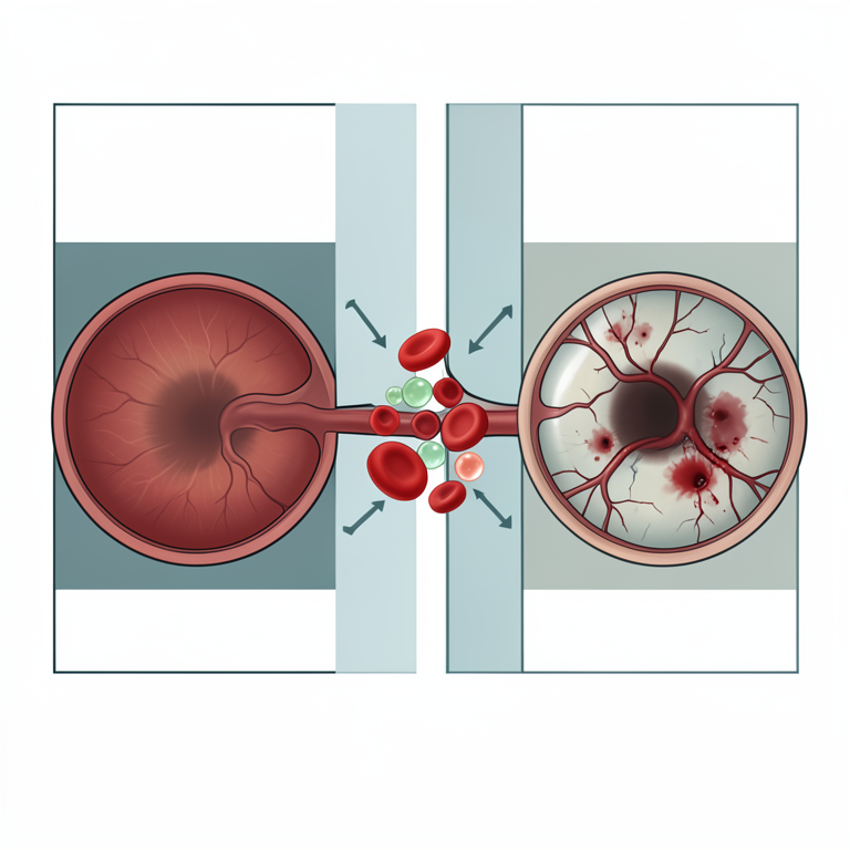
Risk Factors for Diabetic Retinopathy
AI-generated conceptual illustration (human retina and blood vessels). It explains the idea and does not show study data.
View source evidence ↗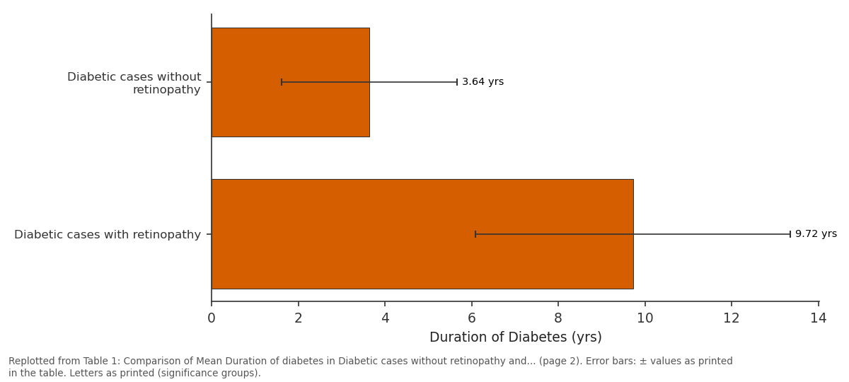
Duration of Diabetes: Diabetic cases without... vs Diabetic cases with...
Replotted from Table 1 (page 2) of the original paper. Every bar is the paper's own table value.
View source evidence ↗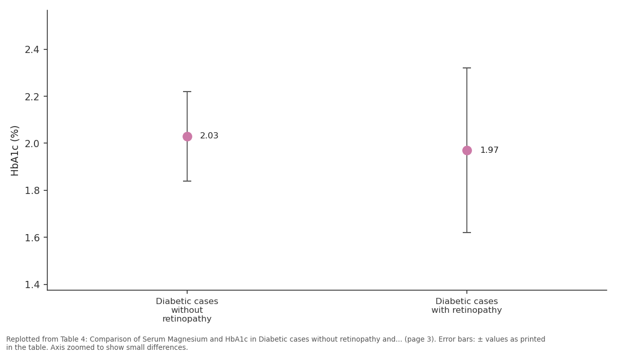
Magnesium (mg/dl): Diabetic cases without... vs Diabetic cases with...
Replotted from Table 4 (page 3) of the original paper. Every bar is the paper's own table value.
View source evidence ↗Figures & tables
From the original paper Table 1
 Table 2
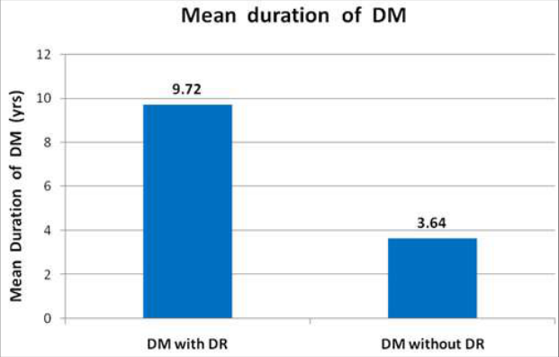 Figure 1
 Table 3
 Table 4
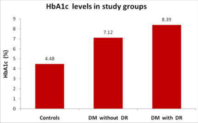 Figure 2
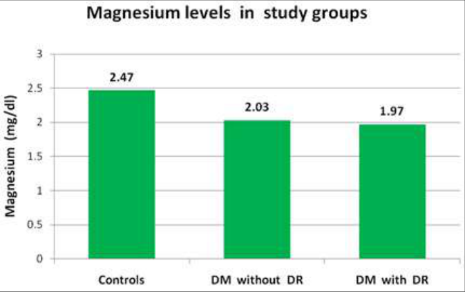 Figure 3
Introduction
Diabetes Mellitus is a complex metabolic infection brought about by hereditary and ecological elements having a variable relationship. It is perhaps the most well-known metabolic issue where either the chemical insulin is missing or the cells of the body are safe from the impacts of insulin [1,2] . It is associated with a high mortality rate due to several complications that appear to worsen over time and carry a significant risk of premature mortality. Irregular insulin discharge, raised blood glucose levels and various entanglements, for example, retinopathy, nephropathy, neuropathy and arteriosclerosis, are the key attributes [1, 3] . Probably the greatest danger to human wellbeing in the 21 st century is Diabetes Mellitus. By 2025, the WHO has anticipated that there will be an ascent to 300 million. The vital variables for the fastest ascent in the commonness of type 2 diabetes mellitus are expanded urbanization and industrialization [4,5] . Diabetic retinopathy (DR) is an intricacy of diabetes mellitus that undermines sight and is one of the main sources of grown-up procured visual deficiency. Compared to average people, the risk of losing sight is around 25 times higher. There is a range of risk factors linked to diabetic retinopathy development and progression, such as diabetes mellitus duration, poor glycemic control, dyslipidemia, hypertension and hypomagnesemia [1,2] . It has long been established that hypomagnesemia is linked with diabetes mellitus. It is said that magnesium depletion has a detrimental effect on the homeostasis of glucose and insulin sensitivity. This connection between diabetes mellitus and magnesium has a wide scope of consequences for the guideline of diabetes and its inconveniences [6,7,8] .
In developing microangiopathy, chronic hyperglycemia and its associated non-enzymatic glycation play an important role. Hyperglycemia leads to the production of advanced end products for glycation, resulting in numerous vascular complications, such as myocardial infarction, etc. [9] Intensive glycemic control, as measured by serum HbA1c levels, has been shown to reduce diabetic complications, especially microvascular diseases, in randomised trials. Several studies have been carried out to study the effect on the progression of retinopathy of these individual risk factors. In diabetic patients with retinopathy, however, very few studies have been conducted to research the association between any of these risk factors. During this investigation, an endeavour was made to discover a connection between the beginning and improvement of diabetic retinopathy between these danger factors.
Material and Methods
80 patients with type II diabetes, 40 patients with clinically analyzed retinopathy, and 40 patients without retinopathy were remembered for the examination. The subjects were chosen from the Ophthalmology Department of the outpatient and inpatient units. The clinical diagnosis was based on the results of history and fundoscopy. Patients ranged in age from 30-60 years. The cases were chosen on the basis of a simple method of random sampling. Chronic diarrhoea, alcoholism, breastfeeding, medications that induce hypomagnesemia (such as diuretics, cisplatin, pentamidine), urinary tract infection, inflammatory disorders such as rheumatoid arthritis, myocardial infarction, recent surgical history, and major trauma were included in the exclusion criteria. After obtaining due consent, the findings were compared with 40 normal healthy randomly selected persons. The controls were balanced by age and sex. Serum magnesium and HbA1c in blood were used in the inquiry. Similarly, the controls were compared.
For the HbA1c calculation, blood obtained in EDTAcontaining vacutainers was used. Serum magnesium was estimated in the autoanalyzer using the Xylidyl blue method using RANDOX KIT-MG 3880. HBA1c was estimated by the inhibition method of Latex agglutination using RANDOX KIT-HA 3830 in the HbA1c analyzer.
Results
The age group was between 30-60 years old. The mean age was 49.53 ± 5.82 years for diabetic patients with retinopathy, 46.77 ± 6.99 years for diabetic patients without retinopathy, and 45.50 ± 6.14 years for controls. Contrasted with diabetic patients without retinopathy, the length of diabetes mellitus in diabetic patients with retinopathy was altogether higher (P<0.001). The outcomes above demonstrate that as the length of diabetes mellitus builds, the recurrence and improvement of retinopathy can increment. There was a substantial increase in mean HbA1c values in non-retinopathic and retinopathic diabetic patients relative to the control group (p<0.001), as shown in Tables 2, 3 & 4.
Likewise, when contrasted with diabetic patients without retinopathy, there was a genuinely huge ascent in mean HbA1c levels in diabetic patients with retinopathy (p<0.001). As appeared in Tables 2, 3 and 4, the mean magnesium levels in diabetic patients without retinopathy and with retinopathy were measurably essentially lower contrasted with the benchmark group (p<0.001). Also, in diabetic patients with retinopathy, there was a measurably huge drop in mean magnesium levels when contrasted with diabetic patients without retinopathy (p<0.05). The above bar graph demonstrates that comparative with diabetic patients without retinopathy and the benchmark groups, the mean magnesium esteems in the serum of diabetic patients with retinopathy are fundamentally lower. Studies have additionally indicated that the danger of diabetic retinopathy increments by 1.89 occasions with each 5-year ascend in the length of diabetes mellitus [10] . Studies conducted by Farhan K H et al. have also shown that the duration of diabetes mellitus will increase the risk of diabetic retinopathy [11] . The mean HbA1c level in controls was 4.48 % and the mean HbA1c levels in diabetics without and with retinopathy were 7.12 % and 8.39%. There was an increase in HbA1c levels in diabetic groups when compared to controls which are statistically significant (p-value < 0.001). Within diabetic groups, HbA1c levels were more in diabetics with retinopathy when compared to diabetics without retinopathy, which is statistically significant (p-value < 0.001). Expanded degrees of HbA1c in diabetic patients with retinopathy and diabetic patients without retinopathy have been noticed [6] . Studies have also shown that every 1 % rise in HbA1c in patients with type 2 diabetes mellitus would result in a 37 % increase in microvascular complications [12] . A significant ascent in the degrees of HbA1c in diabetic patients with retinopathy has appeared by KG Santos et al. [13] The consequences of this study and previous studies show that hyperglycemia is an intense pointer of the advancement of diabetic retinopathy, as appeared by the ascent in HbA1c levels. The potential cause is that hyperglycemia leads to nearly all protein glycation, resulting in the development of advanced end products of glycation. In a few tissues, including blood vessel dividers, these high level glycation final results cause cross-linkage of collagen and other extracellular lattice proteins [14] . Vascular injury caused by hyperglycemia leads to increased glucose flow through the polyol pathway, leading to cellular damage, resulting in numerous micro-vascular and macro-vascular complications [15] . HbA1c is also shown to have a special affinity for oxygen, thus causing tissue anoxia and playing a role in causing micro and macroangiopathy [6] . As mediators of microvascular permeability, ischemia & angiogenesis, the interaction of advanced glycation end products and their receptors has been involved [15] . Mean serum magnesium levels were 2.47 mg/dl in the controls and mean serum magnesium levels were 2.03 mg/dl and 1.97 mg/dl in diabetics without and with retinopathy. Compared to the control group, there was a statistically significant decrease in magnesium levels in the diabetic group (p-value < 0.001). Within the diabetic group, magnesium levels were more in diabetics with retinopathy when compared to diabetics without retinopathy, which is statistically significant (p-value < 0.05).
Discussion
Our results agree with the study conducted by Hatwal et al., Ishrat Kareem et al., who showed that hypomagnesemia is a risk factor for retinopathy formation. The existence of hypomagnesemia in diabetic retinopathy has been shown [6,17] . The exact reason for hypomagnesemia in unexplained instances of diabetes mellitus. The contributory factors may be low food intake, impaired magnesium absorption, increased urinary loss due to hyperglycemia, and osmotic diuresis. For several enzymes that play an important role in the metabolism of glucose, magnesium is essential. It is said that magnesium depletion reduces the sensitivity of insulin, thus raising the risk of secondary complications [18] . The monitoring of plasma magnesium levels in diabetic patients with complications is therefore critical. In these patients, magnesium supplementation may help to reduce the progression of retinopathy [19,20] . The extent of hyperglycemia contributes to the development of advanced end products of glycation. The different microvascular problems result in these advanced glycation end products [15,22] . Also, generalized microangiopathy may be due to retinopathy. K.G Santos et al. have shown that diabetes mellitus length, glycosylated hemoglobin, and albumin excretion concentrations are independently correlated with diabetic retinopathy [13] . Studies have shown that the cumulative effects of different risk factors are likely to be due to increased capillary permeability, microangiopathy, and retinal ischemia [21] .
The beginning and advancement of retinopathy can be limited by early conclusions and ideal treatment in diabetic patients. Further studies of oral supplementation of magnesium in various stages of diabetic retinopathy may be important.
Figures and tables from the paper
| Groups | — | Duration of Diabetes |
|---|---|---|
| Diabetic cases without retinopathy | Mean ± S.D | 3.64 ± 2.03 yrs |
| Diabetic cases with retinopathy | Mean ± S.D | 9.72 ± 3.64 yrs |
| Diabetic cases without retinopathy vs Diabetic cases with retinopathy | p-value | <0.001 |
Table text extracted from the original PDF; superscript letters are shown inline. The original table image is below.
View original table image
| Groups | — | Magnesium (mg/dl) | HbA1c (%) |
|---|---|---|---|
| Controls | Mean ± S.D | 2.47 ± 0.24 | 4.48 ± 0.51 |
| Diabetic cases without retinopathy | Mean ± S.D | 2.03 ± 0.19 | 7.12 ± 0.94 |
| Controls vs. Diabetic cases without retinopathy | p-value | <0.001 | <0.001 |
Table text extracted from the original PDF; superscript letters are shown inline. The original table image is below.
View original table image
| Groups | — | Magnesium (mg/dl) | HbA1c (%) |
|---|---|---|---|
| Controls | Mean ± S.D | 2.47 ± 0.24 | 4.48 ± 0.51 |
| Diabetic cases with retinopathy | Mean ± S.D | 1.97 ± 0.35 | 8.39 ± 1.19 |
| Controls vs. Diabetic cases with retinopathy | p-value | <0.001 | <0.001 |
Table text extracted from the original PDF; superscript letters are shown inline. The original table image is below.
View original table image
| Groups | — | Magnesium (mg/dl) | HbA1c (%) |
|---|---|---|---|
| Diabetic cases without retinopathy | Mean ± S.D | 2.03 ± 0.19 | 7.12 ± 0.94 |
| Diabetic cases with retinopathy | Mean ± S.D | 1.97 ± 0.35 | 8.39 ± 1.19 |
| Diabetic cases without retinopathy vs. Diabetic cases with retinopathy | p-value | <0.05 | <0.001 |
Table text extracted from the original PDF; superscript letters are shown inline. The original table image is below.
View original table image
References
23 in the original paper- Da Silva Correa ZM, Freitas AM, Marcon MM. Risk factors related to the severity of diabetic retinopathy. Arq Bras Oftalmol. 2003;66(6):739-43.
- Manaviat MR, Afkhami M, Shoja MR. Retinopathy and Microalbuminuria in type II diabetic patients. BMC Ophthalmology. 2004;4(1):1-4.
- Chiarelli F, Mohn A, Tumini S, Trotta D, Verrotti A. Screening for vascular complications in children and adolescents with type 1 diabetes mellitus. Horm Res. 2002;57(1):113-116.
- Zimmet PZ. Kelly west lecture 1991 challenges in diabetes epidemiology from west to the rest. Diabetes Care. 1992;15(2):232-52.
- Oe Courten M, Bennet PH, Tuomilehto, Zimmet P. Epidemiology of NIDDM in Non-Europids. In: Alberti KMMM, Zimmet P, DeFronzo RA, Editors. International text book of diabetes mellitus, 2 nd ed. Chichester: John Wiley and Sons. 1997;1:143-70.
- Kareem I, Jaweed SA, Bardapurkar JS, Patil VP. Study of magnesium, glycosylated hemoglobin and lipid profile in Diabetic Retinopathy. Indian Journal of Clinical biochemistry. 2004;19(2):124-127.
- Waltt MK, Zimmermann MB, Spinas GA, Hurrell RF. Low plasma magnesium in type 2 diabetes. Swiss Med Wkly. 2003;133:289-292.
- Ramadass S, Basu S, Srinivasan AR. SERUM magnesium levels as an indicator of status of Diabetes Mellitus type 2. Diabetes Metab Syndr. 2015;9(1):4245.
- Sujatha P, et al. Serum sialic acid, lipid profile and fibrinogen as markers of ST -elevated ECG in myocardial infarction. Jour of Med Sc & Tech. 2012;1(3):29-35.
- Rema M, Pradeepa R. Diabetic Retinopahty: An Indian perspective. Indian J Med Res. 2007;125:297-310.
- Al-Shammari FKH, Al-Meraghi O, Nasif A, AlOtaibi S. The prevalence of diabetic retinopathy and associated risk factors in type 2 diabetes mellitus in AlNaeem area (Kuwait). Middle East Journal of Family Medicine. 2005;3(2):3.
- Tsui EYL, Oi LC, Young R, Young TT. Changing the treatment paradigm to achieve best practice goals for type 2 diabetes mellitus. HK Pract. 2005;27(9):339.
- Santos KG, Tschiedel B, Schneider JR, Souto KEP, Roisenberg I. Prevalence of retinopathy in Caucasian type 2 diabetic patients from the south of Brazil and relationship with clinical and metabolic factors. Braz J Med Biol Res. 2005;38(2):221-225.
- Haffner SM. Management of dyslipidemia in adults with diabetes. Diabetes Care. 1998;21(1):160-177.
- Kotoula Maria G, Koukoulis George, Zintzaras, Karabatsas Constantinos H, Chatzoulis Dimitrios Z. Metabolic control of diabetes is associated with an improved response of diabetic retinopathy to panretinal photocoagulation. Diabetes Care. 2005;28(10):2454-57.
- Hatwal A, Gujral AS, Bhatia RPS, Agarwal JK, Bajpai HS. Association of hypomagnesemia with diabetic retinopathy. Acta Ophthalmol. 1989;67(6):714-716.
- Praveena S, Sujatha P, et al. Study of microelements in Diabetic retinopathy: Journal of Evolution of Medical and Dental Sciences. 2013;2(6):531-8.
- Pham PCT, Pham PMT, Pham SV, Miller JM, Pham PTT. Hypomagnesemia in patients with type 2 diabetes. Clin J Am Soc Nephrol. 2007;2:366-373.
- Ridaura RL, Willett WC, Rimm ED, Liu S, Stampfer MJ, Manson JAE, et al. Magnesium intake and risk of type 2 diabetes in Men and women. Diabetes Care. 2004;27(1):134-140.
- De Vak HW, Hardus PLLJ, Van Rijn HJM, Erkelens DW. Plasma magnesium concentration and progression of retinopathy. Diabetes Care. 1999;22(5):864-865.
- Boelter MC, Gross JL, Canani LH, Costa LA, Lisboa HR, Tres GS et al. Proliferative diabetic retinopathy is associated with microalbuminuria in patients with type 2 diabetes. Braz J Med Biol Res. 2006;39(8):10331039.
- Krowlewski AS, Laffel LMB, Krowlewski M, Quinn M, Warram JH. Glycosylated Hemoglobin and the risk of Microalbuminuria in patients with Insulin dependent diabetes mellitus. N Engl J med. 1995;332(19):1251-5.
- Van Leiden HA, Dekker JM, Moll AC, Nijpels G, Heine RJ, Bouter LM, et al. Blood pressure, lipids and obesity are associated with retinopathy. Diabetes Care. 2002;25(8):1320-1325.
Cited by 5
From OpenAlex, OpenCitations and reference lists read by Scinovex
- Finger millet (Ragi) as an essential dietary supplement with key health benefits: A review ↗
Padmaja Jagati, Indurekha Mahapatra, Debasmita Dash · International Journal of Home Science · 2021
The world geography has so many millet crop producer and India serves as one of the key producers of the millets. Finger millets, a type of small-sized millets, requires larger area of cultivation for growing the crop. Finger millet is considered as a special food supplement because of its high nutritional contents in comparison to other important cereals, namely barley,…
❝ 32PDF - Curcumin loaded deformable drug carrier for the disease of posterior segment of eye: Diabetic retinopathy ↗
Priti Singh, Vandana Gupta · The Pharma Innovation · 2021
Diabetic Retinopathy (DR) is one of the most common complications of diabetes mellitus that affects theblood vessels of the retina, leading to blindness. The current approach of treatment based on anti-inflammatory, anti-angiogenesis drugs and laser photocoagulation are effective but also shows adverse affect in retinal tissues and that can even worsen the visual abilities. Thus, a safe and effective mode…
❝ 5PDF - Comparative evaluation of the treatment of induced ulcer and diabetes with yoghurt and drugs in rats
Momoh AO, Bada AA, Olayiwola AL · International Journal of Horticulture and Food Science · 2022
Stomach ulcer is a break in the lining of the stomach, mostly the first part of the small intestine and occasionally the lower oesophagus. With a gastric ulcer the pain may worsen with eating. Diabetes on the other hand is a group of metabolic disorders in which there are high blood sugar levels over a prolonged period. The effects of yogurt on ulcer induced and diabetes induced in rats was evaluated. The induction of ulcer was done using acetic acid, while diabetes was done using Streptozotocin following standard procedures. The rats were fed with the yogurt (Rev up unsweetened yoghurt) for t
- Antiplatelet and antithrombotic therapy in diabetic patients recent updates ↗
Mohammed Ashfaq, Jaffer Syed, Abdul Mushtaq Mohammed, Shaista Sumayya, S P Srinivas Nayak · International journal of applied research · 2021
Diabetes mellitus (DM) is a metabolic disorder associated with accelerated atherogenesis and an increased risk of atherothrombotic complications. Multiple mechanisms contribute to the pro-thrombotic status which characterizes DM patients underscoring the importance of antiplatelet therapies used for secondary prevention in these patients. For many years, dual antiplatelet therapy (DAPT) with aspirin and the P2Y12 inhibitor clopidogrel has represented the mainstay
❝ 2Found by Scinovex - The effects of mangrove plant leaves (Acanthus ilicifolius) collected in can Gio district on white mice (Mus musculus var. albino) with diabetes ↗
Long Le, Ngoc-Anh Nguyen, Ly-Thai-Uyen Thi, Thi-Tan-Luong Nguyen · Journal of Entomology and Zoology Studies · 2022
This study was conducted to evaluate the hypoglycemic potential of mangrove plant leaves (Acanthus ilicifolius) fluid in white mice with diabetes mellitus with doses of mangrove leaves fluid of 100 mg/kg, 200 mg/kg, 300 mg/kg, 400 mg/kg corresponding to EX1, EX2, EX3, EX4, CT2. Diabetic mice had study results after 7 days. In EX1, EX2 and EX3 there was a…
❝ 0PDF