
Research article · 2022 · Full text
International Journal of Horticulture and Food ScienceComparative evaluation of the treatment of induced ulcer and diabetes with yoghurt and drugs in rats
Volume 4(2) · Pages 55–60 · DOI: 10.33545/26631067.2022.v4.i2a.107 ↗
Research areasHelicobacter pylori-related gastroenterology studiesDiet and metabolism studiesDiet, Metabolism, and Disease
Abstract
Author-writtenStomach ulcer is a break in the lining of the stomach, mostly the first part of the small intestine and occasionally the lower oesophagus. With a gastric ulcer the pain may worsen with eating. Diabetes on the other hand is a group of metabolic disorders in which there are high blood sugar levels over a prolonged period. The effects of yogurt on ulcer induced and diabetes induced in rats was evaluated. The induction of ulcer was done using acetic acid, while diabetes was done using Streptozotocin following standard procedures. The rats were fed with the yogurt (Rev up unsweetened yoghurt) for three (3) weeks, taking the samples of the stomach and pancreas weekly for histopathological examination. The results showed that the 99% acetic acid induced severe gastric ulcer after 72 hrs while Streptozotocin induced type 1 diabetes (235 mg/dl from 90 mg/dl) after 96 hrs. The administration of the yogurt caused cell infiltrations of the stomach wall, distinct ductile without aberrant superior or inferior diverticular of the stomach interlobular lining and well-formed stomach blood vessels. The yoghurt on the other hand caused little positive effect on the pancreas globules after three weeks of administration. The results suggests that consuming unsweetened yoghurts (containing probiotics) may improve glucose metabolism to a degree while it has more gastro-protective effect on ulcer. Therefore, while yoghurt may be recommended for ulcer treatment, it may not be for diabetic patients.
Summary
Source-checkedPlain-language reading of the full paper, prepared with AI and checked against the text. Each point links to the authors' own words.
Why it matters
- Stomach ulcers are characterized by breaks in the lining of the stomach, the lower esophagus, or the first part of the small intestine. View evidence ↗
- Diabetes is a group of metabolic disorders defined by prolonged high blood sugar levels. View evidence ↗
- Because standard medications for these conditions can be expensive and cause side effects, there is a need to evaluate if yogurt could serve as a cost-effective alternative. View evidence ↗
The question
- The study aimed to evaluate the effects of yogurt on rats with induced ulcers and induced diabetes. View evidence ↗
What they did
- Ulcers were induced using acetic acid, and diabetes was induced using Streptozotocin. View evidence ↗
- Rats were treated with Rev up unsweetened yogurt for three weeks, with weekly histopathological sampling of the stomach and pancreas. View evidence ↗
- Daily oral treatments consisted of either yogurt or reference drugs: Melanov for diabetes and Polygel for ulcers. View evidence ↗
What they found
- Induction was successful, with 99% acetic acid causing severe gastric ulcers after 72 hours and Streptozotocin raising blood sugar from 90 mg/dl to 235 mg/dl after 96 hours. View evidence ↗
- Yogurt had a minimal effect on blood sugar, reducing it from 220±6 mg/dl to 212±9 mg/dl over three weeks, whereas the drug significantly reduced it from 205±5 mg/dl to 142±6 mg/dl. View evidence ↗
- By the third week, yogurt treatment showed obvious cell regeneration and healing in the stomach, likely due to probiotics acting as a protective layer. View evidence ↗
- Yogurt treatment for diabetes did not prevent pathological damage to the pancreas, which exhibited poorly formed cells and diffused globular masses. View evidence ↗
What it means
- The authors recommend yogurt containing L. acidophilus and L. plantarum for treating ulcers, but not for treating diabetes due to its limited effectiveness. View evidence ↗
- The authors state that further research is needed to fully evaluate yogurt as a drug alternative, specifically regarding the potential absence of adverse effects. View evidence ↗
Visual summary
2 visualsIllustrations explain the paper; charts are replotted from the paper's own table values.
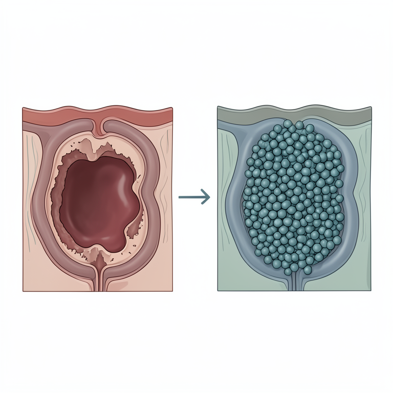
Probiotic Protective Mechanism
AI-generated conceptual illustration (Wistar rats stomach lining). It explains the idea and does not show study data.
View source evidence ↗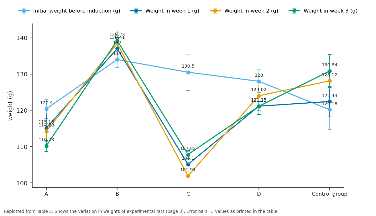
Initial weight before induction (g) and Weight in week 1 (g) and more: A vs B vs C vs D vs Control group
Replotted from Table 2 (page 3) of the original paper. Every bar is the paper's own table value.
View source evidence ↗Figures & tables
From the original paper Table 1
 Table 2
 Table 3
 Figure 1
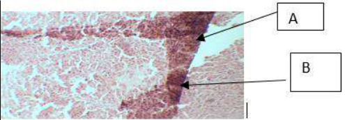 Figure 2
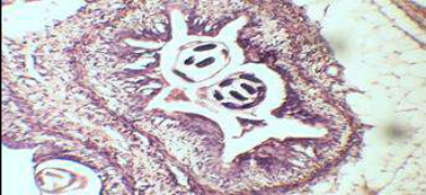 Figure 3
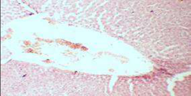 Figure 4
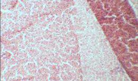 Figure 5
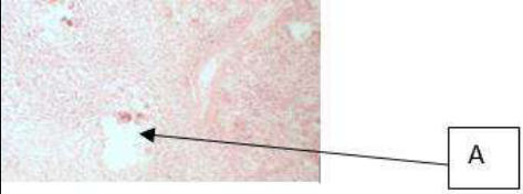 Figure 6
 Figure 7
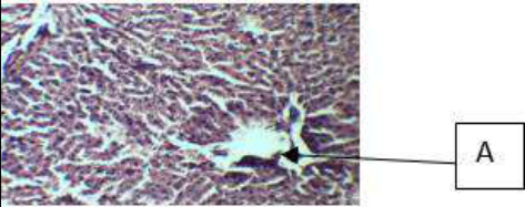 Figure 8
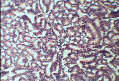 Figure 9
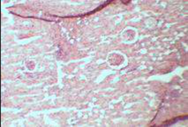 Fig10
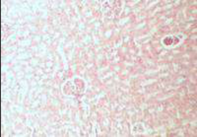 Figure 11
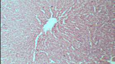 Figure 12
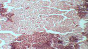 Figure 13
Introducti on
Yogurt is made by the fermentation of lactose (milk sugar) by bacteria l enzymes. This process is anaerobic, meaning that it occurs in the absence of oxygen. Lactose is a compound sugar, made up of the two simple sugars; glucose and galactose. During the making of yogurt, the lactose is broken down by the lactase enzyme (prod uced by bacteria) into these two components (Park 2017) [31] . The beneficial microorganism in yogurt include; Lactobacillus: is a genus of Gram-positive, facultative anaerobic or microaerophilic, rodshaped, non-spore-forming bacteria (Makarova et al., 2006) [26] . Lactobacillus species are normally a major part of the vaginal Microbiota. They form biofilms in the vaginal and gut Microbiota, allowing them to persist during harsh environmental conditions and maintain ample populations (Salas et al., 2016) [17] . Lactobacillus exhibits a mutualistic relationship with the human body as it protects the host against potential invasions by pathogens, and in turn, the host provides a source of nutrients (Martin et al., 2013) [11] . Some species commonly used in yogurt production includes; L. bulgaricus, L. acidophilus, L. casei, L. animalis. Streptococcus thermophiles: also known as Streptococcus salivarius subsp (Tannock et al., 2005) [22] , is a fimbriated, non-motile, gram-positive bacterium, and a fermentative facultative anaerobe of the viridians group that does not produce endospores (Courtin et al., 2003) [3] . Some of the benefits of yogurts include: (1) According to Keyzet, Yogurt can help promote hair growth by getting rid of dandruff, balancing the pH of the sc alp and preventing frizz depending on the added ingredients (Olive oil or Coconut oil). ( 2) It is also used on the face to clean and tighten pores. After all, this treat includes lactic acid -an alpha hydroxyl acid (AHA), which is milder than other exfoliate but still rejuvenates your skin. (3) Yogurt with other ingredient (like crushed Walnuts) can be useful in treating the feet; by relieving dry patches under the feet, giving a soft and moisturized feel. (4) It can also be used as a teeth whitening agent because of it calcium and phosphorus content. Diabetes is a group of metabolic disorders in which there is high blood sugar levels over a prolonged period (WHO, 2014) [25] . Symptoms of high blood sugar include frequent urination, increased thirst, and increased hunger. If left untreated, diabetes can cause many complications. Acute complications can include diabetic ketoacidosis , hyperosmolar hyperglycaemic state, or death (Kitabchi et al., 2009) [8] . Serious long-term complications include cardiovascular disease, stroke, chronic kidney disease, foot ulcers, and damage to the eyes (WHO, 2013) [24] . Diabetes is due to either the pancreas not producing enough insulin or the cells of the body not responding properly to the insulin produced (Shoback et al., 2011) [19] . Stomach ulcer is a break in the lining of the stomach, first part of the small intestine or occasionally the lower oesophagus. An ulcer in the stomach is known as a gastric ulcer while that in the first part of the intestines is known as a duodenal ulcer. In 2014, a total of 422 million adults were reported worldwide to have diabetes, the complications of diabetes can lead to heart attack, stroke, blindness, kidney failure and lower limb amputation. Managing these diseases can be very expensive and the use of medications can lead to uncomfortable side effects. This research will give more exposure and help decide if yogurt can be a cost-effective alternative to treat these diseases rather than expensive medications that have prominent side effects.
Materials and Methods
Materials and Methods
Environment of Study
The study was conducted in Elizade university laboratory, Ilara-Mokin, Ondo state, Nigeria.
Materials
Materials used for this research include Cages, measuring cylinders, conical flask, petri dishes, chloroform, foil paper, cotton wool, paper tape, distilled water, cover slips, slides, specimen bottles, dissecting tools, gloves.
Animals and Feeding
Albino rats were obtained from an animal house in Akure, Nigeria. They were maintained in a customised cage with a mesh top to allow for ventilation at room temperature, the cages were cleaned regularly (twice daily) to prevent infections. Rats' diet consisted of cereals and cereal byproduct (rich in starch and proteins) and definitely water ad libitum.
Experimental design
A total of twenty rats were used for the assay. They were divided into five (5) groups consisting of four (4) rats per group. The groups and their designations are as follows: Group 1-Diabetes induced rats treated with yogurt; Group 2-Diabetes induced rats treated with drug; Group 3-Ulcer induced rats treated with yogurt; Group 4-Ulcer induced rats treated with drug; Group 5-control group.
Confirmatory tests
The lactic acid bacteria present in the yogurt used was isolated and confirmed using MRS agar, microscopy and biochemical tests according to Cheesbrough (2014).
Gram staining test
A drop of distilled water was placed on a clean, grease free slide and a loop full of the organism was added onto the slide and done according Cheesbrough (2014).
Induction of Diabetes
Experimental diabetes mellitus was induced by a single intra-venous injection of Streptozotocin (STZ) dissolved in 0.1M citrate buffer in the adult rats under anaesthetics. After which diabetes was induced in four days.
Induction of Ulcer
The rats were denied food for 24 hrs prior to ulcer induction but had access to water. After which ulcer was induced by oral application of 0.2 ml of pure acetic acid. The animals were returned to their normal diet.
Administration of Treatment
Treatments including yogurt and about 0.5ml of reference drugs for diabetes (Melanov) and ulcer (Polygel) were orally administered to the rats by gavage daily.
Biochemical studies
To monitor the glucose level in diabetic rats, the rats were placed under anaesthetics and about 0.5 ml of blood was collected into an Ethylene Diamine Tetra-acetic Acid (EDTA) bottle from the tail vein and with the use of glucometer the sugar level was determined.
Histopathological test
Each week one rat from each group was sacrificed, from the diabetic group the pancreas was harvested while the stomach for the ulcerated group was harvested and the organs were maintained in specimen bottles containing 20% formalin. The samples were first of all fixed in fixative liquid, trimmed, embedded in paraffin wax, sectioned and stained. Staining was done with the use of Haematoxylin and Eosin and excess stain was removed by rinsing.
Results
Results
Observation of general condition of the experimental rats
Various observation were made on the activities of both groups (Diabetic and ulcerated group). After induction of ulcer; writhing, drastic decrease in body weight, poor intake of food, and fatigue was observed in the rats, while in the diabetic group increased production of urine as compared to the control group was noticed. Table 1 shows that streptozotocin induced diabetes few days after induction. The initial blood sugar after before treatment was as high as 235 mg/dL. An elevation in the blood sugar of the rats can be observed in the first two groups, while the control group maintained blood sugar in the normal range. The group treated with yogurt compared to the group treated with drug didn't show significant reduction in the blood sugar level but there was a little significant variation in the sugar level after treatment compared to the sugar level after diabetes induction. Table 2 shows the elevation and reduction in weight of various experimental group. In the first group the weight is seen to have decreased gradually through the week, the second group shows an increase in the weight, in the third group it was observed that the body weight decreased drastically during the first week of induction and gradually increases through the week, the fourth week showed inconsistency in weight, whereby the weight increased and decreased regularly and finally at the third week there was a drastic reduction in weight is observed compared to the initial weight. The control group however shows increased weight gain from the first week through the third week.
| Groups | Initial weight before induction (g) | Weight in week 1 (g) | Weight in week 2 (g) | Weight in week 3 (g) |
|---|---|---|---|---|
| A | 120.40±2.63 | 115.15±3.81 | 114.28±2.17 | 110.23±1.56 |
| B | 134±2.18 | 137±3.03 | 138.41±2.94 | 139.23±2.60 |
| C | 130.50±5.02 | 105.10±3.48 | 101.91±1.12 | 107.82±1.05 |
| D | 128±3.15 | 121.15±1.30 | 124.02±2.65 | 121.11±2.29 |
| Control group | 120.18±5.42 | 122.43±4.05 | 128.12±2.20 | 130.84±4.54 |
Table text extracted from the original PDF; superscript letters are shown inline. The original table image is below.
View original table image
| Groups | Week one | Week two | Week 3 |
|---|---|---|---|
| Diabetes induced treated with yogurt | 220±6 mg/dl | 216±6 mg/dl | 212±9 mg/dl |
| Diabetes induced treated with drug | 205±5 mg/dl | 180±4 mg/dl | 142±6 mg/dl |
| Control | 90±4 mg/dl | 92±5 mg/dl | 95±6 mg/dl |
Table text extracted from the original PDF; superscript letters are shown inline. The original table image is below.
View original table image
Table 3 shows the results obtained from occult blood test. Two days after induction occult blood test yielded positive result for both the first and second group because of haemorrhage that occurred internally. In week one, also both groups tested positive for occult blood test, in the second week group one showed signs of healing as haemorrhage was not detected and in the third week both groups were negative for occult blood test. The results of the histopathological analysis on the concerned organs of the induced experimental rats showed various pathological features. These features include presence of cell infiltrations, inflammatory cell infiltrations, washing away of stomach sphincter muscles, formation of ulceric vacuoles, traces of dot haemorrhage along the stomach lining spread across the anterior and posterior wall of stomach sphincter, distinct ductile without necrosis on healing granular semi-saccularis, pancreatic tissue with cell infiltrations and washing away of posterior vacuole and diffused globular masses in the anterior part of the tissue.
| Groups | 2 days after induction | Week 1 | Week 2 | Week 3 |
|---|---|---|---|---|
| Induced ulcer treated with yogurt | ₊ | ₊ | - | - |
| Induced ulcer treated with drug | ₊ | ₊ | ₊ | - |
| Control | - | - | - | - |
Table text extracted from the original PDF; superscript letters are shown inline. The original table image is below.
View original table image
Discussion
Results obtained from the confirmatory test carried out on the isolates from yogurt showed that it contained live microorganism (bacteria). These bacteria were probiotics of the Lactobacillus species, and it conformed to their identity with the laid down procedure by Hassan et al., (2014) [27] . According to Makarova et al., (2003), Lactobacillus species are Gram positive rods incapable of producing spores and are catalase positive, as observed in this research.
In table 1, the blood glucose level was elevated to 235 mg/dl compared to the normal 90 mg/dl of the control group after the induction of diabetes. Sugar level of the diabetic group treated with yogurt reduced a little from 220±6 mg/dl to 212±9 mg/dl from the time of induction to the third week of treatment. This may be due to the fact that probiotics appear to slow down digestion of carbohydrates in foods thereby preventing blood sugar spikes, according to Health day, (2016).
In the drug treated group, sugar level reduced from 205±5 mg/dl to 142±6 mg/dl. This shows that the drug had more effect on the reduction of blood sugar than the yogurt. This result is similar to the result obtained by Hassan et al., (2012) [28] in which a particular drug used to treat diabetes caused a reduction in the sugar level of experimental rats. This may be due to the fact that the drugs are well purified and directly targeted at the pancreas. The mechanism by which streptozotocin (STZ) brings about its diabetic state in animals includes selective destruction of pancreatic beta cells thereby causing the cells to be less active, leading to poor sensitivity of insulin for glucose uptake by tissues according to Elisa et al., (2009). The pancreatic sections stained with Haematoxylin and Eosin in this research showed that streptozotocin caused severe destruction of the pancreas. For example, the group induced with diabetes and treated with yogurt showed pathological damages such as poorly formed pancreatic cells with infiltrations and diffused globular masses in the anterior part of the tissue. According to Omoya et al., (2017) [29] , pancreatic cells with infiltrations and diffused globular masses in any animal will definitely make it difficult for such animal to utilize any form of sugar for metabolic activities. Although, there was no necrosis nor haemorrhage in any of the pancreas slide. Alfarag, (2009) stated that the washing away of vacuoles in pancreatic sections shows that the significant content of the pancreas is gone and such pancreas will not be optimal in its metabolic role. This probably explained the reason why the yogurt may not be effective in the treatment of diabetes as seen in this research.
It was observed that the weight of the rats in the first group decreased from 120.40±2.63 g to 110.23 g±1.56 g at the third week (table 2). The second group however showed an increase in body weight of 134±2.18 g to 139.23±2.60 g from week one through week three. In the third group, a drastic decrease in weight was observed i.e. from 130.50±5.02 g to 107.82±1.05 g which may be as a result of loss in appetite and poor food intake. In the fourth group, an initial weight of 128±3.15 g which reduced to 121±2.29 g was observed, the decrease in weight in the third week compared to that of the second week could be as a result of their response to the drug, while the increase in weight in other group is as a result of improved appetite.
Occult blood test was positive in the two groups induced with ulcer few days after induction and during week one, as the week progresses it was observed that occult blood test was negative in all groups. This may be because of the healing effect exerted by the probiotics in the yogurt on the ulcerated areas in the gastrointestinal tract. There is no report or studies on the use of occult blood test in evaluating the healing of ulcers in rats. However, the fact that it was negative by the third week may be an indication that healing has taken place. The results obtained from this research therefore supports the use of yogurt as an alternative for treating ulcer rather than drugs. For instance in the first week of treatment using yogurt infiltration of cells in the stomach wall were observed which is an indication of fast reproduction of the stomach cells or sphincter to replace the ulcerated part.
There was also traces of dot haemorrhage along the stomach lining spread across the wall. However there were areas with distinct stomach wall muscles showing signs of ulcer healing. But in the third week of yogurt treatment there was obvious regeneration of cells which showed healing, this is because the probiotics (Lactobacilli) may have acted as a protective layer in the area of lesions, thereby preventing further inflammation of cells that may result from acid damage (Poonam et al., 2013) [16] . Diabetes induced by Streptozotocin (STZ) injection provided symptoms like excessive urine production and weight loss, while Acetic acid-induced ulcer produced symptoms like; loss of appetite, fatigue and death. According to the data, the increased levels of glucose sugar in STZ-induced diabetic rats were slightly lowered by administration of yogurt, weight gain was observed in the ulcerated group but the most active groups were the control group and the ones treated with yogurt. This research has shown that probiotics can exhibit some kind of protective mechanism on the stomach lining which can be explored further in treatment of some ailments.
The results gathered from this research suggests that consuming unsweetened yogurts (containing probiotics) may improve glucose metabolism to a degree, but is more effective in treating gastrointestinal complications. Probiotics should therefore be introduced into most convenience foods or produced in large scale in form of capsules especially for ulcer patients. I therefore recommend yogurt (Containing probiotics of the species L. acidophilus and L. plantarum) for ulcer treatment, but not for diabetic patients because of the little positive effect observed in this study. Also, further research should be carried out on the use of yogurt as an alternative to drugs because there may be no adverse effects on its use unlike drugs.
Figures and tables from the paper
References
28 in the original paper- Berkeley B, Kizar K, Radha S. Gymnema Sylvestre for Diabetes. Journal of Ethnopharmacology. 2016;30:295-300.
- Chieh-Hsien Lai, Sih-Rong Wu, Jen-Chieh Pang, Latha Ramireddy, Yu-Cheng Chiang, Chien-Ku Lin. et al Designing primers and evaluation of the efficiency of propidium monoazide-Quantitative polymerase chain reaction for counting the viable cells of Lactobacillus gasseri and Lactobacillus salivarius. Journal of Food and Drug Analysis. 2017;25(3):533.
- Courtin P, Rul FO. Interactions between microorganisms in a simple ecosystem: yogurt bacteria as a study model. Le Lait. 2003;84:125-134.
- Mohamed DA, Hassanein MM, El-Messery TM, Fouad MT, El-Said MM, Fouda KA, et al. Amelioration of diabetes in a rat model through yoghurt supplemented with probiotics and olive pomace extract. J Biol Sci. 2017;17(7):320-3.
- Fink G. Stress controversies: post-traumatic stress disorder, hippocampal volume, gastroduodenal ulceration. Journal of neuroendocrinology. 2011 Feb;23(2):107-17.
- Graham ML, Janecek JL, Kittredge JA, Hering BJ, Schuurman HJ. The streptozotocin-induced diabetic nude mouse model: differences between animals from different source. Comp Med. 2011;61(4):356-60.
- Bukhari HM, Abdelghany AH, Nada IS, Header EA. Effect of yoghurt pillared with propolis on hyperglycemic rats. The Egyptian Journal of Hospital Medicine. 2012 Oct 1;49(1):691-704.
- Kitabchi AE, Umpierrez GE, Miles JM, Fisher JN. Hyperglycaemic crises in adult patients with diabetes . Diabetes Care. 2009 Jul;32(7):1335-43. Doi: 10.2337/dc099032. PMC accessible. PMID 19564476
- Buendia JR, Li Y, Hu FB, Cabral HJ, Bradlee ML, Quatromoni PA. Regular yogurt intake and risk of cardiovascular disease among hypertensive adults. American Journal of Hypertension. 2018 Apr;13;31(5):55765. https://doi.org/10.1093/ajh/hpx220.
- Makarova K, Slesarev A, Wolf Y, Sorokin A, Mirkin B, Koonin E, et al. Comparative genomics of the lactic acid bacteria. Proc Natl Acad Sci USA. 2006
- Martin R, Miquel S, Ulmer J, Kechaou N, Langella P, Bermúdez-Humarán LG. Role of commensal and probiotic bacteria in human health: a focus on inflammatory bowel diseas. NCBI. 2013 23 Jul;12(71). Doi: 10.1186/14752859-12-71. PMC 3726476 Freely accessible. PMID 23876056.
- Moroti C, Souza Magri LF, de Rezende Costa M, Cavallini DC, Sivieri K. Effect of the consumption of a new symbiotic shake on glycemia and cholesterol levels in elderly people with type 2 diabetes mellitus. Lipids in health and disease. 2012 Dec;11(1):1-8.
- Mota KS, Dias GE, Pinto ME, Luiz-Ferreira A, Souza-Brito AR, Hiruma-Lima CA, et al. Flavonoids with gastro protective activity. J Basic Med Sci. 2013;16(6):731-742.
- Omoya FO, Momoh AO, Fadare OS. Haematological and Pathological Effects of Bacteria from Vegetable Wastes in Ilara-Mokin, Ondo State. Microbiological Research Journal International (British Microbiology Research Journal). 2018;25(5):1-11.
- Penugonda Anveetha, Vamsi Krishna Chittimoju. Study of HbA1c and serum magnesium in diabetic patients with retinopathy. Int. J Adv. Biochem. Res. 2021;5(1):01-05. DOI: 10.33545/26174693.2021.v5.i1a.55
- Dharmani P, De Simone C, Chadee K. The probiotic mixture VSL# 3 accelerates gastric ulcer healing by stimulating vascular endothelial growth factor. PLoS One. 2013 Mar 6;8(3):e58671. doi.org.10.1371.journal.pone.0058671.
- Salas-Jara MJ, Ilabaca A, Vega M, García A. Biofilm Forming Lactobacillus: New Challenges for the Development of Probiotics. NCBI. 2016 Sept 20 4(3):35. Doi: 10.3390/microorganisms4030035. PMC 5039595 Freely acces s ible. PMID 27681929.
- Salih BA, Abasiyanik MF, Bayyurt N, Sander E. H pylori infection and other risk factors associated with peptic ulcers in Turkish patients: A retrospective study. World Journal of Gastroenterology: WJG. 2007 Jun 21;13(23):3245. PMID 17589905.
- Evivie SE, Huo GC, Igene JO, Bian X. Some current applications, limitations and future perspectives of lactic acid bacteria as probiotics. Food & nutrition research. 2017 Jan 1;61(1):1318034.
- Tannock Gerald W. Probiotics and Prebiotics: Scientific Aspects. Caister Academic Press ; c2005. p. 43. ISBN 1904455-01-8. Retrieved 2014-03-31.
- Uchida M, Shimizu K, Kurakazu K. Yogurt containing Lactobacillus gasseri OLL 2716 (LG21 yogurt) accelerated the healing of acetic acid-induced gastric ulcer in rats. Biosci Biotechnol Biochem. 2010;74:1891-1894.
- Divya Sharma, Dr. Rati Mathur, Dr. Puneet Saxena. Study of significance of serum cystatin-C and vascular endothelial growth factor as an early marker of diabetic nephropathy in type 2 diabetes patients. Int. J Adv. Biochem. Res. 2021;5(1):09-11. DOI: 10.33545/26174693.2021.v5.i1a.57
- WHO. Diabetes Fact sheet N°312. WHO. October 2013. Archived from the original on 26 August 2013. Retrieved 25 March 2014; c2014.
- Makarova K, Slesarev A, Wolf Y, Sorokin A, Mirkin B, Koonin E, et al. Comparative genomics of the lactic acid bacteria. Proceedings of the National Academy of Sciences. 2006 Oct 17;103(42):15611-6.
- Hassan MM, Amin KB, Ahaduzzaman M, Alam M, Faruk MS, Uddin I. Antimicrobial resistance pattern against E. coli and Salmonella in layer poultry. Res. J Vet. Pract. 2014 Jan 1;2(2):30-5.
- Hassan FM, Hadi RA, Kassim TI, Al-Hassany JS. Systematic study of epiphytic algal after restoration of AlHawizah marshes, southern of Iraq. Int. J of Aquatic Science. 2012 Jan 1;3(1):37-57.
- Omoya FO. Co-infection of malaria and typhoid fever among pregnant women attending primary health care centre, Ojo Local Government, Lagos, Nigeria. Health Science Journal. 2017;11(2):0.
- WHO. Diabetes Fact sheet N°312. WHO. October 2013. Archived from the original on 26 Augus t 2013. Retrieved 25 March 2014; c2013.
Cited by 2
From OpenAlex, OpenCitations and reference lists read by Scinovex
- Anti-inflammatory and anti-oxidative effect of pasterurised yoghurt in indomethacin induced gastric ulceration in male Wistar rats ↗
Olushola Ajayi, Blessing Omolaso, Tosan Peter Omayone, Adeoti Gbemisola Adeniran, A.O. Adu, O. J. Olorunfemi · Heliyon · 2024
Background: Yoghurt, a fermented dairy product consumed by diverse cultures for centuries, has garnered significant attention from the scientific community due to its potential health benefits and remarkable versatility. This study investigated the anti-inflammatory and anti-oxidative effects of pre-treatment with pasteurized yoghurt in indomethacin induced gastric ulceration. Method: Thirty male Wistar rats were randomly assigned into five groups. Groups 1…
❝ 6PDF - Anti-Inflammatory and Anti-Oxidative Effect of Pasterurised Yoghurt in Indomethacin Induced Gastric Ulceration in Male Wistar Rats ↗
Oluwadamilola Victor Ajayi, Blessing Omolaso, Tosan Peter Omayone, Adegoke Gbemisola Adeniran, Adu Oluwanifemi Adeyemisi, Olorunfemi Esther Oluwafeyikemi · SSRN Electronic Journal · 2024
❝ 0PDF