
Research article · 2019 · Full text
International Journal of Agriculture and Food ScienceIsolation and characterization of microorganisms associated with soft cheese produced from sheep milk using different coagulants
Volume 1(4) · Pages 33–38 · DOI: 10.33545/2664844x.2019.v1.i4a.26 ↗
Research areasProbiotics and Fermented FoodsEnzyme Production and CharacterizationMilk Quality and Mastitis in Dairy Cows
Abstract
Author-writtenMilk is an ideal medium for the growth of microorganisms because it is highly nutritious. Cow milk is the major milk used for the production of cheese although milk can also be gotten from other animals such as sheep, goat, camels and buffalo. Soft cheese was produced from sheep milk and the microorganisms associated with the soft cheese were isolated and identified using colonial, morphological and biochemical characterization. Different coagulants such as Calotropis procera, Carica papaya, lemon juice and steep water from maize, millet and sorghum were used for the production. The result revealed that six different bacteria were isolated and identified and they include Bacillus cereus, Bacillus subtilis, Lactococcus lactis, Lactobacillus casei, Pseudomonas aeruginosa, Staphylococcus aureus and 8 different fungi species isolated and identified include Aspergillus niger, Aspergillus fumigatus, Rhizopus stolonifer, Penicillium chrysogenium. Mucor hiemalis, Candida albicans, Fusarium verticillioides and Saccharomyces sp. The presence of these microorganisms indicated that the soft cheese produced was not sterile and highly contaminated and it can pose serious health risk to consumers. It is therefore recommended that soft cheese should be produced under hygienic condition since it’s a ready to eat food.
Summary
Source-checkedPlain-language reading of the full paper, prepared with AI and checked against the text. Each point links to the authors' own words.
Why it matters
- Sheep milk is considered a high-quality raw material for the dairy industry, particularly for making cheese. View evidence ↗
- Consuming raw milk and its derivatives can be dangerous, as it is linked to various infections such as salmonellosis and Staphylococcal poisoning. View evidence ↗
- Microbial contamination in soft cheese often stems from environmental factors or the use of basic production equipment. View evidence ↗
The question
- The study aimed to produce soft cheese from sheep milk and identify the associated microorganisms through colonial, morphological, and biochemical analysis. View evidence ↗
What they did
- The researchers used several coagulants for cheese production: lemon juice, Calotropis procera, Carica papaya, and steep water derived from sorghum, maize, and millet. View evidence ↗
- Production involved adding 4ml of the chosen coagulant to warm milk, which was then heated to 45-50°C with stirring until coagulation occurred. View evidence ↗
- Microorganisms were isolated using a modified pour plate method, starting with a 1g cheese sample diluted in 10mL of sterile distilled water. View evidence ↗
- Bacterial plates were incubated at 37°C for 18-24 hours, while fungal plates (PDA) were incubated at 28±2°C for 72-120 hours. View evidence ↗
What they found
- The analysis identified six different bacterial species and eight different fungal species in the soft cheese samples. View evidence ↗
- Among the bacteria, Lactobacillus casei was the most frequent (28.3%), while Pseudomonas aeruginosa was the least frequent (8.4%). View evidence ↗
- Lactobacillus casei was specifically found in cheese produced using lemon juice and steep water from maize and sorghum. View evidence ↗
- Bacillus cereus was exclusively detected in the cheese sample that used Carica papaya as the coagulant. View evidence ↗
- Cheese coagulated with lemon juice showed a lower microbial load, which the authors attribute to the juice's acidity. View evidence ↗
- The microbial load in the finished cheese was higher than in the raw milk, suggesting that the coagulants themselves may have been sources of contamination. View evidence ↗
What it means
- The authors conclude that the produced soft cheese was highly contaminated and not sterile, posing a serious health risk to consumers. View evidence ↗
- To prevent contamination, the authors recommend strict hygiene, regular sterilization of equipment, and thorough washing of utensils during production. View evidence ↗
Visual summary
1 visualsIllustrations explain the paper; they do not show study data.
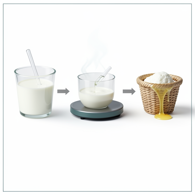
Soft Cheese Production Process
AI-generated conceptual illustration (sheep milk and soft cheese). It explains the idea and does not show study data.
View source evidence ↗Figures & tables
From the original paper Table 1
 Table 2
 Table 3
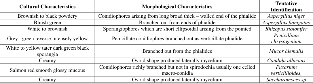 Table 4
 Table 5
 Table 6
Introduction
Milk is secreted by the mammary glands of mammals. It provides not only the nutritional requirements but added protection to new born. Milk is got from a healthy udder and it is sterile but may be contaminated by bacteria present in the tubules, from where milk flows and the storage space called "Cistern"(Otoikhan, 2012). Cow"s milk is the major milk used for the production of cheese although milk can also be gotten from other animals such as bufallo, goat, sheep, camel e.t.c. Sheep milk is an excellent raw material for the milk processing industry especially in cheese production (Park et al., 2007) [23] . Sheep milk has higher specific gravity, viscosity, refractive index, titratable acidity, and lower freezing point than average cow milk (Haenlein and Wendorff, 2006) [12] . Milk is usually coagulated with Calotropis procera, other coagulants such as Carica papaya, lemon juice and steep water from cereals (maize, millet and sorghum) can also be used (Ogunlade et al., 2020) [16] . Soft cheese is fresh cheese, that is, soft, moist curd that has been cut and drained of the whey but never ripened. The soft cheese produced in Nigerian farms especially in the northern part makes use of local ingredients. The vegetable rennet used for the production is made from a native plant Calotropis procera (Sodom apple), which can be cultivated, all year round (FAO, 1994). The presence of microorganisms in soft cheese can be from the rudimentary equipment used for the production of the cheese and also from environmental factors. Raw milk easily becomes sour when it is stored for a long period at high ambient temperatures prevalent in tropical and subtropical countries (Orhevba and Taiwo, 2016) [20] . This is because the inherent lactic acid bacteria and contaminating microorganisms from storage vessels or the environment break down the lactose in milk into lactic acid. Fresh milk drawn from a healthy cow normally contains a low microbial activity particularly with bacterial load of less than 10 3 cfu/mL (Chatterjee et al., 2006 [7] , Lingathurai et al., 2009) [14] , but the load may increase up to 100 fold or more once it is stored for sometime at ambient temperature (Lingathurai et al., 2009) [14] . Some of the factors that increase the bacterial activity in raw milk and its products include health of the animal, cleanliness of the housing area, the nature of feed, the water used at farm, the milk vessels / utensils for storage and essentially the hygiene of the milker / handler (Chatterjee et al., 2006, Ali and Abdelgadir, 2011 and Salman et al., 2011) [7,26] . The presence of this pathogenic bacteria in raw milk and its products have been reported to be a major threat to human health especially those who still drink raw milk (Mubarack et al., 2010 [15] , Lingathurai and Vellathurai, 2010) [13] . and also reduces the keeping quality of milk (Salman et al., 2011) [26] . Consumption of raw milk and its by-products is considered potentially hazardous and has been associated with several types of infections including brucellosis, tuberculosis, salmonellosis, yersiniosis, Escherichia coli O157 and Staphylococcal entero toxin poisoning (Baylis, 2009) [6] .
Materials and Methods
Materials and Methods
Sample collection
The raw milk sample was collected from sheep from Fulani pastoralists at a local farm settlement, Ado Ekiti. It was collected aseptically and transported to the laboratory for analysis.
Plant collection
The leaves (Carica papaya and Calotropis procera) were collected from The Federal Polytechnic, Ado-Ekiti premises and the Authentications of the Plants were done at the Department of Plant Science and Biotechnology, Ekiti State University, Ado-Ekiti, Nigeria. The voucher specimens of UHAE 2018/022 for Carica papaya and UHAE 2018/023 for Calotropis procera have been deposited at the University herbarium.
Collection of coagulants:
Lemon fruits used were purchased from the market and steep water (effluent from pap produced from maize, sorghum, millet) were produced by steeping the grains in water for 3days after which it was milled and later steeped again for 2days. The steep water was then collected for use as coagulants.
Production of soft cheese
The milk was stirred gently during the heating process with a wooden spoon. About 4ml of the leaf extract of Calotropis procera, Carica papaya, Lemon juice and steep water were added to the warm milk and the mixture was heated the second time with intermittent stirring to about 45-50°C and was kept at this temperature until coagulation was achieved and the heating was stopped after the separation of curd and whey. The sign of coagulation was observed within the range of 10-15 min. It was transferred into a small raffia basket to facilitate whey drainage and characteristic shape, when the cheese was firm enough it was removed from the raffia basket and placed inside a covered plastic container for analysis.
Culture Media and Reagents
The following media were used for the isolation and characterization of the microorganisms from the soft cheese, Nutrient Agar (NA) was used for the isolation and enumeration of total viable mesophilic bacteria count; Plate Count Agar (PCA) for the enumeration of total viable mesophilic count; Potato Dexrose Agar (PDA) for isolation and enumeration of Yeast and Molds. Nutient agar was used for subculturing the bacteria isolates and PDA for Yeast and Molds. Each of the media used was prepared and sterilized according to manufacturer's specifications. The reagent used for chemical analysis and biochemical characterization was prepared according to the requirements of each analysis. The media were sterilized at 121 o C for 15 minutes in an autoclave at 1.0Nm -2 .
Isolation of microorganisms
This was done using the modified pour plate method (Olutiola et al., 2000) [19] . One gram (1 gram) of the cheese sample was weighed and introduced into 10mL of sterile distilled water to carry out a ten-fold serial dilution. Molten nutrient agar (NA), plate count agar (PCA) and Potato dextrose agar (PDA) which have been prepared and autoclaved, were allowed to cool to about 45 o C before they were dispensed aseptically into sterile Petri dishes containing 1mL of introduced inoculums from the dilutions. The plates were gently swirled and allowed to set. The inoculated plates were incubated in an incubator (Gallenkomp 9052A, England) at 37 o C for 18-24hours for bacteria while PDA plates were incubated at 28±2 o C for 72-120hrs for fungi. All the plates were observed for growth and counted after incubation.
Subculturing method
Pure colony was obtained from streak plate. The microbial mixture was transferred to the edge of an agar plate with an inoculating loop and then streaked out over the surface of fresh agar plate. Pure colonies obtained were maintained on agar slants at 4 o C and subsequently sub cultured before use. Colonies of fungal growth observed were sub cultured onto fresh potato dextrose agar (PDA) until pure cultures of the fungal isolates were obtained.
Cultural and Biochemical tests
The test carried out on the isolates include Gram's staining reaction, motility, spore staining, catalase, indole production, methyl red, Voges-Proskauer test, citrate utilization, urease test, oxidase and sugar fermentation tests according to the methods of Olutiola et al., 2000 [19] .
Identification of Bacteria
Appearance of the colony of each isolate on the agar media was studied. Characteristics observed include shape, edge, colour, elevation and texture after 24 h of incubation. Staining reaction and other results of biochemical tests were interpreted for the tentative identification of bacterial isolates to specie level according to Cowan and Steel (1993) [8] .
Morphological and Microscopic Identification of Fungi Isolates
Fungal mycelium (48 h) old was aseptically taken with needle and placed gently on a clean slide containing a drop of lactophenol blue, covered with cover slip and examined under the microscope. Microscopic identification was done according to (Barnett et al., 2000 [5] . and Samson et al., 2010) [27] . The identified fungi were maintained on PDA slants at 4 o C in refrigerator for subsequent use.
Results
Tables 1, 2, 3, and 4 shows the Cultural, Morphological and Biochemical characteristics of bacteria and fungi isolated from the soft cheese. Six bacterial species and eight different fungi species were isolated from the samples. The Bacteria includes Bacillus cereus, Bacillus subtilis, Lactococcus lactis, Lactobacillus casei, Pseudomonas aeruginosa, Staphylococcus aureus and the fungi species includes Aspergillus niger, Aspergillus fumigatus, Rhizopus stolonifer, Penicillium chrysogenium. Mucor hiemalis, Candida albicans, Fusarium verticillioides and Saccharomyces sp. Penicillium chrysogenium was isolated from cheese coagulated with Carica papaya (SCPR) and steep water from sorghum (SSO) and maize (SMA). Mucor hiemalis was isolated from cheese coagulated with steep water from sorghum (SSO), millet (SMI) and maize (SMA). Candida albicans was isolated from cheese coagulated with Carica papaya (SCP) and steep water from maize (SMA). Fusarium verticillioides was isolated from all the cheese samples except the ones coagulated with lemon juice (SLJ) and Calotropis procera (SCPR) while Saccharomyces sp was found only in two samples which are cheese samples coagulated with lemon juice (CLJ) and Carica papaya. (SCP) [29] . in their work on nono and wara. Olatunji et al.
(2006) [18] . had earlier reported that cheese harbors bacteria including LAB such as Lactobacillus, Streptococcus as well as yeast and moulds. Bacillus cereus which is known to be highly resistant to environmental stress due to its ability to produce spores was isolated and it is known to be of public health importance since it is pathogenic. The detection of Staphylococcus aureus is also of public health importance because of its ability to cause a wide range of infections especially food-borne intoxication. This organism was equally isolated by Olasupo et al. (2002) [17] . from cheese and kunun-zaki, a cereal based, non-alcoholic beverage.
The high bacterial load found in the cheese samples investigated during the present study is supported by findings of Elkhider et al. (2011) [9] . who reported that cheese samples collected from different producers in rural areas of Eastern Sudan indicate that the level of hygiene and production methods, source of raw milk and its handling could be the main factors responsible for the high microbial loads which might affect the quality of cheese. The decrease in the microbial load of cheese coagulated with lemon juice may be as a result of the acidic nature of the juice. Adetunji et al. (2007) [7] . had earlier reported a decrease in the microbial load of cheese produced with lemon juice. The increase observed in the microbial load of the cheese were higher when compared with those isolated from the raw milk and the coagulants used might serve as source of the contaminants (Adegoke et al., 1992) [1] . A similar increase in microbial counts after addition of coagulants was reported by Adetunji et al. (2006) [3] . Therefore some preventive measures should be taken at this stage such as washing the leaves thoroughly before extraction of the juice, covering the steep water so as to reduce contamination, monitoring the temperature of the milk and controlling the development of acidity. The result is different from the results obtained by Adegoke et al. (1992) [1] . who observed reduction in population of total aerobes at the curdling point during the manufacture of the cheese. The presence of Lactic acid bacteria (LAB) and yeast indicates that the milk was not sterile. LAB are the predominant microorganisms in milk (Sawsan and Maymouna, 2010) [28] . Lactobacillus casei had the highest percentage occurrence (28.3%) followed by Lactococcus lactis (20%), Staphylococcus aureus (18.3%), Bacillus cereus (13.3%), Bacillus subtilis (11.7%) and Pseudomonas aeruginosa (8.4%). This is in line with the work of Guessas and Kihal (2004) [11] . who isolated Lactobacillus and Leuconostoc from raw goat milk. LAB improves food quality and also plays an important role in preventing the growth of undesirable bacteria. LAB creates an acidic environment conducive for the proliferation of yeasts, while yeasts provide growth factors such as vitamins and amino acids for LAB. Axelsson (2004) [2] . reported that LAB is generally associated with habitats rich in nutrients. It is well known that LAB produces a variety of antimicrobial substances with potential importance for food preservation. The existence of mold and yeast in cheese observed in this study was not unusual. Other researchers have isolated Penicillium and Aspergillus spp. from West African cheese (Adegoke et al., 1992) [1] . Oyeleke et al. (2006) [22] . in their study on 'nono', 'wara' 'maishanu', 'fufu' and 'kamu' isolated Lactobacillus bulgaricus, L. lactis, L. acidophilus, Streptococcus thermophillus and S. cremoris. Aspergillus species which was isolated is a known spore-former. This means that it can easily contaminate the dairy products which are usually exposed during processing, storage and hawking. They are the major spoilage organisms of carbohydrate food. However, their growth can result in the production and accumulation of mycotoxins which are of public health and economic importance (Rhodes and Fletcher, 1966). The yeast and moulds counts in this study were lower than those reported by Roostita and Fleet (1998) [23] . who stated that yeast population greater than 6 log cfu/mL were found in 54 % out of 85 cheese samples examined in New South Wales, Australia.
Conclusion
In conclusion, result from this work showed that the presence of both bacteria and fungi isolated from the soft cheese produced indicated that the soft cheese was not sterile. Therefore regular sterilization of dairy equipments, washing of utensils and proper hygiene during production are some of the preventive measures that could be applied to avoid contamination of the soft cheese.
Figures and tables from the paper
| Isolates | Colony colour/Shape | Cell Shape | Gram’s reaction | Spore | Motility |
|---|---|---|---|---|---|
| 1 | White to Yellow | Long Rod | + | + | + |
| 2 | White Spreading | Long rod | + | + | + |
| 3 | Amber | Rod in chains | + | - | - |
| 4 | Grey beaded | Cocci in chains | + | - | - |
| 5 | Greenish blue-brown | Short rod | - | - | + |
| 6 | Golden Yellow | Cocci in clusters | + | - | - |
Table text extracted from the original PDF; superscript letters are shown inline. The original table image is below.
View original table image
| Isolates | Shape | Elevation | Opacity | Emulsification | colour |
|---|---|---|---|---|---|
| 1 | Rod | Flat | Translucent | E | White to yellow |
| 2 | Irregular Rod | Flat | Translucent | E | Cream |
| 3 | Rod | riased | Opaque | E | Amber |
| 4 | Cocci short chains | Flat | Translucent | E | Grey |
| 5 | Rod | Flat | Transparent | E | Greenish Blue-brown |
| 6 | Cocci in clusters | Flat | Translucent | E | Golden yellow |
Table text extracted from the original PDF; superscript letters are shown inline. The original table image is below.
View original table image
| Iso | Ind | Cit | Nit | M/R | Vp | Oxid | Catal | H2S | Glu | Malt | Suc | Lact | Man | Suspected Orgs |
|---|---|---|---|---|---|---|---|---|---|---|---|---|---|---|
| 1 | - | + | + | + | + | - | + | - | + | + | - | - | - | Bacillus cereus |
| 2 | - | + | + | - | + | - | + | - | + | + | + | - | - | Bacillus subtilis |
| 3 | - | - | + | - | - | - | - | - | + | + | + | + | + | Lactobacillus casei |
| 4 | - | - | - | - | - | - | - | - | + | + | + | + | + | Lactococcus lactis |
| 5 | + | + | - | - | - | + | + | - | - | - | - | - | - | Pseudomonas aeruginosa |
| 6 | - | + | - | - | - | - | + | - | + | + | + | - | + | Staph. aureus |
Table text extracted from the original PDF; superscript letters are shown inline. The original table image is below.
View original table image
| Cultural Characteristics | Morphological Characteristics | Tentative Identification |
|---|---|---|
| Brownish to black powdery | Conidiophores arising from long broad thick – walled end of the phialide | Aspergillus niger |
| Bluish green | Branched out from ends of phialide | Aspergillus fumigatus |
| White to brownish | Sporangiophores which are short ellipsoidal arising from the pointed | Rhizopus stolonifer |
| Grey –green reverse intensely yellow | Penicillate conidiophres branched out as verticillate phialide | Penicillium chrysogenium |
| White to yellow tater dark green black sporangia | Branched out from the phialides | Mucor hiemalis |
| Creamy | Ovoid shape produced laterally mycelium | Candida albicans |
| Salmon red smooth glossy mucous | Conidiophores richly branched but not in spirodochia usually one celled macro-conidia | Fusarium verticillioides, |
| Creamy | Ovoid shape produced laterally mycelium | Saccharomyces sp |
Table text extracted from the original PDF; superscript letters are shown inline. The original table image is below.
View original table image
| Sample Code | Lactobacillus casei | Lactococcus lactis | Staphy aureus | Bacillus cereus | Bacillus subtilis | Pseudomonas aeruginosa |
|---|---|---|---|---|---|---|
| SSO | + | + | + | - | - | - |
| SMA | + | + | + | - | - | - |
| SMI | - | + | + | - | + | - |
| SLJ | + | + | - | - | - | - |
| SCPR | - | - | + | - | + | + |
| SCP | - | + | - | + | + | + |
Table text extracted from the original PDF; superscript letters are shown inline. The original table image is below.
View original table image
| Sample Code | A. niger | A. fumigatus | R. stolonifer | P. chrysogenium | M. hiemalis | C. albicans | F. verticill | Sacch.sp |
|---|---|---|---|---|---|---|---|---|
| SSO | - | + | - | + | + | - | + | - |
| SMA | - | + | - | + | + | + | + | - |
| SMI | + | - | + | - | + | - | + | - |
| SLJ | - | + | - | - | - | - | - | + |
| SCPR | + | + | - | - | - | - | - | - |
| SCP | - | - | - | + | - | + | + | + |
Table text extracted from the original PDF; superscript letters are shown inline. The original table image is below.
View original table image
References
29 in the original paper- Adegoke GO, Nse EN, Akanni OO. Effects of heat, processing time and pH on the microflora, aflatoxin content and storability of "wara", a soft, white cheese. Die Nahrung. 1992; 36:259-264.
- Axelsson L. Lactic Acid Bacteria: Classification and physiology. In: Lactic Acid Bacteria, Microbiological and Functional Aspects. Salminen, A.V. and A.O. Wright (Eds.) ouwehand. Marcel Dekker, New York, 1992, 1-66.
- Adetunji VO, Alonge DO, Chen J. The survivability of Listeria monocytogenes and spoilage microorganisms during processing and storage of wara, a southwestern Nigerian soft cheese. International. Assn. Food Protection. 93 rd Annual Management Programme Abstract Book. 2006; 103:13-16
- Adetunji VO, Alonge RK Singh, Chen J. Production of wara, a West African soft cheese using lemon juice as a coagulant. Journal Food Science and Technology. 2007; 41(2), 331-336.
- Barnett JA, Payne RW, Yarrow D. Yeasts: Characteristics and Identification, 3rd edn. Cambridge University Press, Cambridge, 2000.
- Baylis CL. Raw milk and raw milk cheese as vehicles for infection by verocytotoxin-producing Escherichia coli. International Journal of Dairy Technology. 2000; 62:293307.
- Chatterjee SN, Bhattacharjee I, Chatterjee SK, Chandra G. Microbiological examination of milk in Tarakeswar, India with special reference to coliforms. African Journal of Biotechnology. 2006; 5(15):1383-1385.
- Cowan ST, Steel KJ. Manual for the Identification of medical bacteria. Third Edition-Cambridge University Press. 1993, 34-89
- Elkhider IAE, El Zubeir IEM, Basheir AA, Fadlelmoula AA. Composition and hygienic quality of Sudanese white cheese produced by small scale in rural area of eastern Sudan. Annals of Food Science and Technology. 2011; 12(2):186-192.
- FAO. Production Yearbook. 2011; 48:281.
- African Journal of Biotechnology. 2004; 3(6):339-342
- Haenlein GFW, Wendorff WL. Sheep milk production and utilization of sheep milk. In: Park, Y.W. and G. F. W. Haenlein, (Eds.), Handbook of Milk of Non-Bovine Mammals. Blackwell Publishing Professional, Oxford, UK and Ames, Iowa, USA, 2006, 137-194.
- Webmed Central. Microbiology. 2010; 1(10):1-10.
- Lingathurai S, Vellathurai P, Vendan SE, Prem AA. A comparative study on the microbiological and chemical composition of cow milk from different locations in Madurai, Tamil Nadu. Indian Journal of Science and Technology. 2009; 2(2):5154.
- Mubarack MH, Doss A, Dhanabalan R, Balachander S. Microbial quality of raw milk samples Collected from different villages of Coimbatore district, Tamilnadu, South India. Indian Journal of Science and Technology. 2010; 3(1):61-63.
- Ogunlade AO, Oyetayo VO, Ojokoh AO. Influence of Different Coagulants on the Amino acid Content of Soft cheese (wara) Produced from Sheep milk. Journal of Research in Agriculture and Animal Science. 2020; 6(1):3539
- Olasupo NA, Smith SI, Akinsinde KA. Examination of the microbial status of selected indigenous fermented foods in Nigeria. Journal of Food Safety. 2002; 22:85-93.
- Olatunji EA, Shiawoya EL, Ijah UJJ. Bacterial assessment and keeping quality of local cheese (wara) in whey and salt solution. Journal of Sustainable Tropical Agricultural Research. 2006; 17:125-130.
- Olutiola PO, Famurewa O, Sonntag HG. Introduction to General Microbiology: Practical Approach 2 nd Edition. Publishing and Medical Consultants, Nigeria, 2000, 112-178
- Orhevba BA, Taiwo AD.Comparative assessment of "Wara" (Local Cheese) produced using three natural additives as coagulants. Research Journal of Food Science and Nutrition. 2016; 1:49-55.
- Otoikhian CSO. Biochemical Evaluation of Local Milk Product (Fura-de nunu). Nigerian Society for Experimental Biology. African Scientist. 2012; 13(1):49-56.
- Oyeleke SB, Faruk AK, Oyewole OA, Nabara HY. Occurence of Lactic acid bacteria in some locally fermented food products sold in Minna markets. Nigerian Journal of Microbiology. 2006; 20(2):927-930.
- Park Y, Ju´arez M, Ramos M, Haenlein G. "Physicochemical characteristics of goat and sheep milk,"Small Ruminant Research. 2007; 68(1):88-113.
- Rhodes A, Flecher DL. Principles of industrial Microbiology, Pergamam press, Oxford, 1996, 119.
- Roostita R, Fleet GH. The occurrence and growth of yeasts in camembert and blue-veined cheese. Journal of Dairy Science. 1998; 81(5):1205-1213.
- Salman A, Adil MA, Hamad IM. Enumeration and identification of coliform bacteria from raw milk in Khartoum State. Sudan Journal of Cell and Animal Biology. 2011; 5(7):121-128.
- Biodiversity Centre, Utrecht, The Netherlands, 2010.
- Sawsan AA, Maymouna MO. Isolation and Identification of Lactic Acid Bacteria from Raw Cow Milk, White Cheese and Rob in Sudan. Pakistan Journal of Nutrition. 2010;
- Uzeh RE, Regina EO, Ayodiji KR. Microbiological and Nutritional Qualities of Dairy Products: Nono an Wara. Nature and Science. 2006; 4(3):37-40.