
Research article · 2019 · Full text
International Journal of Agriculture and NutritionComparative morphological and anatomical studies between a native species (Prosopis cineraria (l.) Druce) and an introduced species (Prosopis juliflora (sw.) dc.) in south west region of Arabian Peninsula
Volume 1(4) · Pages 15–23 · DOI: 10.33545/26646064.2019.v1.i4a.18 ↗
Research areasPolysaccharides Composition and ApplicationsAnimal Diversity and Health StudiesBotany, Ecology, and Taxonomy Studies
Abstract
Author-writtenThis study shows the similarity and differentiation features between Prosopis cineraria and Prosopis juliflora based on morphological and anatomical characters. Prosopis cineraria is a native species while Prosopis juliflora is an introduced one. The data includes qualitative and quantitative characters. It assures on the importance of plant morphology and anatomy in determining the identification, classification and phylogeny between species.
Summary
Source-checkedPlain-language reading of the full paper, prepared with AI and checked against the text. Each point links to the authors' own words.
Why it matters
- Prosopis cineraria is a native species valued for its versatility, providing food, traditional medicine for various ailments, and materials for fuel and construction. View evidence ↗
- Prosopis juliflora is an introduced species from the Americas that can negatively impact food security by destroying natural pastures and reducing grazing potential. View evidence ↗
- Morphological and anatomical studies are essential for the accurate identification, classification, and understanding of the phylogeny between different plant species. View evidence ↗
The question
- The study aims to clarify the morphological and anatomical differences between the native P. cineraria and the introduced P. juliflora to better understand the processes behind plant invasions. View evidence ↗
What they did
- Plant specimens were collected from Jazan in Saudi Arabia and Aden in Yemen. View evidence ↗
- Morphological characterization included recording plant height, stem and leaflet morphology, and observations of flowering and fruiting. View evidence ↗
- Histological analysis was performed on three individuals per species using rotary microtome sections stained with fast-green safranin. View evidence ↗
- A calibrated ocular scale was used to measure the size of various anatomical parts, including stomata, epidermis, and vascular bundles. View evidence ↗
What they found
- Morphologically, P. cineraria has prickly internodes and leafy stipules, whereas P. juliflora has non-prickly internodes and spiny stipules. View evidence ↗
- P. juliflora exhibits significantly longer petioles (44-103 mm) compared to P. cineraria (9-28 mm). View evidence ↗
- The flowers of P. cineraria are whitish with a calyx of 10-15 mm, while P. juliflora flowers are yellowish with a smaller calyx of 2-3 mm. View evidence ↗
- The stem cuticle of P. juliflora (2 µm) is thicker than that of P. cineraria (1 µm). View evidence ↗
- P. juliflora has a higher stomata frequency (9.36%) compared to P. cineraria (5.97%). View evidence ↗
- The species differ in mesophyll type, with P. cineraria being equifacial and P. juliflora being dorsiventral. View evidence ↗
- P. juliflora possesses more spongy layers (5-7) in the leaf than P. cineraria (1-2). View evidence ↗
- Glandular hairs are present in the leaf base of P. juliflora but absent in P. cineraria. View evidence ↗
What it means
- The authors conclude that morphological and anatomical characters are vital for the identification, classification, and phylogenetic understanding of these species. View evidence ↗
- The identified diagnostic features can be used to distinguish between introduced and native Prosopis taxa. View evidence ↗
Visual summary
2 visualsIllustrations explain the paper; charts are replotted from the paper's own table values.
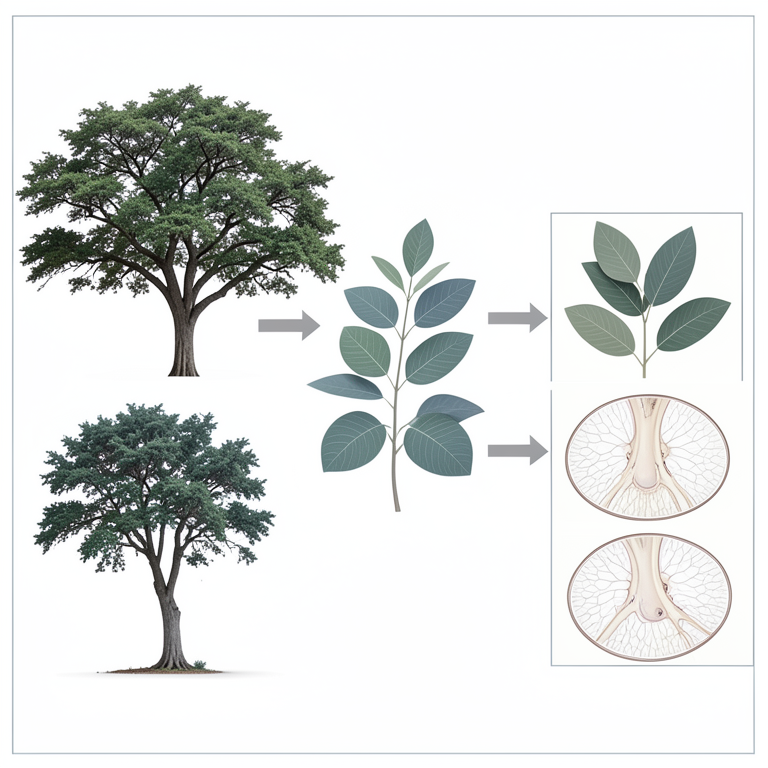
Comparative Study Framework
AI-generated conceptual illustration (Prosopis cineraria and Prosopis juliflora trees). It explains the idea and does not show study data.
View source evidence ↗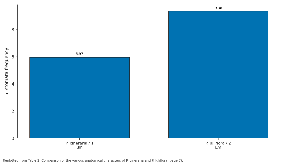
5. stomata frequency: P. cineraria / 1 µm vs P. juliflora / 2 µm
Replotted from Table 2 (page 7) of the original paper. Every bar is the paper's own table value.
View source evidence ↗Figures & tables
From the original paper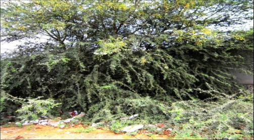 Figure 1Page 2
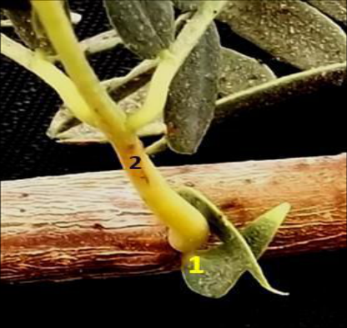 Figure 2Page 2
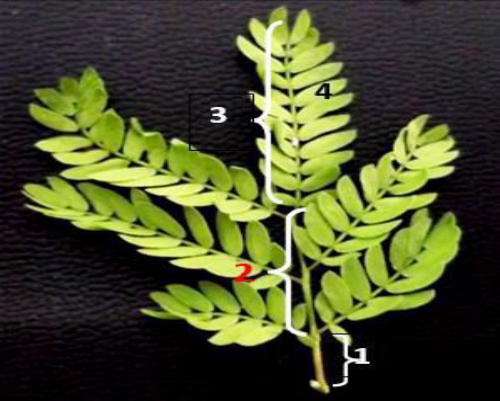 Figure 3Page 2
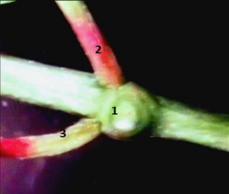 Figure 4Page 3
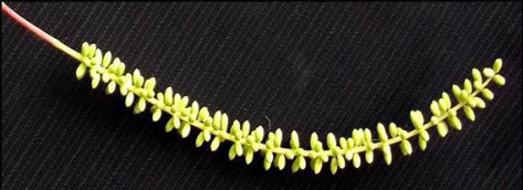 Figure 5Page 3
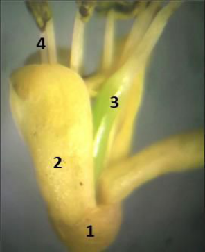 Figure 6Page 3
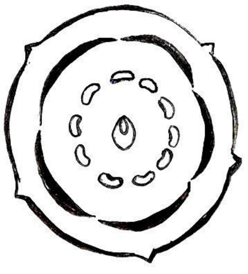 Figure 7Page 3
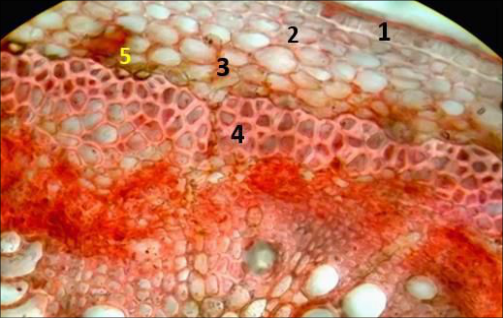 Figure 8Page 3
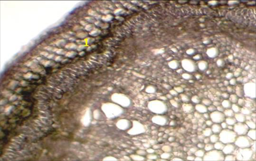 Figure 9Page 4
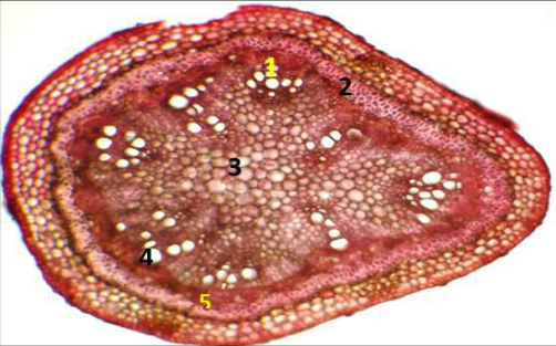 Figure 10Page 4
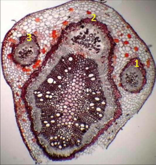 Figure 11Page 4
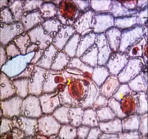 Figure 12Page 4
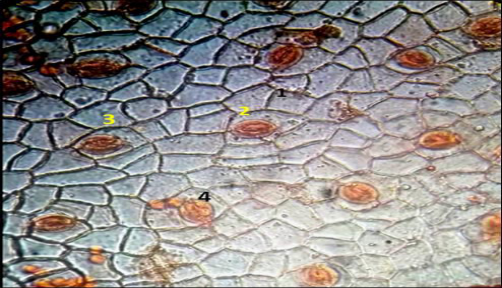 Figure 13Page 5
 Figure 14Page 5
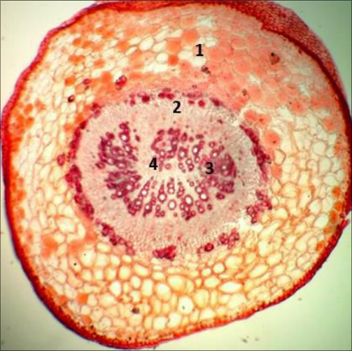 Figure 15Page 5
 Table 1Page 6
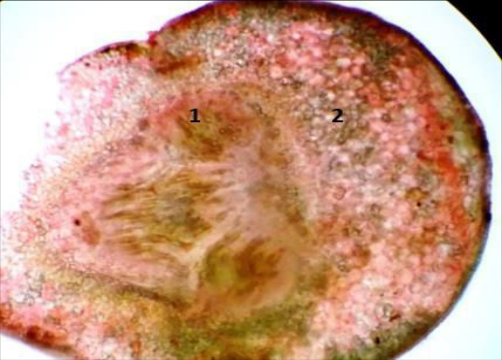 Figure 16Page 6
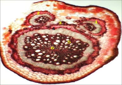 Figure 17Page 6
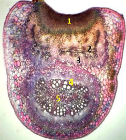 Figure 18Page 6
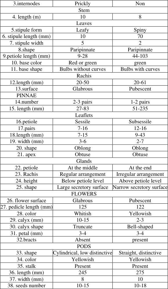 Table 2Page 7
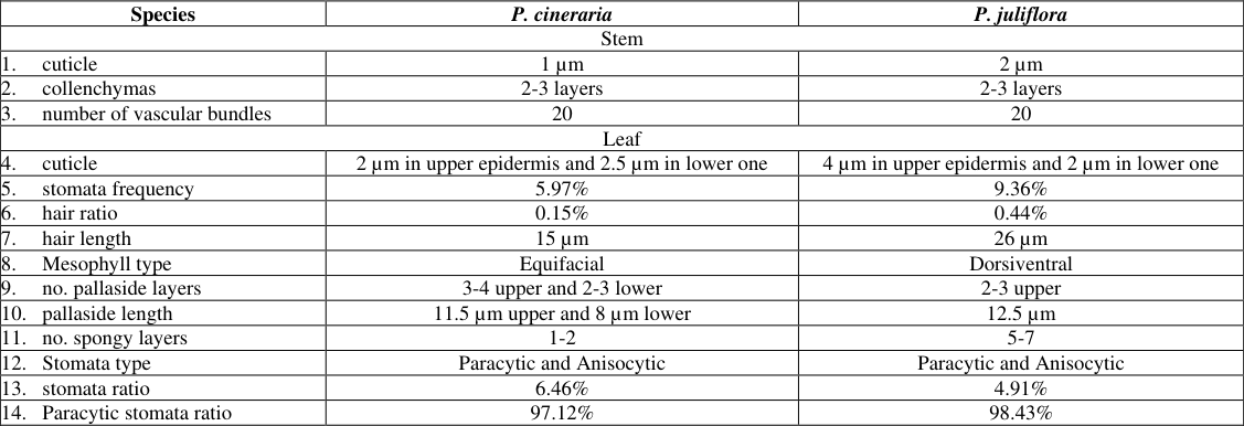 Table 2Page 7
Introduction
The genus Prosopis L. belongs to the family Leguminosae, subfamily Mimosoideae, tribe Mimosae (Burkart, 1976; Sherry et al., 2011) [3,28] . It comprises 44 species and five sections based on observed morphological differences among studied taxa (Burkart, 1976) [3] . The five sections included: Prosopis; Anonychium; Strombocarpa; Monilicarpa and Algarobia (Burkart, 1976; Landeras et al., 2005; Elmeer and Almalki, 2011) [3, 17, 7] . Section Prosopis included 3 species which have internodal prickles. Each of section Anonychium and Monilicarpa included 1 species. Section Strombocarpa included 9 species which have spiny stipules. Section Algarobia included 30 species, which have axillary thorns (Burkart, 1976) [3] . The genus Prosopis L. is thought to have originated in tropical Africa at the end of the Mesozoic or the beginning of the Tertiary period (Burkart, 1976) [3] . Owing to its fast growth and drought hardiness with extraordinary ecological amplitude (Tewari et al., 2000) [29] , Prosopis has a wide distribution, occurring in South, North and Central America, Africa and Western Asia (Burkart, 1976; Llanes et al., 2011; Sherry et al., 2011) [3,18,28] . Prosopis species were introduced to various areas primarily to combat desertification and improve the quality and quantity of the arid regions (El Fadl and Lukznen, 2003; Mosweu1 et al., 2013) [6,20] . Some of the species are of major economic importance; pods are utilized as food and fodder and wood as fuel and timber (Pasiecznik et al., 2001; Sherry et al., 2011) [24,28] . Introduced alien species can destroy natural pasture and reduce grazing potential of natural rangeland and can directly or indirectly affect the food security (Van Wilgen et al., 2008; Van den Berg et al., 2013) [31,30] . P. cineraria is native to dry and arid regions of Arabia and India (Khan, 1999) [1] . Its main population is center on the Thar Desert of India and Pakistan, with less dense populations occur in the Arabian Peninsula, Iran, and Afghanistan (Pasiecznik et al., 2004) [25] . It is considered the national tree of the United Arab Emirates (Jongbloed et al., 2003) [14] . P. cineraria is known as Ghaf in Arabic, Khejri in Indian, and Jand in Pakistan. Prosopis cineraria has been valued by different communities and cultures for the versatility of all its parts which and have been used in many ways as food, i.e., flour, drink, vegetable, and gum (Liu et al., 2012) [19] . Prosopis cineraria extensively used in traditional medicine to cure many diseases such as ailments like leprosy, dysentery, asthma, leucoderma, dyspepsia and earache (Al-Aboudi and Afifi, 2010) [2] . Barks are used for non-nutritional purposes, i.e., wood, tanning, fuel, firewood and charcoal (Garg and Mittal, 2013) [9] . Prosopis juliflora is an introduced plant that presents as a native plant from America to Peru (GISP 2004) [10] . It was first introduced in India as an introduced plant and spread to Pakistan, Myanmar, Sri Lanka, Thailand, Cambodia, Brunei, Vietnam, Philippines and Indonesia and reached the Arabian peninsula in the 1950s in Yemen, Bahrain, United Arab Emirates, Qatar, Saudi Arabia, Kuwait, Iraq and Jordan (Burkart, 1976; Ahmad et al 1996) [3,1] . It was cultivated by man during the last 100-150 years for the perceived value of the trees' products as a rich and delicious flour from pods, a major honey source in Bolivia, Jamaica and Pakistan, a good firewood with excellent charcoal, a source for fibre in the production of paper, for furniture and crafts, used for preparing various medicinal syrups, particularly for expectorants, used to arrest wind erosion in addition to stabilize sand dunes on coastal areas and for ornamentation (Hunde and Thulin, 1989) [13] , (Habit and Saavedra, 1990) [12] , (Poynton, 1990) [27] , (Felker and Moss, 1996) [8] and (Perry, 1998). It is considered as a "Royal tree" in India (Muthana and Arora, 1983) [26] . Prosopis species have been shown differences in their leaves and fruits due to exposure to variable stresses (Villagra et al., 2010) [32] . The introduced and naturalized or invasive species devise different strategies for environmental management (Nel et al., 2004) [22] . The recorded taxonomic identity of introduced and invasive Prosopis taxa remains questionable (Zimmermann, 1991) [33] .
International Journal of Agriculture and Nutrition
The aim of this work will be useful for clarifying the differences between introduced and native Prosopis taxa (Prosopis juliflora and P. cineraria respectively) through morphological and anatomical characterizations for understanding plant invasions and the processes underlining their success.
Materials and Methods
The site selected for the collection of Prosopis sp. is located in the city of Jazan, Kingdom of Saudi Arabia and Aden of Yemen. Plant specimens were identified at the herbarium of Science faculty -Jazan University. The plant materials were washed with distilled water and used or fixed in FAA (Formalin-Acetic Acid-70% ethanol) solution until processing.
Morphological Studies
Characterization and observations on morphology of the two studied plant species including plant height leaflet and stem morphology as well as flowering and fruiting were recorded and discussed.
Anatomical Studies:
The epidermis analysis It was performed by using fragments of the middle third of leaves fixed in 70% alcohol. The selected fragments were boiled in a 10% nitric acid solution (Ghouse and Yunus, 1972) [11] up to the dissociation of the epidermis. Subsequently, the epidermis was washed 3 × in distilled water and placed in a 50% sodium hypochlorite solution for clarification; washed3 × in distilled water and in a solution of distilled water and acetic acid 1:500 (v/v) and mounted between slide and cover slip in 50% glycerin medium.
The histological analysis
The fourth node of the stem and leaf blade (midrib and intercostal region) and proximal, medial and distal petiole samples were used and three individuals from both species were selected. The materials were fixed in 2.5% glutaraldehyde in phosphate buffer pH 7.2, 0.1 M (Karnovsky, 1965) [15] and later preserved in 70%alcohol. The sections obtained by a rotary microtome at and were stained with fast-green safranin (Dizeo de Strittmatter, 1979) [5] . Excess stain was washed off with water before a drop of glycerin was added. Slides were observed with an Olympus microscope and photographs were taken with a digitized camera (Nikkon) (O'Brien et al., 1965) [23] .
In addition to the general observations, size of parts or cell length and width or layers were recorded or measured using a calibrated ocular scale in stomata length and width and pore size, epidermis, mesophyll, ground tissue and vascular bundle.
Results and Discussion
Results and Discussion
Stem and Leaf Morphology
The two studied Prosopis are ever green tree, up to 10m, green or brown red in the apical stems and brown to gray in the lower parts, with spikes arise from the epidermis (Fig. 1). They have alternate leaves, stipulate with small size with dimensions 10 x 5 mm (Fig. 2), swollen leaf base with pale green, petiolate 9-28 mm in length with red green color, paripannate 20-50 mm in length, composed of 2-3 pair leaves 27-83mm in length, each pair is composed of 7-16 pair leaflets with dimensions 7-15 mm in length and 3-6 mm in width, dark green, smooth, oblong, entire, mucronate, the distance between leaflets 6.1-8mm (Fig. 3). Secretory gland is present in the middle of leaf petiole 11mm from leaf base, wide conical like shape, wide secretory opening and there are number of glands between leaflets (Fig. 4). Inflorescence and Fruit Morphology Inflorescence is spike, raceme, 125 mm in length, red color in half spike stalk, little separated florets (Fig. 5). Flower is ebractate, hermaphrodite, actinomorphic, smooth. Calyx is five united sepals, imbricate, opposite sepal edges. Corolla is five united petals, imbricate, opposite petal edges. Androecium is 10 free stamens, nectar gland presents on apical lobe of stamen. Gynoecium is one carpel, marginal placentation (Fig. 6). Floral symbols (Fig. 7) are:
Fruit is smooth legume indehiscent, simple depression between seed positions, yellowish white, 245 mm in length and 8 mm in width.
Stem Anatomy
Epidermis has one layer of polygon parenchyma cells covered with a layer of cuticle 1 µm in thickness. Cortex: It is made up of 6-7 cell layers with 2-3 laminar collenchyma present under the epidermis directly, the rest are round large parenchyma cells with intracellular spaces, some of them are storage, there are two layers of cells contain chloroplasts and rhombic crystals at the end of cortex, endodermis (starch sheath) is composed of one layer of cells containing number of starch granules and some rhombic crystals. Sclerenchyma tissue: lignified thick tissue, composed of fibers present in the outer part of phloem as connected strips (Fig. 8) and (Fig. 9). Pith is star like shape, composed of storage parenchyma cells with thin walls, enlarge in size towards the center, intracellular spaces present without any crystals (Fig. 10).
-phloem
-phloem
Stem Node Anatomy
There is no difference between node and stem in structure. It is composed of the same tissues (epidermis, cortex, vascular tissues and pith). The only difference is present appendages of bundles in the node out from vascular cylinder of the stem which regards as leaf trace. Vascular cylinder of the node has 9 essential vascular bundles and two secondary vascular bundles, variety in size, arranged in a connected cylinder, composed of sclerenchyma, phloem, xylem and pith. These bundles are arranged inside stem cylinder. In addition to the cylinder, there are three essential vascular bundles with large cylindrical in shape exit little from the stem cylinder. This feature reflects Developmental and structural between stem and lateral extensions (Leaves). This extension of vascular tissue from a plant's stem into a nearby leaf, resulting in a leaf gap in the plant's stele is Called leaf trace. The node has rich of rhombic crystals especially in the fibers around vascular cylinders (Fig. 11).
Leaf Anatomy
Upper epidermis has one layer of rectangle parenchyma contains secretory substances, nearly irregular size thick wall, covered with cuticle with 2µm in thickness, glabrous. Lower epidermis: one thick layer of rectangle parenchyma with the same size. It's covered with a thick cuticle 2.5 µm. There are some solitary unicellular trichomes with medium in length 15 µm and 0.15% density. The both epidermis contain number of stomata with high density at the upper one which reach 5.97% for upper epidermis while 6.46% for lower one. There are two types of stomata (Fig. 12 and 13): Paracytic: the guard cells are accompanied with two or more subsidiary cells while the axis of subsidiary cells is parallel to the axis of guard cells and stomata opening. Its density is high for both epidermises 97.12% Anisocytic: the guard cells are surrounded with three subsidiary cells with irregular size where one is small and the two others are large. Its density is very low for both epidermises 2.44%.
Mesophyll
Mesophyll
Leaf Base At the region far from the stem
It is nearly circular in shape and composed of the epidermis which has cubical closely cells covered with a layer of cuticle 1 µm thick. The cortex which is large in size. It's composed of 6-9 large parenchyma which contain very low number of rhombic crystals.
The vascular cylinder which is composed of vascular tissue which contains 8 basic vascular bundles where nearly one bundle is separated and the rest arranged as U letter with different sizes from xylem and phloem. Finally, the pith which is very narrow, present as U letter and composed of parenchyma without crystals (Fig. 15).
At the adjoining base leaf to the stem or the node
This part is oval in shape resemble the previous description with minor differences; there are 7 basic vascular bundles separated from each other. There are no glandular trichomes but stellate crystals and deposit materials are present (Fig. 16).
Leaf Petiole
The shape of leaf petiole in transverse section is oval and concave something at the upper direction and composed of The epidermis which is composed of a layer of parenchyma polygon in shape covered with a thick cuticle layer with 2.5 µm. The cortex which is composed of two types of external cells with 3-4 chlorenchyma layers with low number of chloroplastids while the second type of internal cells with 5-6 layers of parenchyma have some cells with secretory materials. The vascular cylinder which is composed of the vascular tissues: They are composed of 8 vascular bundles with 6 essential vascular cylinders in the petiole having scattered xylem and 2 bundles in the concave area from the upper direction of the petiole which are surrounded with fibers as incomplete circle shape. There are little nmber of rhombic crystals present in upper vascular fibers. The pith which is like very narrow lamellea shape and composed of 3-4 small parenchyma layers with large interspaces (Fig. 17).
Leaf Gland
It looks like bowl. Its height is little over the petiole height. The internal cavity is little concave or depressed and more enlargement so the secretory surface becomes large (Fig. 18). The two investigated Prosopis species reveal some variations in their morphological and anatomical features. The morphological differentiations are like in internode and stipule forms, rachis and flower surface in addition to pod color and shape. Moreover, the anatomical differentiations are like in leaf petiole and base in addition to midrib anatomy. Mesophyll type is Equifacial for P. cineraria while Dorsiventral for P. juliflora (Table 1 and2). These features show so diagnostic where they can be confined to use in classification, identification or phylogeny between introduced and native species taxa (Clement et al., 2015).
| Species | P. cineraria | P. juliflora |
|---|---|---|
| Habit | ||
| 1.shape | Perennial tree | Perennial tree |
| 2.branching | Non | Zigzag |
Table text extracted from the original PDF; superscript letters are shown inline. The original table image is below.
View original table image
Prosopis cineraria is belonged to the section Prosopis which contains two other species and they are resemble in having prickles like found on roses (Rosa spp.) while Prosopis juliflora is belonged to the section Algarobia which have other twenty nine species having axillary thorns (Burkart, 1976) [3] . P. juliflora is likely to be of sub-tropical North American origin where it survives prolonged cold periods so when it is introduced into other regions particularly in the Middle East, the adaptation of morphological and anatomical features is changed. The complex variation has become referring to biology, ecology, silviculture and management of habits.
Conclusion
The study enhances the importance of plant taxonomy via morphological and anatomical characters which play vital role in the knowledge of species in terms of identification, classification and phylogeny.
Figures and tables from the paper
| 3.internodes | Prickly | Non |
|---|---|---|
| Stem | ||
| 4. length (m) | 10 | 8 |
| Leaves | ||
| 5.stipule form | Leafy | Spiny |
| 6. stipule length (mm) | 10 | 70 |
| 7. stipule width | 5 | 2 |
| 8.shape | Paripinnate | Paripinnate |
| 9.petiole length (mm) | 9-28 | 44-103 |
| 10. base color | Red or green | green |
| 11. base shape | Bulbs without curves | Bulbs with curves |
| Rachis | ||
| 12.length (mm) | 20-50 | 20-61 |
| 13.surface | Glabrous | Pubescent |
| PINNAE | ||
| 14.number | 2-3 pairs | 1-2 pairs |
| 15. length (mm) | 27-83 | 51-235 |
| Leaflets | ||
| 16.petiole | Sessile | Subsessile |
| 17.pairs | 7-16 | 12-16 |
| 18.length (mm) | 7-15 | 9-43 |
| 19. width (mm) | 3-6 | 2-7 |
| 20. shape | Oblong | Oblong |
| 21. apex | Obtuse | Obtuse |
| Glands | ||
| 22. petiole | At the middle | At the end |
| 23. Rachis | Regular arrangement | Irregular arrangement |
| 24. height | Below petiole level | Above petiole level |
| 25. shape | Large secretory surface | Narrow secretory surface |
| FLOWERS | ||
| 26. flower surface | Glabrous | Pubescent |
| 27. pedicle length (mm) | 125 | 122 |
| 28. color | Whitish | Yellowish |
| 29. calyx (mm) | 10-15 | 2-3 |
| 30. calyx shape | Truncate | Bell-shaped |
| 31. petal (mm) | 3-4 | 3-4 |
| 32.bracts | Absent | present |
| PODS | ||
| 33. shape | Cylindrical, low distinctive | Straight, distinctive |
| 34. color | Yellowish | Yellowish |
| 35. stalk | Present | Present |
| 36. length (mm) | 245 | 275 |
| 37. width (mm) | 8 | 10 |
| 38. seeds number | 10-15 | 10-18 |
Table text extracted from the original PDF; superscript letters are shown inline. The original table image is below.
View original table image
| Species | P. cineraria | P. juliflora |
|---|---|---|
| Stem | ||
| 1. cuticle | 1 µm | 2 µm |
| 2. collenchymas | 2-3 layers | 2-3 layers |
| 3. number of vascular bundles | 20 | 20 |
| Leaf | ||
| 4. cuticle | 2 µm in upper epidermis and 2.5 µm in lower one | 4 µm in upper epidermis and 2 µm in lower one |
| 5. stomata frequency | 5.97% | 9.36% |
| 6. hair ratio | 0.15% | 0.44% |
| 7. hair length | 15 µm | 26 µm |
| 8. Mesophyll type | Equifacial | Dorsiventral |
| 9. no. pallaside layers | 3-4 upper and 2-3 lower | 2-3 upper |
| 10. pallaside length | 11.5 µm upper and 8 µm lower | 12.5 µm |
| 11. no. spongy layers | 1-2 | 5-7 |
| 12. Stomata type | Paracytic and Anisocytic | Paracytic and Anisocytic |
| 13. stomata ratio | 6.46% | 4.91% |
| 14. Paracytic stomata ratio | 97.12% | 98.43% |
Table text extracted from the original PDF; superscript letters are shown inline. The original table image is below.
View original table image
References
33 in the original paper- Ahmad R, Ismail S, Khan D. Use of Prosopis in Arab/Gulf states, including possible cultivation with saline water in deserts. Building Consensus for the Disenfranchised. (Eds.). Center for Semi-Arid Resources, Kingsville, Texas, USA, 1996; 1:41-1.52.
- Al-Aboudi A, Afifi FU. Plants used for the treatment of diabetes in Jordan: A review of scientific evidence. Pharmaceutical Biology. 2010; 49:221-239. DOI: 10.3109/13880209. 2010.501802.
- Burkart A. Monograph of the genus Prosopis (Leguminosae, Sub-fam. Mimosoideae). Journal of Arnold Arboretum. 1976 57(3):219-249.
- Clement U Okeke, Chisom F Iroka, Alex I Izundu, Nkumah C Okereke, Chibuike I Onwuasoeze, Bio Louis Nyananyo. et al. Comparative systematic leaf and petiole anatomical studies of the genus Stachytarpheta found in Awka Nigeria. Journal of Medicinal Plants Studies. 2015; 3(4):82-84.
- Dizeo de Strittmatter C. "Modificación de una técnica de coloración safranina-fast-green".Boletín de la Sociedad Argentina de Botánica. 1979; 18(3-4):121-122.
- El Fadl MA, Luukkanen O. Effect of Pruning on Prosopisjuliflora: Considerations for Tropical Dry land Agro-forestry. Journal of Arid Environments. 2003; 53(4):441-455.
- Elmeer K, Almalki A. DNA Finger Printing of Prosopis cineraria and Prosopis juliflora Using ISSR and RAPD Techniques. American Journal of Plant Sciences. 2011; 2:527-534.
- Felker P, Moss J. Prosopis: Semi-arid Fuelwood and Forage Tree: Building Consensus for the Disenfranchised. Center for Semi-Arid Forest Resources, nKingsville, Texas, USA, 1996, 111.
- Garg A, Mittal SK. Review on Prosopis cineraria: A potential herb of Thar desert. Drug Invention Today, 2013, 5:60-65. DOI: 10.1016/j.dit.2013.03.002.
- GISP. Africa invaded: The growing danger of invasive alien species. Global Invasive Species Program, Cape Town, 2004, 34.
- Ghouse AKM, Yunus M. Preparations of epidermal peels from leaves of Gymnosperm by treatment with 60% HNO3. Stain Technology. 1972; 47:322-325.
- Habit MA, Saavedra JC. The Current State of Knowledge on Prosopis juliflora. FAO, Rome, Italy, 1990, 23.
- Hunde A, Thulin M. Flora of Ethiopia: Pittosporaceae to Araliaceae, In: Hedberg I, Edwards S (eds), Addis Ababa and Uppsala University, Ethiopia. 1989; 3:71-98.
- Jongbloed M, Gary F, Benno B, Westren AR. The Comprehensive Guide to the Wild Flowers of the United Arab Emirates. Abu Dhabi, UAE: Environmental Research and Wildlife Development Agency, 2003, p. 576.
- Karnovsky MJ. A formaldehyde-glutaraldehyde fixative of high osmorality for use in electron microscopy. Journal of Cell Biology, 1965, 27:137.
- Khan MAR. The Indigenous Trees of the United Arab Emirates: Dubai Municipality. Dubai, UAE, 1999, 150p.
- Landeras G, Alfonso M, Pasiecznik NM, Harris PJC, Rami RL. Identification of Prosopis juliflora and Prosopis pallida accessions using molecular markers. Biodiversity and Conservation. 2005; 1(1):1-16.
- Llanes A, Bonercarrere V, Capdevielle F, Vidal S, Luna V. Genetic diversity in a naturalpopulation of the halophytic legume Prosopis strombulifera revealed by AFLP fingerprinting. Boletin de la Sociedad Argentina de Botanica. 2011; 46(3-4):305-312.
- Liu Y, Singh D, Nair MG. Pods of Khejri, (Prosopis cineraria) consumed as a vegetable showed functional food properties. Journal of Functional Foods. 2012; 4:116-121. DOI: 10.1016/j.jff.2011.08.006.
- Mosweu1 S, Munyati C, Kabanda T, Setshogo M, Muzila M. Prosopis L. Invasion in the South-Western Region of Botswana: The Perceptions of Rural Communities and Management Options. Natural Resources. 2013; 4:496-505.
- Muthana KD, Arora GD. Prosopis juliflora (Swartz) D. C. a fast growing tree to bloom the desert. CAZRI Monograph No. 22, Central Arid Zone Research Institute, Jodhpur, 1983, 19p.
- Nel JL, Richardson DM, Rouget M, Mgidi TN, Mdzeke N, Le Maitre DC. et al. A proposed classification of invasive alien plant species in South Africa: towards prioritizing species and areas for management action. South African Journal of Science. 2004; 100:53-64.
- O'Brien TP, Feder N, McCully ME. Polychromatic staining of plant cell walls by toluidine blue O. Protoplasma. 1965; 59:368-373.
- Pasiecznik NM, Felker P, Harris PJC, Harsh LN, Cruz G, Tewari JC. et al. The Prosopis juliflora-Prosopis pallid complex: a monograph. Coventry: HDRA, 2001, 162p.
- Pasiecznik NM, Harris PJC, Smith SJ. Identifying Tropical Prosopis Species: A Field Guide. Association HDR. Coventry, UK: HDRA Publishing, 2004, 26 p.
- Perry G. Prosopis. Flora of Australia. 1998; 12:7-13.
- Poynton RJ. The genus Prosopis in southern Africa. South African Forestry Journal. 1990; 152:62-66.
- Sherry M, Smith S, Patel A, Harris P, Hand P, Trenchard L. et al. RAPD and microsatellite transferability studies in selected species of Prosopis (section Algarobia) with emphasison Prosopis juliflora and Prosopis pallida. Journal of Genetics. 2011; 90(2):251-264.
- Tewari IC, Harris PIC, Harsh LN, Cadoret K, Pasiecznik NM. Managing Prosopis juliflora (Vilayati Babool) -A Technical manual. CAZRI, Jodhpur and HDRA. Coventry, UK, 2000, 96 p.
- Van den Berg, Kotze ECI, Beukes H. Detection, quantification and monitoring of Prosopis in the Northern Cape Province of South Africa using Remote Sensing and GIS. South African Journal of Geomatics. 2013; 2(2):86-81.
- Van Wilgen BW, Reyers B, Le Maitre DC, Richards DM, Schonegevel L. et al. A biome-scale assessment of the impact of invasive alien plants on ecosystem services in South Africa. Journal of Environmental Management. 2008; 89:336-349.
- Villagra PE, Vilela A, Giordano CV, Alvarez JA. Ecophysiology of Prosopis species from the arid lands of Argentina: What do we know about adaptation to stressful environments? K.G. Ramawat (Ed.). Desert Plants. Biology and Biotechnology. Springer-Verlag. BerlinHeidelberg, 2010, 322-354.
- Zimmermann HG. Biological control of mesquite, Prosopis spp. (Fabaceae), in South Africa. Agriculture, Ecosystems and Environment. 1991; 37:175-186.
Cited by 1
From OpenAlex, OpenCitations and reference lists read by Scinovex
- Taxonomic characterizations of the genus Commicarpus Standl. (Nyctaginaceae) in Saudi Arabia ↗
Maad S. Ytemi, Hameed Alsamadany, Faraj A. Al-Ghamdi, Abadi M. Mashlawi, Ahmed Eesa El-Shabasy · PLoS ONE · 2026
The genus Commicarpus Standl. is a member of the family Nyctaginaceae. The genus includes about 30-35 species distributed across tropical and subtropical regions worldwide, including Saudi Arabia. Five species of Commicarpus are found through, the field survey, which are primarily concentrated in the western and southwestern regions of Saudi Arabia. The collected species are C. grandiflorus, C. helenae, C. mistus,…
❝ 0PDF