
Research article · 2016 · Full text
International Journal of Veterinary Sciences and Animal HusbandryCurrent Advances in surgical management of Ruminal Disorders of bovine
Volume 1(3) · Pages 32–38
Research areasAnimal health and immunology
Abstract
Author-writtenRuminal disorders in ruminant are numerous and constitute a major clinical problem. Disorders of forestomach in adult cattle can result from a variety of common causes including traumatic reticulo-peritonitis, ruminal acidosis, bloat, simple indigestion and vagal indigestion. The bovine forestomach are affected highly due to ingested foreign bodies which are the subject of attention almost all over the world and also major economic importance due to severe loss of production and productivity and sometimes death of the animal. The animals are stabilized with fluids and electrolytes for the imbalances preoperatively. Preparation of the animal for surgery includes removal of the hair and cleansing of the skin around the surgical site. Rumenotomy and diaphragmatic hernia repair are surgeries performed for treating many ruminal disorders in bovine. Anaesthetic techniques required for rumenotomy are para-vertebral nerve block by use of lignocaine hydrochloride. Ultrasound diagnosis, completely siphon out of the fluid, visceral blockade and refilling of rumen with refilling agents are among the current advances in surgical managements of ruminal disorders. A long vertical skin incision starting about three to four centimeters below the transverse process of the lumbar vertebrae is made. The abdominal muscles and peritoneum are also incised corresponding to the skin incision. The rumen incision is closed by double row of continuous inverting sutures using chromic catgut. The commonest complications are wound dehiscence and hemorrhage and seroma formation.
Summary
Source-checkedPlain-language reading of the full paper, prepared with AI and checked against the text. Each point links to the authors' own words.
Why it matters
- Bovine forestomach disorders, often caused by ingested foreign bodies, are a global concern due to their potential to cause significant economic losses through reduced productivity or animal death. View evidence ↗
- In Ethiopia, which has over 38,749,320 cattle, ruminal disorders represent a primary cause of economic loss for livestock owners. View evidence ↗
- Common causes of forestomach disorders in adult cattle include bloat, ruminal acidosis, simple indigestion, vagal indigestion, and traumatic reticuloperitonitis. View evidence ↗
What they did
- Rumenotomy and diaphragmatic hernia repair are the primary surgical procedures used to treat various ruminal disorders. View evidence ↗
- Para-vertebral nerve blocks using lignocaine hydrochloride are the recommended anesthetic technique for rumenotomy. View evidence ↗
- The standard left mid flank laparotomy involves a vertical skin incision of 18-20 cm, starting 3-4 cm below the lumbar vertebrae's transverse process. View evidence ↗
- For rumen impaction and tympany, two-thirds of contents are evacuated and replaced with a specific mixture of sorghum stover (2 kg), rice bran (3 kg), jaggary (1 kg), yeast boluses (8), and sodium bicarbonate (100 g) to restore the ecosystem. View evidence ↗
What they found
- In cases of traumatic reticuloperitonitis, 87% of affected animals were dairy cattle and 93% were older than 2 years. View evidence ↗
- Ruminal acidosis is characterized by a rumen pH falling below 5.5, compared to a normal range of 6.5 to 7.0. View evidence ↗
- Bradycardia occurs in 25-40% of vagal indigestion cases, resulting from decreased feed intake. View evidence ↗
- The most frequent complications following rumenotomy are hemorrhage, wound dehiscence, and the formation of seromas. View evidence ↗
- A poor prognosis is given when a foreign body penetrates the diaphragm, as this can lead to thoracic abscesses, myocarditis, and septic pericarditis. View evidence ↗
What it means
- The authors conclude that laparoscopy, ultrasonography, and radiography are the advanced diagnostic tools, while rumenotomy and diaphragmatic herniorrhaphy are the advanced surgical treatments for bovine ruminal disorders. View evidence ↗
- The authors recommend increasing awareness among livestock owners to avoid overfeeding sugarcane, concentrates, and poor roughage to prevent gastrointestinal obstructions. View evidence ↗
- The authors state that further research is needed to develop newer techniques for controlling visceral pain during rumenotomy. View evidence ↗
Visual summary
1 visualsIllustrations explain the paper; they do not show study data.
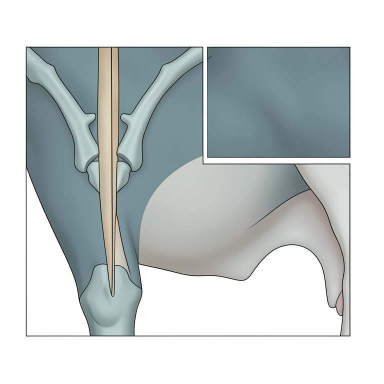
Surgical Incision Site
AI-generated conceptual illustration (bovine flank). It explains the idea and does not show study data.
View source evidence ↗Introduction
Ethiopia has the largest livestock inventories in Africa including more than 38,749,320 cattle. Ruminal disorders are the major causes of economic loss related to their productivity to livestock holders. Rumenotomy is a routine procedure for many diseases in cattle, such as, traumatic reticuloperitonitis, acute and recurrent bloat, ingestion of toxic plants, chemicals, spoiled roughage, or fetal membranes after parturition; placement of a temporary or permanent rumen cannula to relieve bloat; creation of a permanent rumen fistula; and impactions. Other reasons include ingestion of foreign bodies, such as, baling twine, nylon ropes or plastic bags that are obstructing the reticulo-omasal orifice, foreign bodies lodged in the distal esophagus, and carbohydrate engorgement (Fubini and Ducharme, 2004) [7] . In majority of the surgical maladies of the gastrointestinal tract like ruminal tympany, ruminal impaction, diaphragmatic hernia, traumatic reticulitis, abomasal impaction, intussusceptions and caecal dilatation have multifactorial etiologies constitute the most common cause for surgical disorders in dairy cattle (Makhdoomi et al., 1995) [17] . Ruminal surgeries in bovine presently may create challenges for the large animal surgeon. Therefore, understanding what causes ruminal disorders and their surgical managements in cattle, not only reduces the economic loss but also save life of the animal. From the above introduction the following objectives are proposed: To review the major surgical ruminal disorders in bovines. To critically assess the current advances in the surgical management of ruminal disorders in bovine. To highlight the complications of the surgical interventions and their management International Journal of Veterinary Sciences and Animal Husbandry 2. Literature Review 2.1. Ruminal Disorders Rosenberger et al. (1979) [25] reported that the stomach of ruminants was closely associated anatomically and functionally, and diseases of one usually affect the other. Bacterial digestion and fermentation, and physical maceration by contraction of the stomach walls were the two main functions of the forestomachs and the two are interdependent. Thus abnormality of one leads to abnormality of the other and of the two the motility is most readily examinable. Krishnamurthy et al. (1983) [15] reported that the inherent weak spot at the transitional zone of musculo-tendinous junction of the diaphragm caused diaphragmatic hernia and the condition was aggravated in cases of violent fall, tympany, and pregnancy and straining at the time of parturition. Ducharme (1990) [5] reported that the disorders of forestomach compartments in adult cattle were resulted from dietary, inflammatory and / or mechanical factors. The other causes were trichobezoar, ingestion of plastic bags and placenta resulting in mechanical obstruction by lodging in the reticulo-omasal orifice. Tyagi and Singh (1993) [29] opined that the ingestion and lodgment of foreign bodies were common in the bovine primarily due to indiscriminate feeding habits. In addition, industrialization and mechanization of agriculture had further increased the incidence of foreign bodies in these animals. Animals with nutritional deficiencies might also ingest various types of foreign bodies deliberately. The indiscriminate feeding habits and mineral deficiency made them susceptible to inadvertent ingestion of foreign materials. Rumen impaction due to plastics, ropes and leather in the rumen had led to anorexia, decreased production and progressive loss of body condition. Ramprabhu et al. (2003) [22] reported that the bovine forestomach affected highly due to ingested foreign bodies which were the subject of attention almost all over the world and also major economic importance due to severe loss of production and production ability and sometimes death due to anima. Clinical rumen indigestible foreign body impaction was characterized by pale mucous membrane, reduction of milk yield, complete cessation of rumination, impacted rumen, atony, reduced rumen motility, feces became scant and inappetance Rebhun (1995) [23] reported that traumatic reticuloperitonitis (TRP), was a relatively common disease in adult cattle caused by the ingestion and migration of a foreign body in the reticulum. Cattle were more likely to ingest foreign bodies than small ruminants since the latter do not use their lips for prehension and were more likely to eat a chopped feed. The typical foreign body was a metallic object, such as a piece of wire or a nail, often greater than 2.5 cm in length. Hawkins (2002) [11] suggested the classic signs associated with TRP were consistent with an acute, localized peritonitis and included anorexia, fever, tachypnea, and an arched stance with abducted elbows (indicating cranial abdominal pain). If the foreign body had penetrated the diaphragm and pericardium, affected cattle also can had muffled heart sounds, jugular pulses, and brisket edema secondary to congestive heart failure caused by pericarditis. However, not all cattle develop acute peritonitis; a significant population of affected cattle develops chronic or subclinical TRP that was not as easily diagnosed as acute TRP. Clinical signs associated with chronic peritonitis included anorexia, unthriftiness, decreased milk production, rumen hypomotility, and a change in manure consistency. Fubini and Ducharme (2004) [7] reported that the majority of affected cattle (87%) were dairy cattle and 93% were older than 2 years of age. A large number of adult dairy cattle had metallic foreign bodies in their reticulum without signs of clinical disease. It was likely that a predisposing factor in otherwise normal cows, such as tenesmus or a gravid uterus, causes migration of the foreign body into the reticular wall.
Traumatic reticuloperitonitis
Traumatic reticuloperitonitis
Ruminal Acidosis
Carter and Grovum (1990) [3] suggested ruminal acidosis was a metabolic disease of cattle. Like most metabolic diseases it is important to remember that for every cow that had showed clinical signs, there were several more which were affected sub-clinically. Acidosis was said to occur when the pH of the rumen failed to less than 5.5 (normal is 6.5 to 7.0). In many cases the pH can be even lower. The fallen in pH had two effects. Firstly, the rumen stopped moving, became atonic. This depressed appetite and production. Secondly, the change in acidity changed the rumen flora, with acid-producing bacteria taking over. They produce more acid, making the acidosis worse. The increased acid was then absorbed through the rumen wall, causing metabolic acidosis, which in severe cases can lead to shock and death. Owens et al. (1998) [20] reported that the primary cause of acidosis was feeding a high level of rapidly digestible carbohydrate, such as barley and other cereals. Acute acidosis, often resulting in death, was most commonly seen in 'barley beef' animals where cattle had obtained access to excess feed. In dairy cattle, a milder form, sub-acute acidosis, was seen as a result of feeding increased concentrates compared to forage. Krause and Oetzel (2006) [14] reported that acute acidosis often results in death, although illness and liver abscesses might be seen before hand. Cattle might become depressed, go off feed and had an elevated heart rate or diarrhea. Sub-acute acidosis also results in reduced feed intake, poor body condition and weight loss, unexplained diarrhea, lethargy, temperature, pulse rate and respiratory rate may rise.
Bloat
Smith (1998) [28] suggested prodigious volumes of gas were continually generated in the rumen through the process of microbial fermentation. Normally, the bulk of this which ruminants spend a lot of time doing gas was eliminated by eructation or belching. Regardless of whether bloat is of the frothy or free gas type, distention of the rumen compresses thoracic and abdominal organs. Blood flow in abdominal organs was compromised, and pressure on the diaphragm interferes with lung function. The cause of death was usually asphyxia due to pulmonary failure. In animals that are not observed frequently, bloat was commonly manifested as sudden death, reinforcing the concept that this is an acute disease with a short course. Cheng et al. (1998) [4] reported that two types of bloat were observed, corresponding to different mechanisms which prevent normal eructation of gases. Frothy bloat (primary tympany) results when fermentation gases were trapped in stable, persistent foam which was not readily eructated. As quantities of this foam build up, the rumen became progressively distended and bloat occurs particularly in animals on pasture, containing alfalfa or clover (pasture bloat). These legumes were rapidly digested in the rumen, which seems to results in a high concentration of fine particles that trap gas bubbles. Free gas bloat (secondary tympany) occurs when the animal was unable to eructate free gas in the rumen. The cause of this problem was often not discovered, but conditions that partially obstruct the esophagus (foreign bodies, abscesses, tumors) or interfere with ruminoreticular motility (i.e. reticular adhesions, damage to innervations of the rumen) clearly could be involved. Schwartzkopf-Genswein et al. ( 2004) [26] suggested bloat on pasture was frequently associated with "interrupted feeding"animals that were taken off pasture, and then put back on, or turned out on pasture for the first time in the spring. Diagnosis of bloat was typically straightforward, and the clinical picture largely reflects how long the condition had existed. Signs include abdominal distension: the rumen was on the left side, and hence, distention was typically most prominent on that side. As distention continues, the entire abdomen might become distended. Reluctance to move and cessation of feeding, signs of distress: anxiety and vocalization, respiratory distress: rapid breathing, neck extended with protruding tongue, staggering and recumbency: once an animal with bloat was recumbent, death occurs rapidly.
Simple indigestion
Erdman (1988) [6] reported that Simple indigestion was caused by atony of the forestomachs and had characterized clinically by anorexia, lack of ruminal movement and constipation. The disease was common in dairy cattle because of the variability in quality and the large amounts of the feed consumed. The common causes were dietary abnormalities of minor degree including indigestible roughage, particularly when the protein intake was low, moldy, over-heated and frosted feeds and moderate excesses of grain and concentrate intake. Fubini et al. (1989) [8] reported indigestion was more common when heavily fed cows were fed a little more concentrate than they can digest properly. Sudden change to a new source of grain, especially from oats to wheat or barley might have the same effect. Indigestible roughage might include straw, bedding or scrub fed during drought periods. Carter and Grovum (1990) [3] reported that changes in the pH of its contents markedly affect the motility of the rumen and in cases caused by overeating on grain an increase in acidity is probably of importance. High protein diets including the feeding of excessively large quantities of legumes or urea, also depress motility because of the sharp increase in alkalinity which results. The simple accumulation of indigestible food might physically impede ruminal activity. Hawkins (2002) [11] suggested that affected cattle usually show a pronounced fall in milk yield caused probably by the sharp fall in volatile fatty acid production in the atonic rumen. A reduction in appetite was the first sign and that followed closely in milking cows by a slight drop in milk production. Both occur suddenly, the anorexia might be partial or complete but the fall in milk yield was relatively slight. Rumination ceases and there was constipation with scanty, firm feces in most cases although diarrhea might occur on damaged feeds. Secondary ruminal atony occurs in many diseases, especially when septicaemia or toxaemia was present but there were usually additional clinical signs to indicate their presence. Rosenberger et al. (1979) [25] suggested vagal indigestion syndrome was characterized by the gradual development of abdominal distention secondary to rumenoreticular distention. The distention was originally thought to be the result of lesions affecting the ventral vagus nerve. Diseases that result in injury, inflammation, or pressure on the vagus nerve can result in clinical signs of vagal indigestion syndrome. However, vagal nerve damage had not presented in most cases of vagus indigestion, and the most common cause was traumatic reticuloperitonitis. Conditions resulting in mechanical obstruction of the cardia or reticulo-omasal orifice (e.g. papillomas or ingested placenta) could also result in vagal indigestion if ruminoreticular distention was present and the condition was sub-acute to chronic. Erdman (1988) [6] suggested there were four types of vagal indigestion described based on the purported site of the functional obstruction. Type I was failure of eructation or free-gas bloat, type II was a failure of omasal transport, type III was secondary abomasal impaction, and type IV was indigestion of late gestation. Type I and IV were rare. Type I vagal indigestion, or failure of eructation, results in free-gas bloat and had been attributed to inflammatory lesions in the vicinity of the vagus nerve, such as localized peritonitis, adhesions (usually after an episode of traumatic reticuloperitonitis), or chronic pneumonia with anterior mediastinitis. Other potential causes for type I vagal indigestion include pharyngeal trauma, which affects a more proximal part of the vagus nerve, and esophageal compression by abscesses or neoplasia, such as lymphosarcoma. Vagal indigestion could develop in cattle after abomasal volvulus without abomasal impaction. These cases would presumably fall into the category of type I vagal indigestion with damage to the vagal nerve near the reticulum and omasum. Fubini et al. (1989) [8] reported that type II vagal indigestion, more correctly termed as failure of omasal transport, develops as a result of any condition that prevents ingesta from passing through the omasal canal into the abomasum. Adhesions and abscesses (reticular or single liver abscesses) were the most common cause of failure of omasal transport and had usually located on the right or medial wall of the reticulum near the route of the vagus nerve. Reticular abscesses and adhesions were almost invariably the result of traumatic reticuloperitonitis. Mechanical obstruction of the omasal canal by ingested material (e.g. plastic bags, rope and placenta) or masses (e.g. lymphosarcoma, squamous cell carcinoma, granulomas, or papillomas) could also cause chronic ruminoreticular distention due to failure of omasal transport. Tyagi and Singh (1993) [29] suggested type III vagal indigestion was a secondary abomasal impaction. Primary abomasal impaction develops due to feeding of dry, course roughage, such as straw, in a chopped or ground form with restricted access to water and usually during extremely cold temperatures. Secondary abomasal impaction was seen most commonly after an episode of traumatic reticuloperitonitis or occasionally as a sequel to abomasal volvulus. Braun et al. (1998) [2] reported that type IV vagal indigestion, or partial forestomach obstruction, was poorly defined. It typically develops in cattle during gestation and was more appropriately termed indigestion of late gestation. The condition had thought to be related to the enlarging uterus shifting the abomasum to a more cranial position, which inhibits normal abomasal emptying. Distention of the dorsal and ventral sacs of the rumen results in an "L-shaped" rumen on rectal examination. Left dorsal and left and right ventral distention of the abdomen causes a "papple" (pear plus apple) shape as viewed from behind. Cattle with vagal indigestion syndrome have a diminished appetite, which typically improves temporarily if distention is relieved. Milk production gradually decreases, fecal output was reduced, and the rumen develops a "splashy" fluid consistency.
Vagal indigestion
Radostits et al. (2000) [21] suggested the feces were characteristically very scant and sticky and may contain longer than normal particles. The strength of rumen contractions was decreased; however, rumen motility is often increased (3 to 4 contractions/min). However, rumen contraction sounds were not audible because the contents had become frothy due to the prolonged contractions and failure of the rumen to empty. Kumar et al. (2003) [16] reported that temperature and respiratory rate were usually normal; however, these can be increased depending on the cause. Bradycardia was present in 25-40% of cases and was due to decreased feed intake rather than a direct stimulation of the vagus nerve. Tachycardia develops as the disease progresses and cattle become dehydrated.
Diagnosis
Wilson and Ferguson (1984) [30] suggested on rectal palpation, the rumen was distended with gas or froth that occupies the entire left abdomen pushes the left kidney to the right of the midline. The ventral sac of the rumen was enlarged and palpable to the right of the midline (the characteristic "Lshaped" rumen). It was important to recognize that diagnosis of vagal indigestion syndrome requires the presence of a markedly increased ruminoreticular volume. Palpation of the lower half of the right side of the abdomen below the costochondral junction might detect an impacted abomasum that feels doughy. Singh et al. (1993) [27] used flexible fiberoptic laparoscope for the diagnosis of intra abdominal disorders in ruminants and reported that it was useful tool in detecting and characterizing the lesions of traumatic reticuloperitonitis and concluded that the laparoscopy was a superior clinical examination and diagnostic tool in ruminants. Braun et al. (1998) [2] diagnosed DH was by radiograph and during rumenotomy. It was repaired under post xiphoid transabdominal approach after 48 h of rumenotomy. A crescent shaped 20 -25 cm length skin incision was made on the post xiphoid region under local infiltration analgesia with 2 percent lignocaine alone or with sedation. The abdomen was entered after incising the muscles and parietal peritoneum. The hernial rent was located and examined for the extent of adhesions. The herniated reticulum was separated from the adhesions bluntly from the diaphragmatic vent and thoracic structures. The firm adhesions were dissected with curved Metzenbaum scissors. Reticulum was withdrawn into the abdominal cavity. The herniorrhaphy was performed by applying continuous lock stitch using black braided silk No 2. The celiotomy incision was closed in three layers as per standard procedure. Kumar et al. (2003) [16] reported that the cows with TRP showed the clinical signs of chronic indigestion, pyrexia, partial anorexia, laboured breathing, an absence or reduced rumen motility, grunt, weight loss and a positive reaction to foreign body test. Displacement of reticulum from the peritoneum or an extensive gas fluid interface in the reticular region on a radiograph and well developed capsule in Ultrasonography suggested reticular abscess in ruminants. Braun (2003) [1] used Ultrasonography on standing non-sedated cattle with a 3.5 MHz linear transducer and assessed the size, position and contents of the abomasum, and intestines with ileus and diagnosed TRP, inflammatory fibrinous changes, reticular abscess and abdominal abscesses. Rohn et al. (2004) [24] recommended diagnosis was based on the presence of sub acute to chronic ruminoreticular and abdominal distention. Lateral radiographs of the reticulum should be taken to identify an opaque linear foreign body or reticular abscess. Ultrasonography of the cranioventral abdomen can indicate the presence of focal peritonitis and the reticular contraction rate.
Preoperative preparations 2.2.1. Preparation of surgical pack
Hofmeyr and Oehme (1988) [13] reported that gross contamination must be removed from the surgical instruments, before sterilization regardless of the sterilization technique used. Soak the soiled instruments in water added with detergent for ten to fifteen minutes, then rinse and dry. Ultrasonic instrument cleaners might also be used, as a latest technique for this purpose. Braun et al. (1998) [2] suggested before placing, the instruments were separated and placed in order of their intended use. The surgical pack wrapper had spread over diamond-wise on the table so that one corner remains towards the left, one corner towards the right, one corner on the near side and the fourth corner to the far side. The items should be assembled on the pack wrappers, in such a way that when the pack was opened the articles might be removed in the order in which they were used. Hawkins (2002) [11] reported that general surgical pack had been sterilized in the autoclave 121 o C, at 15 pounds pressure, for 15 minutes. After removal from the autoclave, packs were allowed to cool and dry on racks individually. The pack after sterilization could be stored in a closed cabinet, marking the date of sterilization. It could be stored for a week. It should be opened only at the top of the instrument table in the operation theater by a sterile team member. If the items had packaged, sterilized and handled properly, they remain sterile unless the package was opened, gets wet or damaged.
Preparation of the operation theatre
Hawkins (2002) [11] recommended that Ultraviolet Lamps must be provided for the sterilization of the theatre and fixed in the ceiling. The ultraviolet lamps had been used to control the airborne infection in the operation theatre. The entire room should be disinfected at least once weekly or immediately after contaminated surgery. Surgery room must be cleaned superficially between each surgery and thoroughly on a daily basis. Surfaces to be disinfected should be cleaned well, before disinfecting.
Preparation of surgical team
Hofmeyr and Oehme (1988) [13] recommended that the surgeon and his/her assistants must scrub well before surgery. The objectives were; mechanical removal of gross dirt and oil from the hands, reduction of transient organism counts and prolonged depressant effect on the resident microbes of the hands and forearms. Washing with soap removes most of the transient bacteria by mechanical friction. Brush fingers & thumb-palm, back of the hand and the forearm, up to elbow and similarly the opposite hand. Scrub for two to three minutes, with an antiseptics-impregnated sponge, between additional surgical procedures. When scrubbing had over, hold the hands and forearms higher than the elbows to allow water to run away from the cleanest area to prevent contamination of the scrubbed area. Fubini and Ducharme (2004) [7] reported that human hairs harbor bacteria. Surgical head covers should cover occipital and temporal hair to prevent falling of hair into the surgical area. Shoe covers effectively exclude bacteria, from street shoes. Face masks prevent droplets and particles of microorganisms shed from the nasopharynx and or oropharynx. Gowns act as a barrier between the skin of the surgical team members and the patient. The gown should be put on after final scrubbing. After final scrubbing and gowning, the gloves should put on the dried hands. Noordsy (1989) [19] recommended excessively soiled patient should be bathed before surgery to avoid contamination of the operation theatre and surgical site. The animals were stabilized with fluids and electrolytes for the imbalances. Food withheld for 12-24 hours before surgery, to minimize the potential for vomiting and aspiration during anesthesia. Water restricted 8-12 hours before pre-medication. Walk the animal to encourage, voiding of urine and motion or an enema might be given. Preparation of the site of operation should perform in the preparation room. Noordsy (1989) [19] reported that skin and hair of the animals were reservoir for bacteria & source of contamination. Skin preparation kills surface bacteria, but do not sterilize the skin. Approximately 20% of bacteria reside in deeper skin structure. Shaving of the surgical site causes multiple small lacerations and skin erosions that were rapidly colonized by bacteria. Hair clipping was the most recommended technique. It causes less skin trauma. Preparations of the animal for surgery include removal of the hair and cleansing of the skin around the surgical site. This should be done with an electric clipper or depilatory rather than a razor. A vacuum cleaner should used to collect hair and debris. Enough area of each side of the proposed incision should be clipped. Initial site preparation was performed outside the operation theatre, in the preparation room. The patient's skin should be scrubbed with a disinfectant. Tyagi and Singh (1993) [29] recommended scrub brushes were avoided because they caused excessive skin trauma. Scrubbing was done by applying gentle pressure in a circular motion. Beginnings at the proposed incision site, the scrubbing action continue outwardly, in enlarging concentric circles, until the outer margins of the clipped area reached. The sponge had then replaced with a clean one, and this process repeated until the dirt had been absent on discarded sponges. Sponge had never returned to the central area once it was contacted a peripheral region. Sufficient water used to ensure production of a good lather, but excess water should be avoided because wetting increase heat loss and moist contamination of surgical drops.
Preparation of the patients
Preparation of the patients
Site preparation
Site preparation
Transfer of the patient to operation theatre and positioning
Noordsy (1989) [19] suggested the patient was transferred to the operating room, without contaminating the prepared area and general anesthesia was given. Position the patient in such a way; it should be comfortable for both the patient and the surgeon. Secure the patient with ropes, to minimize movements during surgery, without any interference to the circulation or respiration. Application of antiseptics to the site: surgical sprit / Povidone-iodine were applied to skin using gauze sponges, held by gloved hand or sterile forceps; again working outwardly from this proposed site.
Aseptic draping
Fubini and Ducharme (2004) [7] recommended draping provides a barrier to bacteria and debris from unsterile area during surgery. Draping isolates the sterile surgical working area for the surgeon. After wearing the gown and gloves, the patient was draped, with sterile shrouds, by the surgeon.
Advance surgical management 3.1. Anaesthesia and control
Noordsy (1989) [19] studied anaesthetic techniques required for rumenotomy were para-vertebral nerve block by use of lignocaine hydrochloride. Standing position was preferable than recumbent, since it creates less stress, more room to move and familiarity but cow may go down. Monitoring the cardiovascular and respiratory systems and body temperature were quite useful.
Surgery 3.2.1. Left mid flank laparotomy
Fubini and Smith (1982) [9] reported that an 18-20 cms long vertical skin incision starting about 3-4 cms below the transverse process of the lumbar vertebrae should be made. The abdominal muscles and peritoneum should also incised corresponding to the skin incision. The skin was incised with a smooth but firm motion. The pressure on the scalpel should be adequate enough to ensure complete penetration of the skin. Dissection of the subcutaneous fascia and oblique muscles continues to expose the glistening aponeurosis of transverse abdominis muscle. Different muscles from outside to inside are incised one by one along with their fascia after grasping them with Allis tissue forceps and separated by inserting Metzenbaum scissors. In the last peritoneum was grasped with allis tissue forceps and then it was incised taking care not to cause any injury to underlying rumen. The length of incision from skin to the peritoneum should be in descending order to facilitate closure. The skin incision should be long enough to allow the surgeon's arm inside the abdomen. Johnston and Morris (1987) [10] recommended the abdominal cavity should be thoroughly explored. Caudal abdominal cavity explored first, the dorsal and ventral sac of rumen, urinary bladder, uterus, left kidney and intestinal masses then right cranial abdomen was reached by passing the arm ventral to the superficial layer of greater omentum and directed cranially to locate the pylorus, body and fundus of the abomasum, the omasum, right wall of the reticulum, and left lobe of the liver (for any abscess). A thorough search was made by inserting hand in the abdominal cavity through the incision and rolling over the rumen on all sides to rule out any herniation, abcessation or foreign bodies. No attempt should be made to break down the firm adhesions if present.
Rumenotomy
Hofmeyr and Oehme (1988) [13] reported that if the rumen had been grossly distended, aspiration was done by piercing a 16 gauge needle on dorsal aspect. After this a fold of rumen was exteriorized. Following laparotomy, a Weingarth ring was fixed to the dorsal commissure of the incision by its thumb screw. The rumen was fixed to the ring. As the rumen wall was incised hooks were placed into cut edges of ruminal wall, pulled out and hooked around the frame until the rumen had been reflected outward all the way around the incision. The rumen was incised and fixed with skin on either side with the help of towel clamps. After fixation, the rumen was incised longitudinally in the vertical direction on the dorsal compartment by repeatedly inserting Metzenbaum scissors. Singh et al. (1993) [27] recommended that transruminal exploration was done to find out the position, size and consistency of rumen, reticulum and abomasum by palpation. Explore the ruminoreticular fold, esophageal orifice and reticuloomasal orifice for lesions and meticulously the reticulum for FBs and remove all of them. Then try to invert the reticulum to determine the presence, location and extent of adhesions. Probe all the cells of reticulum at the site of adhesion in search of FBs. The reticular wall might be swept with a magnet to find out the leftover ferromagnetic FBs. Fubini and Ducharme (2004) [7] suggested enhancing the diagnosis an ultrasound probe may be carried into the reticulum for confirmation of reticular abscess, tumor, etc. The hand might be introduced through the reticuloomasal orifice into the omasal canal to evaluate the omasum and the abomasal lumen and mucosa. After surgical intervention the surgeon rescrubs his/her hands and the edges of rumen incision were thoroughly cleaned and redrapped. Animals confirmed for rumen and reticular obstructive disorders were subjected to rumenotomy. In cases of rumen impaction and ruminal tympany, two-third of the ruminal contents were evacuated and partially refilled with refilling agents viz; 2 kg of 5 cm length chopped sorghum stover, 3 kg of rice bran, 1 kg of jaggary, 8 numbers of yeast bolus and 100 g of sodium bicarbonate to revive the rumen eco system. Ducharme et al. (1990) [5] reported in diaphragmatic hernia (DH), the predominant signs for the early diagnosis of the disease recorded were recurrent ruminal distension, increased ruminal motility, forceful ruminal contraction over the entire ruminal area on the left side, respiratory distress and voiding of hard and small quantity pellety dung. Reticular sounds heard cranial to sixth rib with muffled heart sounds in all the animals. In advanced cases of DH animals showed regurgitation of ruminal contents.
Intra operative management
Johnston and Morris (1987) [10] suggested the Weingarth apparatus prevented intra peritoneal contamination during rumenotomy. In animals of reticulo-ruminal tympany, gases with frothy materials have been recovered from the rumen. In animals of ruminal impaction, the impacted mass was very dry and hard. The partially undigested feed was impacted highly forming layers. In DH animals the highly churned food particles with pungent smell were gushing on opening the rumen. The entire contents of reticulum and rumen were fluid. A three inch diameter corrugated plastic hose had been adequate to completely siphon out the fluid. Mehta et al. (1988) [18] recorded that visceral blockade provided visceral pain control during manipulation and anastomosis of intestine. During ligation, resection and closure of mesentery, the animal evidenced moderate to severe pain. The Fifteen percent lignocaine spray provided visceral pain control during ligation, resection and closure of mesentery.
Rumen and abdomen closure
Tyagi and Singh (1993) [29] recommended after finishing the required operation the ruminal cut edge were thoroughly cleaned after removal of hooks and sutured by a double row of continuous inverting sutures using chromic catgut no. 2 or 3. The abdominal wound was sutured in a routine manner.
Post-operative management
Johnston and Morris (1987) [10] recommended post-operative fluid therapy (oral and intravenous) complemented with administration of analgesics to treat dehydration, shock, electrolyte imbalance and to moderate the vigor of peristalsis of the bowel in intestinal surgery. Rohn et al. (2004) [24] reported that a thorough clinical examination with special emphasis on general condition, liver function and dehydration status were of great importance in determining the prognosis of abdominal surgery in cow.
Post-operative complications and their management
Smith (1998) [28] and Johnston and Morris (1987) [10] suggested intravenous fluids like Ringer's lactate and 5% dextrose in normal saline were used to correct electrolyte loss and dehydration. Fubini and Ducharme (2004) [7] recommended that the commonest complications should be wound dehiscence and hemorrhage. Others included fever, edema, slipped ligature, wound infection, peritonitis, death, intestinal obstruction/adhesion and physiological bloat. Herzog et al. ( 2004) [12] suggested for first two days the animal were maintained with fluid therapy, supportive therapy and antibiotics. The laparotomy wounds were cleaned and dressed daily up to five days and the sutures were removed on clinical union. The animal was allowed to access liquid diet from the third day and easily digestible feeds from the fourth day onwards gradually.
Prognosis
Rosenberger et al. (1979) [25] reported that the prognosis depends on the location of the reticular perforation. If the foreign body had penetrated the diaphragm, a poor prognosis should be given; septic pericarditis, myocarditis, and thoracic abscesses were possible sequelae. If perforation involves the right wall of the reticulum, a guarded prognosis was given; adhesions that involve the ventral branch of the vagus nerve might result in vagal syndrome type II or III. A favorable prognosis was given when the perforation did not affect the thoracic cavity and right side of the reticulum. Single abdominal abscesses (reticulum, liver) also carry a favorable prognosis if they could be drained or resected. Herzog et al. (2004) [12] . suggested extensive adhesions in the cranial abdomen were not necessarily associated with a poor prognosis. If the adhesions did not involve the vagus nerve, ruminal motility did not appear to be greatly impaired by the presence of adhesions. This might be because of the rumen wall was protected from restricting adhesions by the superficial layer of the greater omentum.
Conclusion and Recommendations
The stomachs of ruminants are closely associated anatomically and functionally, and diseases of one usually affect the others. Disorders such as traumatic reticuloperitonitis, ruminal acidosis, simple indigestion, vagal indigestion and bloat have no doubt plagued keepers of ruminant livestock since livestock were domesticated. Radiography, Ultrasonography and laparoscopy are the advanced diagnostic tools for ruminal disorder in bovine. Rumenotomy and diaphragmatic herniorrhaphy are advanced surgical managements performed for the ruminal disorders in bovine. Commonest complications are wound dehiscence, hemorrhage, edema, slipped ligature, wound infection, peritonitis, death, intestinal obstruction/adhesion and physiological bloat. Major economic importance due to severe loss of production and production ability and sometimes death of the animal needs a great attention for ruminal disorders in cattle. Therefore, based on the above conclusion the following recommendations are forwarded: Create awareness among the cattle stockholders to give up excess feeding of poor roughage, concentrates and sugarcane which are the predisposing causes for gastrointestinal obstruction in bovine.
Understanding the causes of ruminal disorders in cattle and implementing current advanced surgical management practices may spare the production and economic losses associated with ruminal disorders. Superior clinical examination and diagnostic tools should give due attention with veterinary professionals in management and control of ruminal disorders before it reaches risk stage. The ruminants' owners should be advised not to allow their ruminants to freely wander in streets especially in the cities and as much as possible to prevent nutritional deficiencies. Further studies should be conducted for controlling of visceral pain during rumenotomy with newer techniques. The postoperative complications encountered in all animals if any should be recorded and managed accordingly.
References
30 in the original paper- Braun U. Ultrasonography in gastrointestinal disease in cattle. Vet. 2003; 166:112-124.
- Braun U, Iselin U, Fluri E. Ultrasonic findings in five cows before and after treatment of reticular abscesses. Vet. Rec., 1998; 42:184-189.
- Carter R, Grovum W. A review of the physiological significance of hypertonic body fluids on feed intake and ruminal function: salivation, motility and microbes. J. Anim. Sci. 1990; 68:2811-2832.
- Cheng K, McAllister T, Popp J, Hristov A, Mir Z, Shin H. A review of bloat in feedlot cattle. J Anim. Sci. 1998; 76:299-308.
- Ducharme N. Surgery of the bovine forestomach compartments. Vet Clin North Am., (Food Anim Pract), 1990; 6:371-397.
- Erdman R. Dietary buffering requirements of the lactating dairy cow: A review. J. Dairy Sci. 1988; 71:3246-3266.
- Fubini S, Ducharme N. Surgery of ruminant forestomach compartment. In Farm Animal Surgery. S.L. Fubini and N.G. Ducharme (Eds), Saunders, Elsevier, 2004, 161240.
- Fubini S, Ducharme N, Erb H, Smith D, Rebhun W.. Failure of omasal transport attributable to perireticular abscess formation in cattle: 29 cases J. Am. Vet. Med. Assoc., 1980-1986, 1989; 194:811-814.
- Fubini S, Smith D. Failure of omasal transport due to traumatic reticuloperitonitis and intraabdominal abscess. Compend. Contin. Educ. Prac. Vet., 1982; 4:492-494.
- Johnston J, Morris D. Comparison of duodenitis, proximal jejunitis and small intestinal obstruction in horses: 68 cases (1977-1985). J Am. Vet. Med. Assoc., 1987; 191:849-854.
- Hawkins L. Bovine Digestive Diseases In: Large Animal Digestive Diseases notes. LAMS 5350, 2002, 22-24.
- Herzog K, Kaske M, Bischoff C, Kehler W, Hoeltershinken M, Starke A et al. Post-surgical development of inflammatory adhesions and reticular function in cows suffering from traumatic reticuloperitonitis. Dtsch Tierarztl Wochenschr, 2004; 111:57-62.
- Hofmeyr F, Oehme F. Textbook of Large Animal Surgery. The digestive system. In: 2 nd ed. Baltimore: Williams & Wilkins, 1988, 448.
- Krause K, Oetzel G. Understanding and preventing sub acute ruminal acidosis in dairy herds: a review. Anim. Feed Sci. Technol. 2006; 126:215-236.
- Krishnamurthy D, Nigam K, Deshpande P, Peshin S, Sharma D. Bovine diaphragmatic hernia-An analysis of 140 clinical cases. Indian Vet J., 1983; 60:1011-1016.
- Kumar R, Lakkawar A, Nair M, Varshney K. Gastrointestinal obstruction in cows. Indian Vet. J. 2003; 80:318-319.
- Makhdoomi D, Singh A, Singh M, Krishnamurthy D. Intestinal obstruction in ruminants: A review. Indian J. Vet. Surg., 1995; 16:81-82.
- Mehta I, Krishnamurthy D, Peshin P. Haematological and bio chemical changes in bovine diaphragmatic hernia: A clinical study. Indian J of Anim. Sci. 1988; 58:764-767.
- Noordsy J. Rumenotomy in Cattle, in Food animal surgery. Lenexa, Kan., Veterinary Medicine 2 nd ed. 1989, 105-109.
- Owens F, Secrist D, Hill W, Gill D. Acidosis in cattle: a review. J Anim. Sci. 1998; 76:275-286.
- Radostits O, Gay C, Blood D, Hinchcliff K. Diseases of alimentary tract -II. In: Veterinary Medicine. 9 th ed. Book Power Publishers, Philadelphia, U.S.A. 2000, 259346.
- Ramprabhu R, Ramasamy V, Vijayakumar G, Rajendran, N, Subramanian M. Left displacement of abomasum in a Holstein Friesian cow: A case report. Indian J. Anim. Hlth., 2003; 42:87-88.
- Rebhun WC. Diseases of Dairy Cattle. Philadelphia, Williams and Wilkins, 1995, 113-116.
- Rohn M, Tenhagen B, Hofmann W. Survival of dairy cows after surgery to correct abomasal displacement: Association of clinical and laboratory parameters with survival in cows with left abomasal displacement. J. Vet. Med. Appl. Physiol. Pathol. Cli. ed., 2004; 51:300-305.
- Rosenberger G, Dirksen H, Grunder E, Grunert K, Stober M, Mack R. Clinical Examination of Cattle. 2 nd ed. Verky Paulparay, Berlin, Humburg, 1979, 203-209.
- Schwartzkopf-Genswein K, Beauchemin K, McAllister T, Gibb D, Streeter M, Kennedy A. Effect of feed delivery fluctuations and feeding time on ruminal acidosis, growth performance, and feeding behavior of feedlot cattle. J Anim. Sci. 2004; 82:3357-3365.
- Singh J, Singh A, Patil D. The digestive system. In: Ruminant Surgery. (Eds) Publisher and Distributors, Delhi, India. 1993, 225-312.
- Smith R. Impact of disease on feedlot performance: a review. J. Anim. Sci. 1998; 76:272-274.
- Tyagi R, Singh J. Ruminant Surgery. 1 st ed. CBS, New Delhi, 1993, 199.
- Wilson A, Ferguson J. Use of flexible fiberoptic laparoscope as a diagnostic tool in cattle. Can. Vet. J, 1984; 25:229-234.
Cited by 1
From OpenAlex, OpenCitations and reference lists read by Scinovex
- Left ventrolateral oblique laparotomy as a means of exploration in undiagnosed gastrointestinal obstruction in cattle
B Dwibedy, AK Sahoo, Samir Ranjan Nayak, I. Nath, SS Behera, Bandita Jena · International Journal of Veterinary Sciences and Animal Husbandry · 2018
Sixteen presented undiagnosed clinical cases of gastrointestinal obstruction in cattle in the periphery of Department of Veterinary Surgery and Radiology, College of Veterinary Science & Animal Husbandry (CVSc & AH) were selected for present study. The animals were divided into 2 groups by the following criteria. Group I included 10 animals in which the diagnosed surgical condition was easily accessed and corrected. Group II included 6 animals in which the diagnosed surgical condition was approached and treated with “some difficulty” or could not be corrected. In all the 16 cases an explorator