
Research article · 2018 · Full text
International Journal of Veterinary Sciences and Animal HusbandryLeft ventrolateral oblique laparotomy as a means of exploration in undiagnosed gastrointestinal obstruction in cattle
Volume 3(3) · Pages 87–95
Research areasAnimal health and immunologyVeterinary Equine Medical ResearchMicrobial infections and disease research
Abstract
Author-writtenSixteen presented undiagnosed clinical cases of gastrointestinal obstruction in cattle in the periphery of Department of Veterinary Surgery and Radiology, College of Veterinary Science & Animal Husbandry (CVSc & AH) were selected for present study. The animals were divided into 2 groups by the following criteria. Group I included 10 animals in which the diagnosed surgical condition was easily accessed and corrected. Group II included 6 animals in which the diagnosed surgical condition was approached and treated with “some difficulty” or could not be corrected. In all the 16 cases an exploratory diagnosis through left ventrolateral oblique laparotomy approach under regional anesthesia was possible and the diagnostic efficiency of this approach was found to be 100%. During systematic exploration, the approachability of different parts of the gastrointestinal system was studied. Based on the palpability and exteriorizability of different parts of the gastro-intestinal tract, the exploratory diagnosis through left ventro-lateral oblique laparotomy approach was confirmed to be 100% for cases under study and also effective for surgical correction of physical obstructions of the GI tract commonly occurring in cattle when a definitive preoperative diagnosis is not possible.
Summary
Source-checkedPlain-language reading of the full paper, prepared with AI and checked against the text. Each point links to the authors' own words.
Why it matters
- Surgical diseases of the digestive tract are a major clinical issue in ruminants, with abdominal surgeries being the most common type of operation. View evidence ↗
- Diagnosing gastrointestinal obstructions in the field is challenging for veterinarians due to limited diagnostic facilities. View evidence ↗
- The standard standing left flank approach for rumenotomy is limited because it does not allow the surgeon to explore the right side of the abdomen. View evidence ↗
The question
- The study aimed to evaluate a novel left ventrolateral oblique laparotomy incision to allow access to both the fore stomachs and the right side of the abdomen in undiagnosed cases. View evidence ↗
What they did
- The study involved 16 cattle of either sex and various ages where a definitive preoperative diagnosis could not be established. View evidence ↗
- Animals were divided into Group I (10 animals) where conditions were easily accessed and corrected, and Group II (6 animals) where conditions were difficult to approach or could not be corrected. View evidence ↗
What they found
- The left ventrolateral oblique laparotomy approach achieved 100% diagnostic efficiency in all 16 cases. View evidence ↗
- In Group I, 10 animals were successfully treated for conditions including omasal impaction (n=2), ruminoreticular foreign bodies (n=2), traumatic reticulitis (n=2), and concurrent omasal impaction and foreign bodies (n=3). View evidence ↗
- In Group II, 6 animals were diagnosed with more complex conditions, including diaphragmatic hernia (n=2), cecal dilation and displacement (n=2), intestinal volvulus (n=1), and abomasal obstruction (n=1). View evidence ↗
- The approach allowed palpation and exteriorization of the rumen, abomasum, most of the jejunum, and two-thirds of the cecum. View evidence ↗
- Certain areas remained non-exteriorizable, including the ileum, the proximal jejunum, the ascending duodenum, and various parts of the colon. View evidence ↗
What it means
- The authors conclude that the left ventrolateral oblique laparotomy is a viable solution for exploring and correcting common gastrointestinal obstructions when a preoperative diagnosis is unavailable. View evidence ↗
- The authors state that because the study only included 16 cases, further investigation with a larger number of animals is needed to validate the technique. View evidence ↗
Visual summary
3 visualsIllustrations explain the paper; charts are replotted from the paper's own table values.
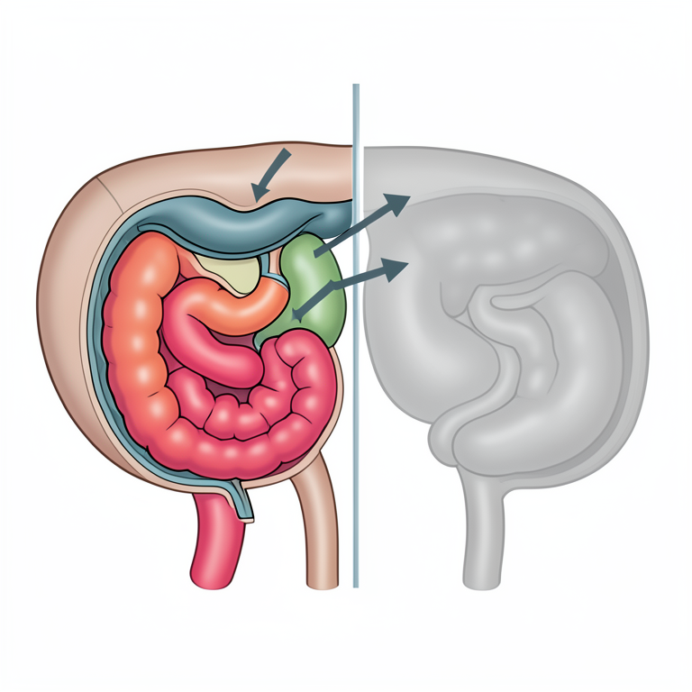
GI Tract Accessibility Map
AI-generated conceptual illustration (adult cow gastrointestinal tract). It explains the idea and does not show study data.
View source evidence ↗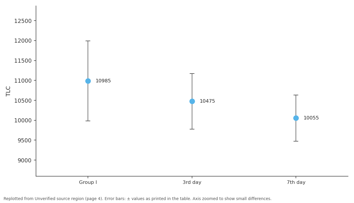
TLC: Group I vs 3rd day vs 7th day
Replotted from a table (page 4) of the original paper. Every bar is the paper's own table value.
View source evidence ↗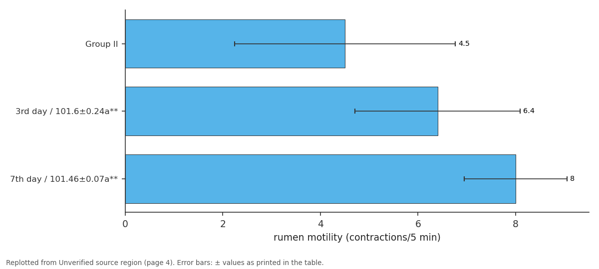
rumen motility (contractions/5 min): Group II vs 3rd day / 101.6±0.24a** vs 7th day / 101.46±0.07a**
Replotted from a table (page 4) of the original paper. Every bar is the paper's own table value.
View source evidence ↗Figures & tables
From the original paper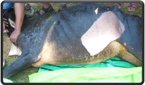 Figure 1
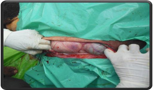 Figure 2
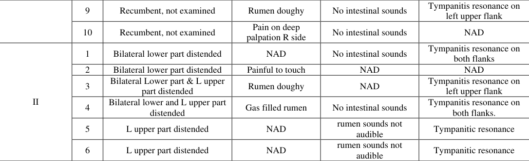 Table 3
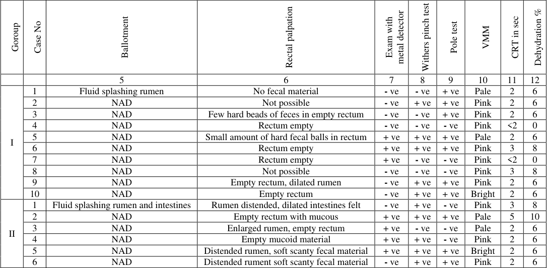 Table 3
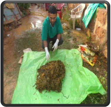 Figure 3
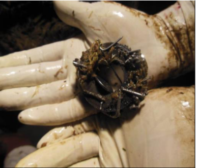 Figure 4
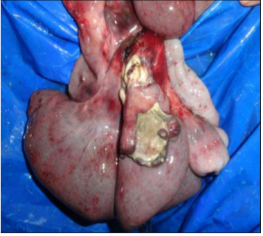 Figure 5
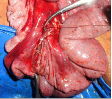 Figure 6
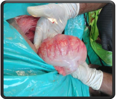 Figure 7
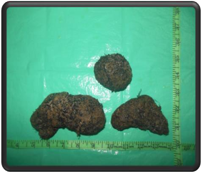 Figure 8
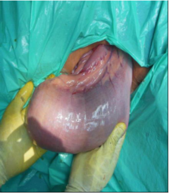 Figure 9
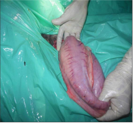 Figure 10
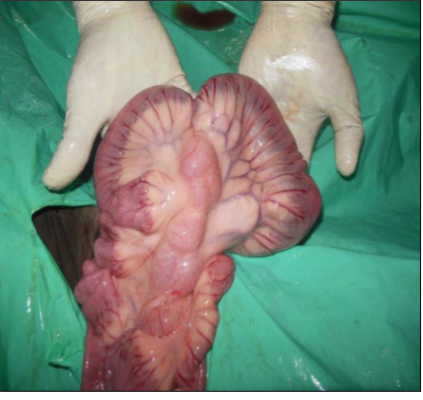 Figure 11
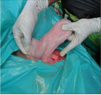 Figure 12
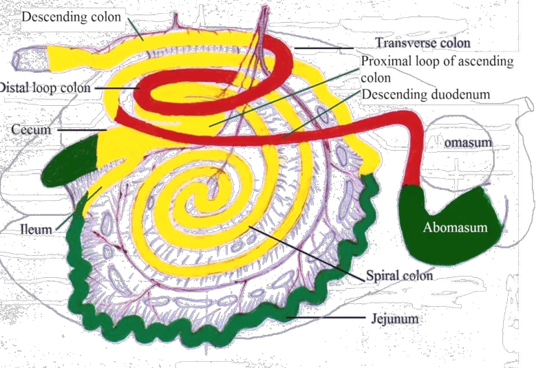 Figure 13
Introduction
Surgical diseases of the digestive tract in ruminants are numerous and represent a major clinical problem and abdominal surgery generally constitutes the single largest group of operations carried out in ruminants [1] . Ruminants with obstructive gastrointestinal diseases show many common clinical symptoms like scanty or total absence of fecal output, complete or partial anorexia and varying degrees of abdominal distension with or without abdominal pain [2] . These conditions pose a significant diagnostic challenge to the veterinarian under field conditions with limited diagnostic facilities. Surgical conditions of the fore stomachs like impaction, obstruction of the reticulo omasal orifice, ruminoreticular foreign bodies or displacement of abomasum exhibit these types of symptoms [3] . The same symptoms are also manifested by obstructive diseases of the small and large intestines: volvulus, intussusception, strangulation, caecal affections, extraluminal (tumours) or intraluminal obstructions (bezoars) [4] . Internal hernias (gut tie, diaphragmatic or omental hernias) although rarely found, can give rise to similar symptoms [5] . Functional obstructions of the gastrointestinal system (vagal indigestion, paralytic ileus) also mimic the same symptoms [6] and need to be carefully distinguished from the physical conditions. Different distention patterns of bovine abdomen (left or right side distension, upper or lower quadrant distension) may give a rough idea of the organ involved but further tests are required to find the exact location of the lesion. There are several methods practiced by veterinarians for diagnosing abdominal disorder such as auscultation, percussion, rectal palpation but none of them give a conclusive diagnosis to the operating surgeon. Though radiography and ultrasonography can also give valuable clues they are purposefully excluded in the present study. In the absence of a confirmed diagnosis, abdominal exploration has been used extensively and a right flank approach is commonly employed. However, if during a right flank exploration the surgeon learns that omasal impaction is the cause of obstruction it has to be approached from a rumenotomy incision through the left flank. In the standing left flank approach for rumenotomy, exploration of the right side of the abdomen is not possible. Hence, to deal with undiagnosed cases of GI obstruction in cattle an alternate novel laparotomy incision site is required through which the fore stomachs can be surgically approached and exploration of the right side of the abdomen can be done. In addition, this approach can facilitate surgical handling of some obstructive conditions. Therefore, a Left ventrolateral oblique laparotomy approach under regional anesthesia was followed in the present study.
Materials and Methods
Materials and Methods
Ethical Approval
The approval from the Institutional Animal Ethics Committee to carry out the current study was not required as all the bovine cases were clinical suitable candidate for surgical intervention and surgery was performed as a remedial measure to cure the animal rather than putting stress on the animals.
Screening of animals and study design
For the purpose of this study, all the bovine cases exhibiting obstructive gastrointestinal symptoms referred to the Department of Veterinary Surgery and Radiology, College of Veterinary Science and Animal Husbandry (CVSc & AH), OUAT for a period of one year were carefully sorted out on a case by case basis. A total of 47 cases were thoroughly examined by the usual diagnostic methods like observing vital signs, auscultation, palpation, percussion and hematobiochemical examination as described below. Radiography and ultrasonography were excluded during the period of study. Based on the clinical examination detailed below in 31 animals (66%) a definitive diagnosis could be done; they were treated by the usual surgical or medical protocols and excluded from the study. In the remaining 16 animals (34%) where a definitive diagnosis could not be established, surgical exploration was attempted through a left ventrolateral oblique laparotomy with regional anesthesia for arriving at a definitive diagnosis and suitable treatment regimen. These 16 animals of either sex and irrespective of age formed the subjects of the present study. Sixteen animals were divided into 2 groups by the following criteria. Group I included 10 animals in which the diagnosed surgical condition was easily accessed and corrected. Complete obstruction of the reticulo-omasal orifice with a large phytobezoar (n=1), omasal impaction (n=2), ruminoreticular foreign bodies (n=2), traumatic reticulitis (n=2) and concurrent omasal impaction and ruminoreticular foreign bodies (n=3) were diagnosed. The phytobezoar and foreign bodies were removed manually or by magnet swapping and omasal impaction was treated by omasal flushing. Group II included 6 animals in which the diagnosed surgical condition was approached and treated with "some difficulty" or could not be corrected. Conditions like cecal dilation and displacement (n=2), intestinal volvulus (n=1), obstruction of the abomasum with phytobezoars (n=1) and diaphragmatic hernia (n=2) were diagnosed.
Signalment and anamnesis
The age sex, breed, pregnancy, lactation status, pedigree, and last date of breeding of all the animals were recorded. A brief and complete history about onset of disease, time of onset, symptoms observed and duration of illness, drop in milk yield, reduction in appetite and food consumption and management practices, previous treatments used, evidence of abdominal pain, nature and volume of the feces voided were all recorded and documented in table 1
Clinical Examination
Thorough clinical examination was carried out by measuring vital signs such as temperature, respiration, pulse rate, visible mucous membrane, capillary refill time, dehydration status [7] , auscultation, percussion. Special examinations of the various abdominal organs are done as follows. Palpation of abdominal organ through the body wall on the left and right side was done as per description of Smith (2015) [8] . Examination of abdominal contour, palpation of left and right hemiabdomen, rumen motility, abdominal auscultation and percussion of both left and right side, abdominal ballottement, detection of foreign body through metal detector was carried out systematically. Various tests were also performed for eliciting signs of pain such wither pinch test and pole test or bar test. Trans-rectal examination was carried out in all the animals to determine presence or absence of fecal material, its colour and consistency. In females, the uterus was examined for pregnancy, stage of gestation, viability of the fetus, disposition of the gravid horns and any other fetal or maternal abnormalities (retained placenta, metritis, pyometra, maceration, mummification, hydrops amni, etc.). The contents and disposition of the caudal and visceral part of rumen were palpated for any obvious abnormalities e.g. ruminal collapse, medial displacement in LDA, L-shaped rumen in vagus indigestion etc. The cecum was searched and palpated for any dilatation, displacement or torsion by palpating apex and palpable portion of the body of the cecum. The accessible portion of the caudal right abdomen was searched for any sausage shaped firm mass (suggestive of intussusceptions) or for a large ingesta or gas filled viscous (suggestive of RDA). Any other obvious abnormalities found were noted.
Rumen Liquor Examination
Collection of rumen fluid was done by introducing a lubricated probang through the mouth into the rumen and to avoid contamination with saliva, a minimum of 100 ml of ruminal fluid was collected by gentle to and fro motion of the probang [9] . Ruminal fluid pH was determined immediately on the spot by wide range pH paper and the results were recorded. Ruminal fluid was examined for density and activity of protozoa by observing a drop of rumen fluid under the low power objective [10] . Rumen liquor was filtered through a double layer muslin cloth and then centrifuged at 3000 rpm for 5 minutes and chloride concentration was estimated using diagnostic kits with the help of an autoanalyzer (Turbochem-100). Hemato-Biochemical tests were performed on the day of operation (day 0), 3 rd and 7 th post-operative days to monitor therapy and evaluate the process of recovery.
Hemato-Biochemical studies
Blood collected from jugular vein was used for estimation of PCV, hemoglobin, differential count and total leukocyte count. Serum was also collected to estimate serum sodium, potassium, calcium and chloride using diagnostic kits with the help of an autoanalyzer (Turbochem-100). All the laboratory tests were performed on the day of operation (day 0), 3 rd and 7 th post-operative days to monitor therapy and evaluate the process of recovery.
Selection of Surgical Site and anesthesia
Since flank approach has limited accessibility, left ventrolateral oblique incision (Figure 1) was chosen in order to have easy approach to a larger area of the abdominal cavity. The proposed incision was given on the lower left abdomen starting from a point just above the level of the stifle and extending obliquely and cranioventrally up to the last rib (Figure 2) [11] . The surgical site was aseptically prepared and draped using sterile plastic drapes. Aggressive and uncooperative animals were given xylazine @ 0.03-0.05 mg/ kg body weight intramuscularly along with local analgesia. Local analgesia was achieved by linear infiltration or paravertebral anesthesia in all the cases and due care was taken not to exceed the toxic dose of lignocaine [12] .
Surgical Exploration, Diagnosis and Treatment
The animals in which no preoperative diagnosis could be made (n=16) were operated by a left ventrolateral oblique laparotomy approach for exploratory diagnosis. They were restrained properly on a soft bed in right lateral recumbency. In this technique after the skin incision, the external most cutaneous trunci muscle and the successive layers of external, internal and transverse abdominal muscles were bluntly separated longitudinally along their fiber direction and the peritoneum was incised. On reaching the abdomen the dorsal sac of the rumen was exteriorized, brought out through the incision, fixed to the rumenotomy ring and incised. All foreign materials present within the rumen were removed and 3/4 th of contents were emptied. Magnet swapping of the reticulum was done and any metallic foreign bodies present were retrieved. In the presence of omasal impaction, omasal flushing was done as per the method described by Nayak et al. (2000) [13] . The cardia and reticulo omasal orifices were explored for any abnormalities like presence of abnormal masses, foreign bodies or immature amphistomes. Twenty yeast tablets were put into the rumen to balance the microflora and fauna. Then the ruminal incision was lavaged with copious saline and closed with No-2 chromic catgut in inverted Cushing pattern. The abomasum was palpated by passing the hand between the floor of the rumen and the ventral body wall to the right side for any abnormalities which was attempted for correction by exteriorizing the abomasum. Then the rumen was slightly deflected cranially with the greater omentum and the small and large intestines were explored for obstruction or any other abnormality. Attempt was made to exteriorize the affected part and effect necessary surgical correction. Laparotomy wound was closed by apposing the muscle layers separately with no-2 chromic catgut as per standard procedure. Skin incision was closed routinely. The results and findings were analyzed in each group. During exploration of the gastrointestinal tract the approachability of different parts were carefully observed and recorded.
Postoperative Care and Management
All the animals were given fluid therapy for 3 to 5 days which was modified according to the need of the patient based on laboratory findings and clinical improvement. Streptopenicillin was given intramuscularly once daily for 5-7 days and Metronidazole inj @ 10 mg/kg IV once daily for 3 days. NSAID's (Meloxicam @ 0.2mg/kg IM or Flunixin meglumine @ 1mg/kg IV) were given daily for 3 days. In some cases, Neostigmine @ 0.02 mg/ kg SC BID for 2 to 3 doses was given to promote intestinal motility. Regular dressing of the wound with fly repellent cream was done till healing. A clean cloth was covered over the wound for 2 to 3 days. The owners were advised to give good nursing care and liquid diet to their animals. One to two liters rumen cud from fresh goat rumens was given to the animals that remained anorectic after evaluation of rumen pH and protozoa and it was repeated when necessary. Feeding was gradually changed from soft to coarse diet. Sling support to recumbent and weak animals was given in order to bring them to foot. Abscess on the suture line was treated on general principles by removing one or two sutures, draining the pus and application of topical antiseptics.
Statistical Analysis
Analysis of variance (ANOVA) was used to compare the means on different days among different groups. Paired "t" test was used to compare the mean values on different days with their respective base value in each group [14] . For nonparametric observations Kruskal-Wallis one-way test was used to compare the mean between the groups on corresponding days [15] .
Results
The mean and standard error value of vital signs, rumen fluid examination, haematological and biochemial parameters were evaluated in sixteen animals on zero day, 3rd and 7 th
Result of Surgical exploration and treatment
As laparotomy was done by grid technique, during blunt dissection the spinal nerves could be retracted safely without the need for trans-section and bleeding was minimal in all cases. In Group I, a 15 to 30 cm long incision was adequate for the purpose of laparotomy and subsequent exploration. The rumen could be exteriorized easily in 6 cases. In 4 cases after carefully packing of the exteriorized part of the rumen, decompression was done by giving a small incision and releasing some of the gas after which complete exteriorization of the site of incision on dorsal sac became easy. In Group II, a 20 to 30 cm incision was adequate for laparotomy and subsequent exploration. In one case extension of the incision was required for abomasotomy for better exteriorization. In two cases the rumen was easily exteriorized and in another two cases the rumen could be exteriorized after removal of some amount of ingesta from a small incision with utmost care not to soil the peritoneal cavity with ruminal contents. In the rest two cases, some amount of frothy material was removed through a small incision and then the dorsal sac was easily exteriorized. Since a preoperative diagnosis could not be achieved in these 16 cases under study, abdominal exploration through a left ventrolateral oblique incision formed the basis for diagnosis and discussed below. The results of exploratory diagnosis and treatment have been summarized in table 4. Diaphragmatic hernia Herniorrhaphy declined *Total typhlectomy was done by a second right flank approach as ileo-caeco-colic orifice was not exteriorizable from the left ventrolateral oblique site.
Group
Group
Postoperative Care
All 15 animals received fluid therapy for 1 st 3 days that consisted of physiologic saline and 5% dextrose in physiologic saline [16] . Additionally, ten animals with hypocalcemia received calcium borogluconate @ 50-200 ml IV daily. Three animals needed an additional infusion of calcium on 4 th day. Based on the serum electrolyte levels fluid was discontinued on 3 rd day in 8 animals. Seven animals having electrolyte deficiency or anorexia were given fluid therapy for 2 more days. Four animals were found to be hypokalemic on 3 rd postoperative day and were given potassium chloride @ 5-10 gm/animal orally for 5 days. Minimum 5 days of antibiotic therapy was continued in all animals. In addition, all animals received metronidazole IV for 3 days to combat anaerobic infection. Meloxicam was given @ 0.2mg/kg IM for 3 days to 13 animals and Flunixin meglumine @ 1 mg/kg IV for 3 days was given to two animals. Four animals were given neostigmine @ 0.02mg/kg SC twice daily for 2 days to promote intestinal motility under close supervision. On the 3 rd postoperative day, 6 animals remained anorectic with low viable protozoal count. One of them had ruminal pH 8 which was corrected by administering 0.5 L of vinegar orally into the rumen. All other animals had normal or slightly acidic pH. All these 6 animals were given 2 liters of rumen cud from fresh goat rumens brought from slaughter house twice on 3 rd and 5 th day. All animals showed dramatic improvement in appetite and rumination following cud transplantation. Sutures were removed in all animals from 12-15 days. Out of 16 animals operated in Group I 8 animals recovered well and 2 animals died whereas 3 animals in Group II survived out of 6 animals indicating the complexity of the surgery done.
Discussion
As this study was conducted on undiagnosed cases of gastrointestinal obstruction in cattle using left ventrolateral oblique laparotomy approach, a systematic exploration of the forestomachs and the small and large intestines were done in every case to establish a diagnosis. During exploration, the approachability of the gastrointestinal tract was carefully observed and noted. Parts of the gastrointestinal tract palpable and exteriorizable through this site are rumen (reticulum and reticulo-omasal orifice accessible from rumenotomy site), abomasums (Figure 10), Jejunum (except a small proximal portion) (Figure 11), Cecum (2/3 rd of its length from the blind end) (Figure 12). Parts of the gastrointestinal tract palpable but that were not exteriorizable are ascending duodenum from caudal flexure to the beginning of jejunum, Proximal portion of jejunum, Ileum, Cecum (1/3 rd of its length from ileocecocolic junction), Colon (proximal loop and spiral loop), Descending colon, Peritoneal part of rectum. Parts of the gastrointestinal tract which were neither palpable nor exteriorizable are descending duodenum up to caudal flexure, Colon (distal loop and transverse colon) (Figure 13).
Conclusion
Since most parts of the gastrointestinal tract (except the ascending duodenum and small part of colon) can be explored from a left ventrolateral oblique laparotomy site, this approach leads to a confident and nearly complete exploration of both parts of the abdomen in undiagnosed cases of gastrointestinal obstruction in cattle. As obstructive diseases of the forestomachs, cecum and jejunum are most common and these organs are easily accessed from this site, a left ventrolateral oblique laparotomy approach can be adopted as a working solution for abdominal exploration and surgical correction of commonly found gastrointestinal obstructions where a confirmatory diagnosis is not possible. Omasal flushing for treatment of omasal impaction is easier from a left ventrolateral oblique laparotomy site than from higher left flank incision in standing position particularly in large sized animals as long hands are required to reach the reticuloomasal orifice in case of the latter approach. This approach can also be used in cattle diagnosed with multiple and concurrent obstructive conditions of the forestomachs, jejunum and/or cecum for effective surgical management from a common site. As this study was conducted in 16 clinical cases, further investigation is required for validation of this technique in more number of animals.
Acknowledgement
AcknowledgmentsThe author is highly obliged to the livestock owners who have enthusiastically supported in carrying out the postoperative evaluation of the clinical cases.
Figures and tables from the paper
| — | 9 | Recumbent, not examined | Rumen doughy | No intestinal sounds | Tympanitis resonance on left upper flank |
|---|---|---|---|---|---|
| 10 | Recumbent, not examined | Pain on deep palpation R side | No intestinal sounds | NAD | |
| II | 1 | Bilateral lower part distended | NAD | No intestinal sounds | Tympanitis resonance on both flanks |
| 2 | Bilateral lower part distended | Painful to touch | NAD | NAD | |
| 3 | Bilateral Lower part & L upper part distended | Rumen doughy | NAD | Tympanitis resonance on left upper flank | |
| 4 | Bilateral lower and L upper part distended | Gas filled rumen | No intestinal sounds | Tympanitis resonance on both flanks. | |
| 5 | L upper part distended | NAD | rumen sounds not audible | Tympanitic resonance | |
| 6 | L upper part distended | NAD | rumen sounds not audible | Tympanitic resonance |
Table text extracted from the original PDF; superscript letters are shown inline. The original table image is below.
View original table image
| Goroup | Case No | Ballotment | Rectal palpation | Exam with metal detector | Withers pinch test | Pole test | VMM | CRT in sec | Dehydration % |
|---|---|---|---|---|---|---|---|---|---|
| 5 | 6 | 7 | 8 | 9 | 10 | 11 | 12 | ||
| I | 1 | Fluid splashing rumen | No fecal material | - ve | - ve | + ve | Pale | 2 | 6 |
| 2 | NAD | Not possible | - ve | + ve | + ve | Pink | 2 | 6 | |
| 3 | NAD | Few hard beads of feces in empty rectum | - ve | - ve | + ve | Pink | 2 | 6 | |
| 4 | NAD | Rectum empty | - ve | - ve | - ve | Pink | <2 | 0 | |
| 5 | NAD | Small amount of hard fecal balls in rectum | + ve | + ve | + ve | Pale | 2 | 6 | |
| 6 | NAD | Rectum empty | + ve | + ve | + ve | Pink | 3 | 8 | |
| 7 | NAD | Rectum empty | + ve | - ve | - ve | Pink | <2 | 0 | |
| 8 | NAD | Not possible | - ve | - ve | - ve | Pink | 3 | 8 | |
| 9 | NAD | Empty rectum, dilated rumen | - ve | + ve | + ve | Pink | 2 | 6 | |
| 10 | NAD | Empty rectum | - ve | + ve | + ve | Bright | 2 | 6 | |
| II | 1 | Fluid splashing rumen and intestines | Rumen distended, dilated intestines felt | - ve | + ve | - ve | Pink | 3 | 8 |
| 2 | NAD | Empty rectum with mucous | + ve | + ve | + ve | Pale | 5 | 10 | |
| 3 | NAD | Enlarged rumen, empty rectum | + ve | - ve | - ve | Pale | 2 | 6 | |
| 4 | NAD | Empty mucoid material | + ve | + ve | - ve | Pink | 2 | 6 | |
| 5 | NAD | Distended rumen, soft scanty fecal material | + ve | + ve | + ve | Bright | 2 | 6 | |
| 6 | NAD | Distended rument soft scanty fecal material | - ve | + ve | + ve | Pink | 2 | 6 |
Table text extracted from the original PDF; superscript letters are shown inline. The original table image is below.
View original table image
References
12 in the original paper- Singh J, Singh AP, Patil DB. The digestive system, in Ruminant surgery by Tyagi RPS, and Singh J, 1st ed, CBS Publishers and Distributors, New Delhi, 1993, 183224.
- Asrat M, Manohar M, Melkamu S. Clinical and Rumen Fluid Evaluation of Ruminal Disorders in Cattle, Journal of Animal Research. 2015; 5(2):359-372.
- Asrat M, Velappa R. Current Advances in surgical management of Ruminal Disorders of bovine, International Journal of Veterinary Sciences and Animal Husbandry. 2016; 1(3):32-38.
- Fubini SL, Ducharme NG. Bovine surgery, in Farm Animal Surgery, St. Louis, MO, Elsevier, 2004, 189-208.
- Divers TJ, Peek SF. Non infectious diseases of the gastrointestinal tract, in Rebhun's diseases of dairy cattle, 2 nd ed, Saunders Elsevier, 2008, 130-199.
- Radostits OM, Gay CC, Hinchcliff KW, Constable PD. Diseases of the alimentary tract II, in Veterinary Medicine-A Textbook of the Diseases of Cattle, Horses, Sheep, Pigs and Goats. 10 th ed. Saunders Elsevier, Philadelphia, 2007, 293-382.
- Fecteau ME. Fluid therapy in ruminants, in Large animal internal medicine, 5 th ed. Elsevier Mosby, St. Louis, Missouri, 2015, 1387-1390.
- Smith BP. Large Animal Internal Medicine, 5th ed. Elsevier Mosby, St. Louis, Missouri, 2015, 739-842.
- Garrett EF, Pereira MN, Nordlund KV. Diagnostic methods for the detection of subacute ruminal acidosis in dairy cows, Journal of Dairy Science. 1999; 82:1170.
- Cockcroft P, Jackson P. Clinical examination of the abdomen in adult cattle, In Practice. 2004; 26:304-317.
- Parish SM, Tyler JW, Ginsky JV. Left oblique celiotomy approach for cesarean section in standing cows, Journal of American Veterinary Medical Association. 1995; 207:751-752.
- Clarke KW, Trim CM, Hall LW. Anesthesia in cattle, in Veterinary Anaesthesia, 11 th ed, Elsevier, 2014, 313-343.