
Research article · 2026 · Full text
International Journal of Biology SciencesIsolation, characterization, molecular identification and abiotic stress tolerance profiling of nodule associated bacteria from Clitoria ternatea L.
Volume 8(9) · Pages 21–30 · DOI: 10.33545/26649926.2026.v8.i9a.986 ↗
Research areasPlant-Microbe Interactions and ImmunityLegume Nitrogen Fixing SymbiosisMicrobial Community Ecology and Physiology
Abstract
Author-writtenThis study was conducted to identify and characterize the rhizobacteria found in the root nodules of Clitoria ternatea L. Seeds of Clitoria ternatea L. (blue single petal and blue double petal varieties) were grown in normal soil, and four types of bacteria were isolated. Morphological and biochemical analyses were performed to identify and characterize these bacteria. The ability of each bacterium to tolerate various abiotic stresses (such as pH, salinity, and temperature) was also examined. Their phylogenetic identification was determined based on 16S rRNA analysis. The four isolated bacteria were found to belong to the species Bacillus velezensis and Bacillus zanthoxyli.
Summary
Source-checkedPlain-language reading of the full paper, prepared with AI and checked against the text. Each point links to the authors' own words.
Why it matters
- Biological nitrogen fixation is essential for life, and leguminous plants succeed largely due to their symbiotic relationship with nitrogen-fixing Rhizobia bacteria. View evidence ↗
- Beyond true Rhizobium, other non-rhizobial bacteria like Bacillus and Streptomyces can exist as endophytes within root nodules. View evidence ↗
- The nodule flora of the legume Clitoria ternatea L. has not been previously identified. View evidence ↗
The question
- The study aimed to identify and characterize the morphological, biochemical, molecular, and stress-tolerance properties of bacteria found in the root nodules of Clitoria ternatea L. View evidence ↗
What they did
- The researchers used blue-flowered single-petal and double-petal varieties of Clitoria ternatea L. grown in normal soil. View evidence ↗
- Root nodules were surface-sterilized using 70% alcohol and 0.1% mercuric chloride for 3 minutes each before bacterial isolation. View evidence ↗
- Experiments were conducted in triplicate and analyzed using one-way ANOVA, with significance set at p ≤ 0.05. View evidence ↗
What they found
- Four Gram-positive bacterial strains were isolated: two from the single-petal variety (CTspbNSI, CTspbNSII) and two from the double-petal variety (CTdpbNSI, CTdpbNSII). View evidence ↗
- The isolated bacteria were mesophilic, tolerating temperatures from 20°C to 44°C with an optimum growth temperature of 28°C. View evidence ↗
- All four isolates showed broad pH tolerance, growing in ranges from pH 4 to pH 10. View evidence ↗
- The isolates demonstrated salinity tolerance up to 5% NaCl. View evidence ↗
- Statistical analysis confirmed that pH (p = 0.048), temperature (p < 0.001), and the specific strain (p = 0.005) significantly influenced bacterial growth. View evidence ↗
What it means
- The authors conclude that these diverse, stress-tolerant strains can be used to enhance nitrogen fixation in legumes, supporting sustainable food security. View evidence ↗
Visual summary
2 visualsIllustrations explain the paper; charts are replotted from the paper's own table values.
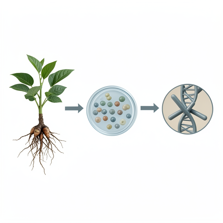
Workflow of Bacterial Identification
AI-generated conceptual illustration (Clitoria ternatea L. root nodules and isolated bacteria). It explains the idea and does not show study data.
View source evidence ↗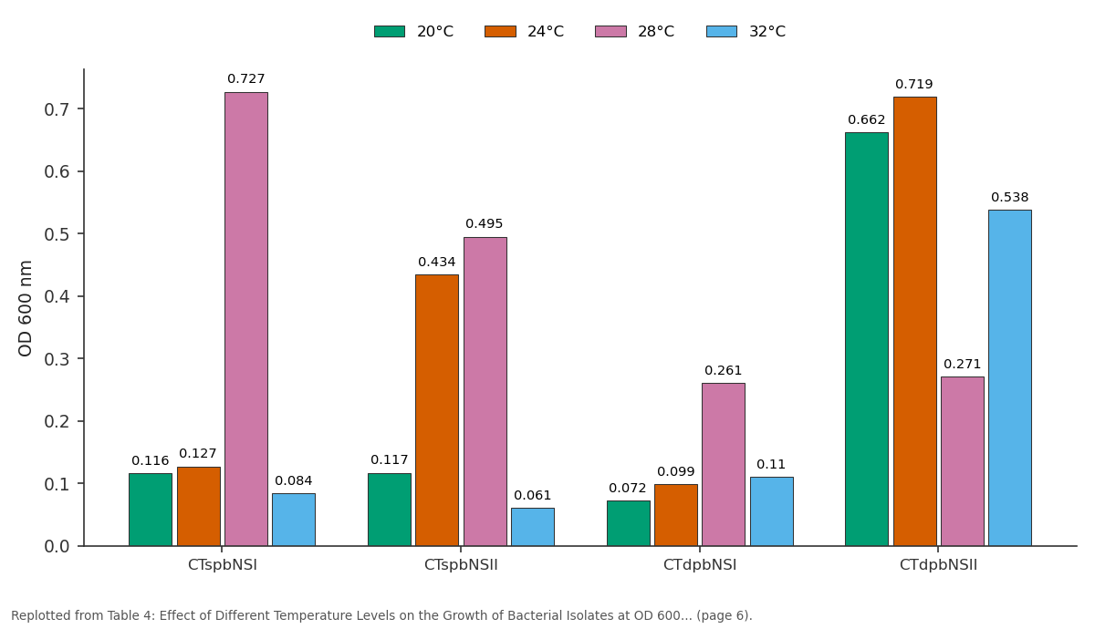
20°C and 24°C and more: CTspbNSI vs CTspbNSII vs CTdpbNSI vs CTdpbNSII
Replotted from Table 4 (page 6) of the original paper. Every bar is the paper's own table value.
View source evidence ↗Figures & tables
From the original paper Table 1
 Table 2
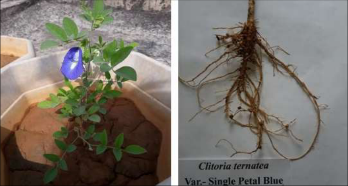 Figure 1
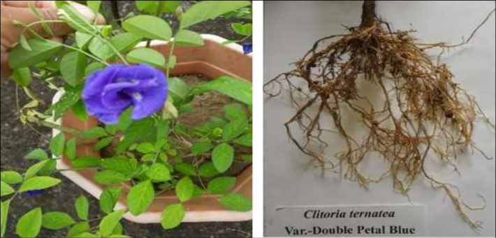 Figure 2
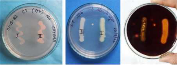 Figure 2
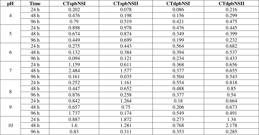 Table 3
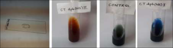 Figure 3
 Table 4
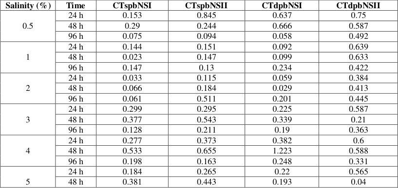 Table 5
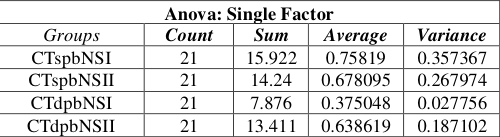 Table 6
 Table 7
 Table 8
 Table 8
 Table 8
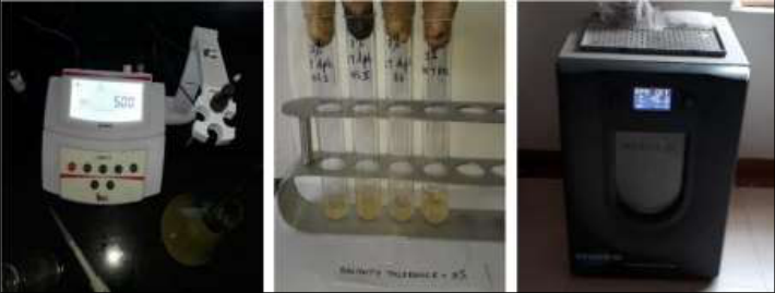 Figure 4
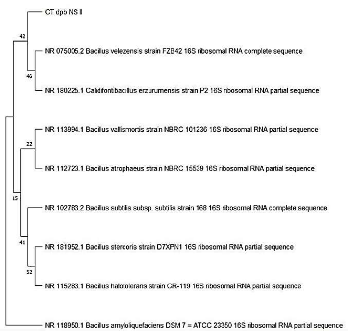 Figure 5
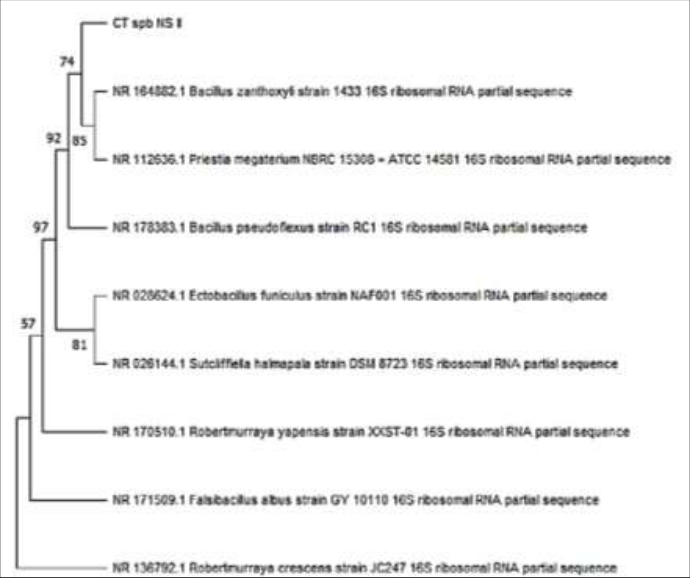 Figure 6
Introduction
Nitrogen (N 2 ) is an essential element for all forms of life. It is found in amino acids and proteins, and the process of nitrogen fixation leads to the formation of numerous other organic compounds [1] . N 2 is the most abundant gas in Earth's atmosphere; however, it is not highly reactive [2] . Biological nitrogen fixation is a vital component of microbial processes [3] . The Leguminosae family is the largest plant family on Earth. A key reason for the success of
Material and Methods
Material and Methods
Seed collection
Seeds of the blue-flowered, single-petal Clitoria ternatea L. were purchased online from 'Jai Shree Radhe' (New Delhi) and blue flowered, double petal was purchased from Banrujjedar Orchid Bagan Private Limited, Kolkata, West Bengal and grown in pots containing normal soil.
Collection and surface sterilization of root nodules
After flowering, nodules were harvested from the roots of freshly uprooted plants. The roots were thoroughly washed with tap water to remove impurities. Healthy nodules were selected for bacterial isolation. Initially, the harvested nodules were washed under running tap water to remove soil particles adhering to their surfaces. Subsequently, the surfaces of nodules were sterilized using 70% alcohol for 3 minutes, followed by 0.1% mercuric chloride for another 3 minutes, and finally rinsed multiple times with sterilized distilled water under aseptic conditions [25,26,27,28,29,30] .
Bacterial isolation and purification
The nodules were crushed using a sterilized mortar and pestle to obtain a milky suspension of bacteroid. This suspension was spread onto Yeast Extract Mannitol Agar (YEMA) medium. The plates were then incubated at 28 ± 2°C for 24 hours. After 24 hours, colonies were selected for sub-culturing to obtain pure cultures.
Characterization of cultures
The isolated bacteria were characterized based on their size, form, elevation, margin, color (pigmentation), Gram staining reaction, and appearance on YEMA medium.
Morphological characterization
Size, form, elevation, margin, pigmentation, and appearance were observed on YEMA medium. (Vincent, 1970): This method was used to classify bacteria into Gram-positive and Gram-negative. Using a clean slide and a sterilized wire loop, a loopful of normal saline was placed in the center of the slide, and the loop was re-sterilized. A sample was collected using the wire loop, and a circular smear was prepared. After preparing the smear, the slide was gently heat-fixed by passing it over a Bunsen flame. Crystal violet was applied to the smear for 30 seconds and then washed off with water. Iodine solution was added and left for one minute, followed by washing with water. Subsequently, acetone or ethanol was applied for decolorization and immediately rinsed off with water. Counter-staining was performed using safranin within one minute. After drying, the slide was examined under a compound microscope. Gram-negative cells appear pinkish-red, while Gram-positive cells appear violet.
Gram's reaction
Gram's reaction
Biochemical characterization
The bacterial isolates were characterized by various biochemical methods [31,32,33,34,35] . a) Casein test: Milk agar medium was prepared and inoculated with the isolated strains, then incubated at 28±2°C for 24 hours. dispensed into test tubes, and allowed to solidify. The isolated strain was inoculated and incubated at 28±2°C for 24 hours. The colour of both the butt (bottom section) and the slant (sloped section) was observed. j) Congo red test: The strain culture was plated on CRYEMA medium and incubated at 28°C for 24 hours. After this period, the dye-absorbing capacity of the bacteria was observed.
Physiological characterization 1. pH Tolerance:
The pH tolerance capacity of all bacterial isolates was tested using Yeast Mannitol (YEM) broth adjusted to various pH levels (e.g., 4, 5, 6, 7, 9, and 10). The pH of the YEM broth was adjusted using HCl and NaOH. Each isolate was inoculated into YEM broth and incubated at 28°C. The effect of pH on the growth of each isolate was recorded at 24, 48, and 96 hours.
Temperature Tolerance:
The effect of temperature on the growth of each bacterial isolate was assessed using YEM broth. Each isolate was inoculated into YEM broth and incubated at various temperatures (e.g., 20, 24, 28, 32, 36, 40, and 45°C), and the growth pattern of each isolate was recorded at 24-hour intervals. 3. Salinity tolerance: Tolerance to sodium chloride (NaCl) was tested for each bacterial isolate using YEM broth supplemented with varying concentrations of NaCl (e.g., 0.5%, 1%, 2%, 3%, 4%, and 5%). The effect of salinity on the growth of each isolate was recorded at 20, 48, and 96 hours.
The growth of bacterial isolates were recorded using spectrophotometer by measuring the optical density (OD) at 600 nm.
Molecular identification: All four bacterial isolates were selected for molecular characterization. DNA was extracted from the isolates, and its quality was assessed on a 1.0% agarose gel. Amplification of the rRNA gene was performed using LCO 1490 and HCO 2198 primers. Upon resolution on an agarose gel, a single, distinct PCR amplicon band of 1500 bp was observed. Forward and reverse DNA sequencing reactions of the PCR amplicons were conducted using the BDT v3.1 cycle sequencing kit on an ABI 3730xl Genetic Analyzer. The rRNA gene sequences were used to perform BLAST searches against the NCBI GenBank database to identify the bacterial strains. The top ten sequences based on identity scores were selected and aligned using the multiple alignment software program Clustal W. Phylogenetic trees were constructed using the Maximum Likelihood method based on the Kimura 2parameter model within the MEGA 11 software package [36,37] .
2.9. Statistical Analysis -All experiments were performed in triplicate, and the results were expressed as mean ± standard deviation (SD). Statistical analysis was performed using appropriate statistical software. Differences among treatments were analyzed using one-way analysis of variance (ANOVA). Differences were considered statistically significant at p≤0.05. The relationship between phytochemical constituents and antioxidant activity was statistically evaluated using correlation analysis. The experimental data were presented graphically using mean values with appropriate error bars.
Result Isolation and morphological characterization
A total of four strains were isolated from the root nodules of Clitoria ternatea L. (two from the blue single-petal plant, designated CTspbNSI and CTspbNSII, and two bacterial isolates from the blue double-petal plant, designated CTdpbNSI and CTdpbNSII). Gram staining was performed on all isolates, and microscopic examination revealed that all four studied isolates were Gram-positive. The nitrogenfixing bacteria were isolated on YEMA selective medium, and all samples showed good growth. The colony morphology of the isolates was observed; all isolates exhibited creamy-white colonies (Table 1).
| S. No. | Isolates | Morphological characterization of bacterial isolates from Clitoria ternatea L | — | — | — | — | — | — |
|---|---|---|---|---|---|---|---|---|
| Size | Form | Elevation | Margin | Pigmentation | Gram staining | CRYEMA Media | ||
| 1 | CTspbNSI | Small, Moderate | Circular | Raised | Entire | Off white | +ve | Pale yellow |
| 2 | CTspbNSI I | Small, Moderate | Rhizoid | Flat | Undulate | White | +ve | Pale pink |
| 3 | CTdpbNS I | Pin point, small | Circular | Raised | Entire | White | +ve | Pale Pink |
| 4 | CTdpbNS II | Small, Moderate | Circular | Flat | Entire | White | +ve | Pale Pink |
Table text extracted from the original PDF; superscript letters are shown inline. The original table image is below.
View original table image
Cultural and metabolic characterization
Results from qualitative tests on YEMA + Congo Red medium showed that three isolates (CTspbNSII, CTdpbNSI, and CTdpbNSII) appeared pale pink, while one isolate (CTspbNSI) appeared pale yellow. This indicated that the isolates did not absorb the red dye from the Congo Red indicator present in the medium.
Biochemical characterization
Several biochemical tests were conducted to identify and characterize the isolates in accordance with Bergey's Manual. The purity of the test isolates was re-confirmed through various biochemical tests on different media. The biochemical behavior of the test strains is presented in the table (Table 2.).
| S. No. | — | 1. | 2. | 3. | 4. |
|---|---|---|---|---|---|
| Isolates | CTspbNSI | CTspbNSII | CTdpbNSI | CTdpbNSII | |
| Biochemical Test | Casein Test | - | - | - | - |
| Catalase test | + | + | + | + | |
| Citrate utilization Test | + | + | + | + | |
| Gelatin Hydrolysis test | - | - | - | - | |
| Indole test | - | - | - | - |
Table text extracted from the original PDF; superscript letters are shown inline. The original table image is below.
View original table image
Physiological Characterisation
In this study, most isolates were able to tolerate temperatures ranging from 20°C to 44°C, with optimal growth observed at 28°C (Table 3). The results of this test align with previous studies indicating that root nodule bacteria are mesophilic, with an optimal growth temperature of 28°C for most isolates.
| pH | Time | CTspbNSI | CTspbNSII | CTdpbNSI | CTdpbNSII |
|---|---|---|---|---|---|
| 4 | 24 h | 0.202 | 0.078 | 0.086 | 0.216 |
| 48 h | 0.476 | 0.198 | 0.156 | 0.299 | |
| 96 h | 0.79 | 0.519 | 0.421 | 0.475 | |
| 5 | 24 h | 0.898 | 0.978 | 0.476 | 0.445 |
| 48 h | 0.674 | 0.874 | 0.349 | 0.399 | |
| 96 h | 0.449 | 0.699 | 0.199 | 0.232 | |
| 6 | 24 h | 0.275 | 0.443 | 0.564 | 0.682 |
| 48 h | 0.132 | 0.384 | 0.394 | 0.537 | |
| 96 h | 0.094 | 0.121 | 0.234 | 0.433 | |
| 7 | 24 h | 1.159 | 0.611 | 0.368 | 0.656 |
| 48 h | 2.484 | 1.577 | 0.377 | 0.655 | |
| 96 h | 0.161 | 0.035 | 0.504 | 0.543 | |
| 8 | 24 h | 0.252 | 1.161 | 0.554 | 0.818 |
| 48 h | 0.447 | 0.652 | 0.488 | 0.85 | |
| 96 h | 0.876 | 0.258 | 0.377 | 0.54 | |
| 9 | 24 h | 0.842 | 1.264 | 0.18 | 0.664 |
| 48 h | 0.657 | 0.75 | 0.206 | 0.673 | |
| 96 h | 1.737 | 0.174 | 0.549 | 0.491 | |
| 10 | 24 h | 0.887 | 1.872 | 0.273 | 1.34 |
| 48 h | 1.6 | 1.281 | 0.768 | 2.178 | |
| 96 h | 0.83 | 0.311 | 0.353 | 0.285 |
Table text extracted from the original PDF; superscript letters are shown inline. The original table image is below.
View original table image
All the bacterial isolates tested demonstrated a broad pH tolerance, capable of growing across acidic and alkaline ranges from pH 4 to 10 ((Table 4). The optimal pH for the growth of root nodule bacteria typically falls between 6 and 7.
| S. No. | Isolates | 20°C | 24°C | 28°C | 32°C | 37°C | 40°C | 45°C |
|---|---|---|---|---|---|---|---|---|
| 1 | CTspbNSI | 0.116 | 0.127 | 0.727 | 0.084 | 0.227 | 0.443 | 0.138 |
| 2 | CTspbNSII | 0.117 | 0.434 | 0.495 | 0.061 | 0.216 | 0.324 | 0.11 |
| 3 | CTdpbNSI | 0.072 | 0.099 | 0.261 | 0.11 | 0.046 | 0.023 | 0.106 |
| 4 | CTdpbNSII | 0.662 | 0.719 | 0.271 | 0.538 | 0.404 | 0.348 | 0.394 |
Table text extracted from the original PDF; superscript letters are shown inline. The original table image is below.
View original table image
All isolates are capable of tolerating high salt concentrations (up to 5% NaCl) (Table 5).
| Salinity (%) | Time | CTspbNSI | CTspbNSII | CTdpbNSI | CTdpbNSII |
|---|---|---|---|---|---|
| 0.5 | 24 h | 0.153 | 0.845 | 0.637 | 0.75 |
| 48 h | 0.29 | 0.244 | 0.666 | 0.587 | |
| 96 h | 0.075 | 0.094 | 0.058 | 0.492 | |
| 1 | 24 h | 0.144 | 0.151 | 0.092 | 0.639 |
| 48 h | 0.023 | 0.147 | 0.099 | 0.633 | |
| 96 h | 0.147 | 0.13 | 0.234 | 0.422 | |
| 2 | 24 h | 0.033 | 0.115 | 0.059 | 0.384 |
| 48 h | 0.066 | 0.184 | 0.029 | 0.413 | |
| 96 h | 0.061 | 0.511 | 0.201 | 0.445 | |
| 3 | 24 h | 0.299 | 0.295 | 0.225 | 0.587 |
| 48 h | 0.377 | 0.543 | 0.339 | 0.21 | |
| 96 h | 0.128 | 0.211 | 0.19 | 0.363 | |
| 4 | 24 h | 0.277 | 0.373 | 0.382 | 0.6 |
| 48 h | 0.533 | 0.655 | 1.223 | 0.588 | |
| 96 h | 0.198 | 0.163 | 0.248 | 0.331 | |
| 5 | 24 h | 0.184 | 0.265 | 0.22 | 0.565 |
| 48 h | 0.381 | 0.443 | 0.193 | 0.04 |
Table text extracted from the original PDF; superscript letters are shown inline. The original table image is below.
View original table image
Statistical analysis
One-way ANOVA revealed that the tested conditions had a significant effect on the four strains of Clitoria ternatea L. For Table 6, the effect of pH was significant (F = 2.752, p = 0.048), while Table 7 showed a highly significant effect of temperature (F = 89.275, p<0.001). Similarly, Table 8 showed a significant difference among the strains (F = 4.692, p = 0.005). Overall, the results indicate that pH, temperature, and the tested conditions significantly influenced the growth/response of the strains.
| Anova: Single Factor | — | — | — | — |
|---|---|---|---|---|
| Groups | Count | Sum | Average | Variance |
| CTspbNSI | 17 | 3.369 | 0.198176 | 0.020279 |
| CTspbNSII | 17 | 5.369 | 0.315824 | 0.04649 |
| CTdpbNSI | 17 | 5.095 | 0.299706 | 0.089618 |
| CTdpbNSII | 17 | 8.049 | 0.473471 | 0.031118 |
Table text extracted from the original PDF; superscript letters are shown inline. The original table image is below.
View original table image
| ANOVA | — | — | — | — | — | — |
|---|---|---|---|---|---|---|
| Source of Variation | SS | df | MS | F | P-value | F crit |
| Between Groups | 5739.097 | 4 | 1434.774 | 89.27495 | 0.000000000000000324 | 2.689628 |
| Within Groups | 482.1422 | 30 | 16.07141 | |||
| Total | 6221.239 | 34 |
Table text extracted from the original PDF; superscript letters are shown inline. The original table image is below.
View original table image
| Anova: Single Factor | — | — | — | — |
|---|---|---|---|---|
| Groups | Count | Sum | Average | Variance |
| Temperature | 7 | 226 | 32.28571 | 80.2381 |
| CTspbNSI | 7 | 1.862 | 0.266 | 0.056117 |
| CTspbNSII | 7 | 1.757 | 0.251 | 0.028919 |
| CTdpbNSI | 7 | 0.717 | 0.102429 | 0.005941 |
| CTdpbNSII | 7 | 3.336 | 0.476571 | 0.027967 |
Table text extracted from the original PDF; superscript letters are shown inline. The original table image is below.
View original table image
| Anova | — | — | — | — | — | — |
|---|---|---|---|---|---|---|
| Source of Variation | SS | df | MS | F | P-value | F crit |
| Between Groups | 1.73448 | 3 | 0.57816 | 2.752491 | 0.047973 | 2.718785 |
| Within Groups | 16.80398 | 80 | 0.21005 | |||
| Total | 18.53846 | 83 |
Table text extracted from the original PDF; superscript letters are shown inline. The original table image is below.
View original table image
| Anova: Single Factor | — | — | — | — |
|---|---|---|---|---|
| Groups | Count | Sum | Average | Variance |
| CTspbNSI | 21 | 15.922 | 0.75819 | 0.357367 |
| CTspbNSII | 21 | 14.24 | 0.678095 | 0.267974 |
| CTdpbNSI | 21 | 7.876 | 0.375048 | 0.027756 |
| CTdpbNSII | 21 | 13.411 | 0.638619 | 0.187102 |
Table text extracted from the original PDF; superscript letters are shown inline. The original table image is below.
View original table image
Identification and phylogenetic analysis of isolates
Four rhizobacteria were identified using 16S rRNA sequencing. BLAST and phylogenetic tree analysis revealed that three strains (CTspbNSI, CTdpbNSI, and CTdpbNSII) shared 97.03% to 98.79% similarity with Bacillus velezensis, while one strain (CTspbNSII) showed 90.96% similarity with Bacillus zanthoxyli.
The nucleotide sequences of all four isolates (i.e., CTspbNSI, CTspbNSII, CTdpbNSI, and CTdpbNSII) were submitted to NCBI, and accession numbers PZ879549, PZ879550, PZ879549, and PZ879549 respectively were obtained.
Discussion
This study is expected to reveal the diversity of certain rare native bacterial strains. Isolating native bacterial populations and understanding their characteristics could provide a valuable biochemical resource for utilizing new bacteria to enhance agricultural productivity. The initial phase involved isolating and purifying bacterial strains from Clitoria ternatea L. plants grown in ordinary soil. The test strains were identified and classified based on their cultural characteristics, biochemical properties, and other factors. The bacterial strains exhibited variations across several characteristics. The growth rates of the strains and their behavior on different media were recorded. A total of four isolates were obtained from the root nodules of Clitoria ternatea L. (two each from the single-petal blue and double-petal blue varieties). The morphological characteristics of all four isolates were observed on YEMA media plates. Bacterial colony sizes ranged from pin-point to small and medium. The isolates appeared white to off-white and translucent, with round and rhizoidal shapes; colony diameters ranged from 2 to 3 mm, featuring entire margins, convex or flat profiles, and a mucoid (sticky) texture. All four isolates were found to be Gram-positive.
The isolates exhibited light pink and light yellow colors on Congo Red YEMA medium; the pink coloration resulted from the poor absorption of the Congo Red dye present in the medium. The results of the biochemical characterization of the isolates are presented in Tables 1 and2
Conclusion
This study revealed that bacterial isolates obtained from the root nodules of Clitoria ternatea L. are genetically and physiologically diverse. These isolates exhibited varying degrees of tolerance to different stress factors (pH, salinity, and temperature). Phenotypic analysis enabled the selection of strains capable of growing under diverse stress conditions. Consequently, this research facilitates the selection of superior strains that can enhance nitrogen fixation in legumes and contribute to food and nutritional security in a highly sustainable manner.
Acknowledgement
AcknowledgementsThe authors are grateful to Mr. Biologist, Pune, for 16S rRNA gene sequencing, and to the Head, University Department of Botany, Ranchi University, Ranchi, Jharkhand, for providing laboratory facilities to conduct this research.
Figures and tables from the paper
References
37 in the original paper- Abdalla MH, Issa AA, Ohyama T. Impact of harsh environmental conditions on nodule formation and dinitrogen fixation of legumes. In: Advances in Biology and Ecology of Nitrogen Fixation. 2014. ISBN:978953.
- Egamberdieva D, Kucharova Z. Cropping effects on microbial population and nitrogenase activity in saline arid soil. Turkish Journal of Biology. 2008;32(2):85-90.
- Matiru VN, Dakora FD. Potential use of rhizobial bacteria as promoters of plant growth for increased yield in landraces of African cereal crops. African Journal of Biotechnology. 2004;3:1-7.
- Fisher RF, Long SR. Rhizobium-plant signal exchange. Nature. 1992;357:655-660.
- Wanore, Sebaro D, Bachore MA, Eromo EK, Nure RK, Bidiko GB, et al. Isolation and characterization of nitrogen fixing bacteria from rhizosphere of chickpea and pea nodules, Hossana, Ethiopia. Uttar Pradesh Journal of Zoology. 2023;44(15):97-107. DOI:10.56557/upjoz/2023/v44i153574.
- Doyle JJ. Phylogeny of the legume family: an approach to understanding the origins of nodulation. Annual Review of Ecology and Systematics. 1994;25:325-349.
- Lerouge P, Roche P, Faucher C, Maillet F, Truchet G, Promé JC, et al. Symbiotic host-specificity of Rhizobium meliloti is determined by a sulphated and acylated glucosamine oligosaccharide signal. Nature. 1990;344:781-784.
- Schultze M, Kondorosi A. Regulation of symbiotic root nodule development. Annual Review of Genetics. 1998;32:33-57.
- Duangkhet M, Chikoti Y, Thepsukhon A, Thapanapongworakul P, Chungopast S, Tajima M. Isolation and characterization of rhizobia from nodules of Clitoria ternatea in Thailand. Plant Biotechnology. 2018;35:123-129. DOI:10.5511/plantbiotechnology.18.0402a.
- Zakhia F, Jeder H, Willems A, Gillis M, Dreyfus B, de Lajudie P. Diverse bacteria associated with root nodules of spontaneous legumes in Tunisia and first report for nifH-like gene within the genera Microbacterium and Starkeya. Microbial Ecology. 2006;51:375-393. DOI:10.1007/s00248-006-9025-0.
- Selvakumar G, Kundu S, Gupta AD, Shouche YS, Gupta HS. Isolation and characterization of nonrhizobial plant growth promoting bacteria from nodules of Kudzu (Pueraria thunbergiana) and their effect on wheat seedling growth. Current Microbiology. 2008;56(2):134-139. DOI:10.1007/s00284-007-9062-z.
- Li JH, Wang ET, Chen WF, Chen WX. Genetic diversity and potential for promotion of plant growth detected in nodule endophytic bacteria of soybean grown in Heilongjiang Province of China. Soil Biology and Biochemistry. 2008;40:238-246. DOI:10.1016/j.soilbio.2007.08.014.
- Palaniappan P, Chauhan PS, Saravanan VS, Anandham R, Sa TM. Isolation and characterization of plant growth promoting endophytic bacterial isolates from root nodule of Lespedeza sp. Biology and Fertility of Soils. 2010;46:807-816. DOI:10.1007/s00374-0100485-5.
- Deng ZS, Zhao LF, Kong ZY, Yang WQ, Lindstrom K, Wang ET, et al. Diversity of endophytic bacteria within nodules of Sphaerophysa salsula in different regions of Loess Plateau in China. FEMS Microbiology Ecology. 2011;76:463-475. DOI:10.1111/j.1574-6941.2011.01063.x.
- Aeron A, Chauhan PS, Dubey RC, Maheshwari DK, Bajpai VK. Root nodule bacteria from Clitoria ternatea L. are putative invasive nonrhizobial endophytes. Canadian Journal of Microbiology. 2015;61(2):131142. DOI:10.1139/cjm-2014-0483.
- Dhanasekaran S, Rajesh A, Mathimani T, Melvin Samuel S, Shanmuganathan R, Brindhadevi K. Efficacy of crude extracts of Clitoria ternatea for antibacterial activity against gram-negative bacterium (Proteus mirabilis). Biocatalysis and Agricultural Biotechnology. 2019;21:101328.
- Raghu KS, Shamprasad BR, Kabekkodu SP, Paladhi P, Joshi MB, Valiathan MS, et al. Age-dependent neuroprotective effects of medhya rasayana prepared from Clitoria ternatea Linn. in stress-induced rat brain. Journal of Ethnopharmacology. 2017;197:173-183.
- Jain RA, Shukla SH. Pharmacognostic evaluation and phytochemical studies on stem of Clitoria ternatea Linn. Pharmacognosy Journal. 2011;3:62-66.
- Taur DJ, Taware SB, Patil RN, Patil RY, Kharya MD. Pharmacognostical and preliminary phytochemical evaluation of Clitoria ternatea leaves. Pharmacognosy Journal. 2010;2:260-265.
- Nithianantham K, Ping KY, Latha LY, Jothy SL, Darah I, Chen Y, et al. Evaluation of hepatoprotective effect of methanolic extract of Clitoria ternatea (Linn.) flower against acetaminophen-induced liver damage. Asian Pacific Journal of Tropical Disease. 2013;3:314-319.
- Linggam K, Ramanathan S, Sasidharan S, Mansor S. Evaluation of antinociceptive effect of methanolic leaf and root extracts of Clitoria ternatea Linn. in rats. Indian Journal of Pharmacology. 2014;46:515-520.
- Adhikary R, Sultana S, Bishayi B. Clitoria ternatea flower petals: effect on TNFR1 neutralization via downregulation of synovial matrix metalloproteases. Journal of Ethnopharmacology. 2018;210:209-222.
- Kalyan BV, Kothandam H, Palaniyappan V, Praveen AR. Hypoglycaemic activity of seed extract of Clitoria ternatea Linn. in streptozotocin-induced diabetic rats. Pharmacognosy Journal. 2011;3:45-47.
- Phrueksanan W, Yibchok-anun S, Adisakwattana S. Protection of Clitoria ternatea flower petal extract against free radical-induced hemolysis and oxidative damage in canine erythrocytes. Research in Veterinary Science. 2014;97:357-363.
- Dekak A, Chabi R, Menasria T, Benhizia Y. Phenotypic characterization of rhizobia nodulating legumes Genista microcephala and Argyrolobium uniflorum growing under arid conditions. Journal of Advanced Research. 2018;14:35-42. DOI:10.1016/j.jare.2018.06.001.
- Vincent JM. The manual for the principal study of root nodule bacteria. Oxford: Blackwell Scientific Publications; 1970.
- Shahriar A, Kobra A, Shomi F, Dutta M, Emran T, Mallick J. Presumptive correlation between phenotypic, genotypic and symbiotic diversities with antibiotic susceptibility traits of rhizobial strains from plant legumes. Journal of Advanced Biotechnology and Experimental Therapeutics. 2020;3:59-69. DOI:10.5455/jabet.2020.d108.
- Beck DP, Materon LA, Afandi F. Practical RhizobiumLegume Technology Manual. Syria: ICARDA; 1993.
- Boakye EY, Lawson IY, Danso SK, Offei SK. Characterization and diversity of rhizobia nodulating selected tree legumes in Ghana. Symbiosis. 2016;69:8999. DOI:10.1007/s13199-016-0383-1.
- Somasegaran P, Hoben HJ. Handbook for rhizobia: methods in legume-Rhizobium technology. Heidelberg: Springer-Verlag; 1994.
- Oraon V, Singh K. Assessment of phenotypic and genotypic diversity of Rhizobium bacteria in Madhya Pradesh. Journal of Biology, Agriculture and Healthcare. 2015;5(7).
- Deshmukh VV, Mane SS, Gade RM, Ingle RW, Joshi MS. Biochemical studies of Bradyrhizobium japonicum isolates. American International Journal of Research in Formal, Applied and Natural Sciences. 2013;4(1):5357.
- Shoaib M, Muzammil I, Hammad M, Bhutta ZA, Yaseen I. A mini-review on commonly used biochemical tests for identification of bacteria. International Journal of Research Publications. 2020;54(1).
- Singh B, Kaur R, Singh K. Characterization of Rhizobium strain isolated from the roots of Trigonella foenum-graecum (fenugreek). African Journal of Biotechnology. 2008;7(20):3671-3676.
- Datta A, Singh RK, Tabassum S. Isolation, characterization and growth of Rhizobium strains under optimum conditions. International Journal of Pharmaceutical Sciences Review and Research. 2015;32(1):199-208.
- Kimura M. A simple method for estimating evolutionary rate of base substitutions through comparative studies of nucleotide sequences. Journal of Molecular Evolution. 1980;16:111-120.
- Kumar S, Stecher G, Tamura K. MEGA7: Molecular Evolutionary Genetics Analysis version 7.0 for bigger datasets. Molecular Biology and Evolution. 2016;33(7):1870-1874.