
Research article · 2026 · Full text
International Journal of Biology SciencesFight against the Resistance of Salmonella Spp. to Antibiotics: Case of the use of Plant Extracts from the Ivorian Pharmacopea
Volume 8(9) · Pages 06–14 · DOI: 10.33545/26649926.2026.v8.i9a.984 ↗
Research areasEssential Oils and Antimicrobial ActivityMedicinal Plant ResearchEthnobotanical and Medicinal Plants Studies
Abstract
Author-writtenThe importance of the emergence of antimicrobial resistance has led to the search for alternative solutions, including phytotherapy. The aim of this study is to find plants from the Ivorian pharmacopoeia with antibacterial powers on Salmonella spp. strains, with a view to their use in the fight against salmonellosis. The antibacterial activity of the aqueous and ethanolic (70%) extracts of the identified plants was carried out In vitro on 3 strains of Salmonella (multi) resistant, of avian and human origin, by the method of diffusion in agar medium. The minimal inhibitory (MIC) and bactericidal (MBC) concentrations were determined by macro dilution and inoculation methods on solid medium. The results obtained show that the aqueous and ethanolic extracts of Mallotus oppositifolius and Kalanchoe crenata, and the ethanolic extract of Heterotis rotundifolia, have the best antimicrobial activities, with inhibition zone diameters ranging from 12±1 mm to 31±1 mm, MICs between 6.25 mg/mL and 50 mg/mL and MBCs ≥ 25 mg/mL. In view of these results, these plants could constitute an alternative to the use of antibiotics for the control of salmonellosis.
Summary
Source-checkedPlain-language reading of the full paper, prepared with AI and checked against the text. Each point links to the authors' own words.
Why it matters
- Salmonella is identified as the most frequent cause of bacterial infections transmitted through food. View evidence ↗
- The high occurrence of salmonellosis and the rise of multidrug-resistant strains make this a primary public health concern. View evidence ↗
- Medicinal plants (phytobiotics) are being explored as promising alternatives to antibiotics to prevent the impact of enteric pathogens. View evidence ↗
The question
- The study aims to identify plants from the Ivorian pharmacopoeia that possess antibacterial properties against Salmonella spp. strains. View evidence ↗
What they did
- The researchers collected eight plant species known for anti-inflammatory or anti-diarrheal properties: Abrus precatorius, Boerhavia diffusa, Heterotis rotundifolia, Kalanchoe crenata, Mallotus oppositifolius, Plectranthus monostachyus, Sida acuta, and Tectona grandis. View evidence ↗
- Aqueous and 70% ethanolic extracts were tested in vitro against three multi-resistant Salmonella strains of human and avian origin using agar diffusion. View evidence ↗
- The Minimum Inhibitory Concentration (MIC) and Minimum Bactericidal Concentration (MBC) were determined using macro dilution and solid medium inoculation. View evidence ↗
What they found
- The most effective antimicrobial activities were found in the aqueous and ethanolic extracts of Mallotus oppositifolius and Kalanchoe crenata, and the ethanolic extract of Heterotis rotundifolia, with inhibition zones between 12±1 mm and 31±1 mm. View evidence ↗
- The ethanolic extract of Mallotus oppositifolius showed the highest overall activity, reaching an inhibition diameter of 31 ± 1 mm against Salmonella Typhi. View evidence ↗
- The lowest Minimum Inhibitory Concentration (MIC) was 6.25 mg/mL, observed for the ethanolic extract of Mallotus oppositifolius. View evidence ↗
- Ethanolic extracts of Mallotus oppositifolius, Kalanchoe crenata, and Heterotis rotundifolia were able to kill Salmonella strains at a concentration of 50 mg/mL. View evidence ↗
- Both aqueous and ethanolic extracts of Boerhavia diffusa showed no antibacterial activity against any of the tested strains. View evidence ↗
- All tested extracts demonstrated bactericidal power against the Salmonella enterica serogroup O: 21 strain. View evidence ↗
What it means
- The authors conclude that the strong antibacterial activities of these plant extracts make them plausible alternatives to traditional antibiotic molecules. View evidence ↗
- The authors suggest that replacing classic antibiotics in breeding with these plant extracts could help mitigate the emergence of multidrug-resistant bacteria and the overuse of antibiotics. View evidence ↗
Visual summary
2 visualsIllustrations explain the paper; charts are replotted from the paper's own table values.
Agar Diffusion Method Concept
AI-generated conceptual illustration (Salmonella bacteria on agar plates with plant extract discs). It explains the idea and does not show study data.
View source evidence ↗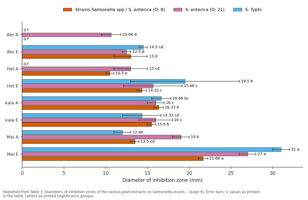
Strains Salmonella spp / S. enterica (O: 8) and S. enterica (O: 21) and more: Abr A vs Abr E vs Het A vs Het E vs kala A
Replotted from Table 3 (page 6) of the original paper. Every bar is the paper's own table value.
View source evidence ↗Figures & tables
From the original paper Table 1
 Table 2
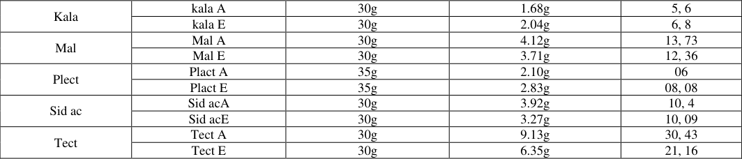 Table 3
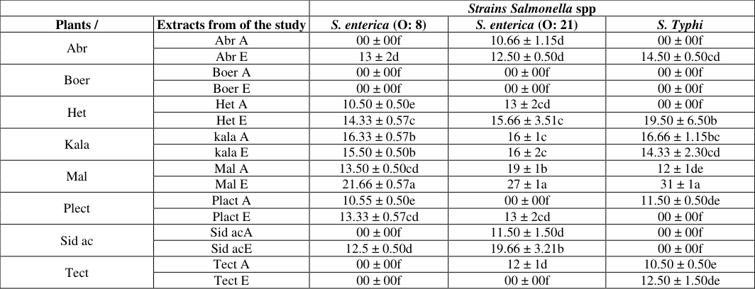 Table 3
 Table 4
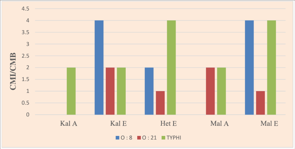 Figure 1
Introduction
Salmonella is the most common cause of foodborne bacterial infections (Angulo et al., 2000). Human contamination, which is of intrinsic or secondary origin, can occur mainly through the consumption of contaminated food (Bell & Kyriakides, 2002) [7] . Salmonella is an evolving hazard with a strong adaptive capacity. In the last decades, two major phenomena illustrate this (Rabsch & Tschäpe, 2001) [23] . Thus, in the 1980s, the Enteritidis serotype emerged in the United States and Europe, particularly associated with avian species and which has the capacity to cause a trans-ovarian infection and consequently to contaminate the eggs. The epidemic quickly becomes a global pandemic among human cases (Rabsch & Tschäpe, 2001) [23] . The significant incidence of salmonellosis as well as the emergence of multidrug resistance and resistance to critical antibiotics in human therapy, in a context where the danger of antibiotic resistance is the subject of all attention, make it a health issue first class (Ouattara et al., 2013) [20] . To date, the only way to treat health and economic problems result to avian pathogens remain antibiotic treatment. However, the use of these molecules knows its limits, due to the appearance and the propagation of multidrug resistance bacterial strains. An effective solution to this problem of antibiotic resistance would be to limit or avoid the use of antibiotics, by looking for alternative ways to the use of these molecules. Recently, means of prevention have been proposed as alternatives to the use of antibiotic molecules, in order to reduce the impact of enteric pathogens. Among these means, the use of phytobiotics, such as medicinal plants, would seem to offer promising results (Mehrnaz et al., 2010) [16] with a view to ensuring control of the host's digestive microbiota, whether animal or human, and to limit the appearance of enteropathogenic bacteria such as Salmonella sp. Medicinal plants are a potential source of antimicrobial compounds and / or inhibitors of antibiotic resistance mechanisms, acting according to several mechanisms: (i) formation of complexes with macromolecules such as proteins and polysaccharides, thus inhibiting their functions (polyphenols); (ii) rupture of microbial membranes (lipophilic flavonoids, terpenoids, defensins) and (iii) inhibition of the adhesion of microbial proteins to the polysaccharide receptors of the host (polypeptides) (Okusa et al., 2009) [18] . These compounds also act as efflux pump inhibitors (flavanoids, terpenoids, alkaloids), inhibitors of penicillin binding proteins (PLP), quinones, terpenoids, causing the permeability of bacterial membranes (terpenoids) and are also inhibitors of betalactamases (alkyl gallates). The active element of medicinal plants is grouped into several chemical families, namely sulfur compounds: thiosulfonates, aldehydes, phenols, terpenes and tannins. These substances work by blocking the multiplication of the microorganism or by destroying its wall (Rhayour et al., 2003) [24] . If today the different properties of phyto-molecules observable In vitro are recognized (antibacterial, immunomodulatory, antioxidant action), none of these molecules has yet demonstrated a real potential to replace antibiotics. The purpose of this work is to select plant with high antimicrobial activitiy against Salmonella spp for their use against salmonellosis.
Materials and Methods
The bacterial strains are composed of three (3) Salmonella sp. with known antibiotic profiles, (
Plant Collection
The plants used in this study were collected from traditional therapists at the medicinal plant sales market in the commune of Bingerville. Eight (08) species of plants were collected; these plants, known for their anti-diarrheal and/or anti-inflammatory properties, were sent to the herbarium of the National Floristic Center of the Félix Houphouët-Boigny University of Abidjan (Côte d'Ivoire), for their identification. They are Abrus precatorius, Boerhavia diffusa, Heterotis rotundifolia, Kalanchoe crenata, Mallotus oppositifolius, Plectranthus monostachyus, Sida acuta and Tectona grandis.
Preparation of Aqueous and Hydroethanolic Extracts of the Different Plants Collected
The preparation of the aqueous and hydroethanolic extracts of the various plants was carried out according to the method described by Zirihi et al. (2003) [29] . To do this, the leaves of the various freshly harvested plants are washed, cleaned, weighed and then dried in the dark. After grinding the dried leaves, 100 g of each powder are dissolved respectively in 1000 ml of sterile distilled water and in 1000 ml of hydroethanolic solution (70%). The various solutions obtained are filtered successively through a fine mesh sieve, percal fabric and hydrophilic cotton. The filtrate obtained is dried in an oven at 55 ° C. for 48 hours, to obtain the total aqueous extract (ETA). As for the hydroethanolic extract (EE70), it is obtained after drying the filtrate in an oven at 40 ° C. for 48 hours. The various extracts obtained are stored at 4 ° C. for the antibacterial tests
Yield
The yield, expressed as a percentage relative to the initial mass of the various plant powders subjected to extraction, was determined according to the following formula.
Preparation of Bacterial Inocula
The strains of Salmonella sp. are revived in Muller-Hinton broth, then an aliquot of each bacterial suspension is seeded on Muller-Hinton agar and incubated at 37 ° C for 24 H, to obtain young colonies. From a few young colonies, the turbidity of each bacterial suspension is adjusted so that its opacity is equivalent to Mac Farland's 0.5 control., estimated at 10 8 CFU / mL (Bauer et al., 1966) [6] ; Baker et al., 1983) [4] ; (Moosdeen et al., 1988) [17] . Subsequently, diffusion tests in agar medium are carried out as well as the determination of the Minimum Inhibitory Concentration (MIC) and the Minimum Bactericidal Concentration (CMB), in order to assess the anti-bacterial power of each extract.
Antibacterial Activity of Plant Extracts Sensitivity Test
Stock solutions concentrated at 200 mg / mL are prepared from each plant extract. Whatman No. 1 papers of 9 mm diameters impregnated with plant extracts were used as antibiotic disc for the diffusion tests in agar medium as described by Bauer et al. (2003) [5] . A bacterial suspension of 10 6 CFU/ml of the test strain was inoculated with a swab on Mueller-Hinton agar (Bio-Rad, France) agar. The bacterial suspension (10 6 CFU/ ml) was obtained by diluting an inoculum of 108 CFU/ml with sterile distilled water. The inoculum was obtained by homogenizing fresh colonies of the strain in sterile distilled water and then adjusting the turbidity to 0.5 Mc Farland scale of 10 8 CFU/ml. After plating, sterile Whatman No. 1 paper of 9 mm diameter were placed on the cultured plates. Synthetic antibiotic discs were used as positive control and the negative control was sterile distilled water. The plates were then left for 2 hours at room temperature (25°C ± 2°C) for pre-diffusion of the substances before being incubated at 37°C. The antibacterial activity of the extracts is determined by measuring the diameter of the zone of inhibition around each disc (Doughari et al., 2007) [9] . For each extract, the experiment was triplicated.
Determination of the Minimum Inhibitory Concentration (MIC)
The determination of the antibacterial parameters (MIC) was carried out according to the method of dilution in liquid medium, as carried out by Kouadio et al. (2015) [15] . Thus, in hemolysis tubes, one (1) milliliter with a concentration range of 100 to 3.12 mg / mL was prepared by the double dilution method. For each bacterial strain, a bacterial inoculum whose turbidity corresponds to the opacity of 0.5 Mc Farland is prepared in sterile distilled water, then diluted to 1/100 in two times concentrated Mueller Hinton broth, for the obtaining a bacterial inoculum estimated at 10 6 CFU / mL. Subsequently, a ½ dilution by adding one (1) mL of different concentrations of extracts, ranging from 50 to 1.56 mg / mL, is carried out. After 24 h of incubation at 37 °C, the turbidity of the medium reflects bacterial growth. The MIC of an extract for a given bacterial strain is the smallest concentration of extract, showing no visible growth to the naked eye of the bacteria.
Determination of the Minimum Bactericidal Concentration (MBC)
After seeding the concentration range, a count of the viable colonies is carried out in order to determine the CMB. Using a 10 µL calibrated loop, a 5 cm strip seeding of the bacterial inoculum, used for inoculating the concentration range and its dilutions ranging from 10-1 to 10-4 is carried out on Mueller-Hinton agar (BioRad, France), to constitute the bactericidal control. Subsequently, subcultures of the tubes without visible growth are carried out on Mueller-Hinton agar (BioRad, France). After 24 hours of incubation at 37 ° C., the observable colonies are compared to the bactericidal control. MBC is the smallest concentration whose subculture shows germ growth less than or equal to 0.0001% of survivors.
Determination of the Modality of Action of Different Plant Extracts
The antibacterial effect of the various plant extracts tested is judged to be bactericidal or bacteriostatic according to the MBC / MIC ratio (Fauchere & Avril, 2002), if the report: MBC / CMI ≤ 2, the substance is said to be bactericidal; MBC / CMI > 2, the substance is said to bebacteriostatic.
Statistical Analysis
The means of the inhibition diameters were calculated for each extract relative to each bacterial strain using the EXCEL 2013 software, at the end of the various tests. The STATISTICA 2019 software was used for the one-way analysis of variance (ANOVA 1). It was used to compare the means of the diameters of inhibition of the different extracts on each bacterial isolate, at the probability threshold p less than 5%. The results obtained were expressed in the form of the mean ± Standard deviation.
Results
Results
The Yield
The yield depending on the plant species, the organ used, the drying conditions, the content of each plant species in metabolites and the nature of the solvent used in the extraction. The different yields of the extractions of the different plants are reported in Table 2.
| Plants / | Extracts from of the study | Mass before extraction (g) | Mass after extraction (g) | Yield (%) |
|---|---|---|---|---|
| Abr | Abr A | 30g | 2.84g | 9, 46 |
| Abr E | 30g | 3.14g | 10, 46 | |
| Boer | Boer A | 30g | 4.5g | 15 |
| Boer E | 30g | 2.30g | 7, 6 | |
| Het | Het A | 17g | 1.34g | 7, 88 |
| Het E | 17g | 1.04g | 6;11 |
Table text extracted from the original PDF; superscript letters are shown inline. The original table image is below.
View original table image
Antibacterial Activity
The antibacterial activity of plant extracts differs from one plant to another and / or from one type of extract to another, on each of the strains studied (Photographs 1 and 2). 1).
Antibacterial Activity of Plant Extracts on
Antibacterial Activity of Plant Extracts on
Discussion
The results of this study showed that an aqueous and/or ethanolic extracts of the selected plants have antibacterial activity against (multi) resistant strains of Salmonella from different origins. These medicinal plants are used in traditional medicine for the treatment of bacterials infections. Some of these plants, like many others, have been the subject of several studies in order to demonstrate their antibacterial activities. From these previous studies, we can quote [26] in Côte d'Ivoire on In vitro evaluation of the antifungal activity of extracts from Mallotus oppositifolius leaves against two phytopathogenic fungi. Dougnon et al. (2017) [10] on antibacterial activity of various plants including Heterotis rotundifolia against enteropathogenic bacteria in Benin. Kouadio et al. (2015) [15] in Côte d'Ivoire on activity of Mallotus oppositifolius leaves on multidrug-resistant bacteria. The antibacterial activity of these plants is thought to be due to the presence of certain chemical compounds contained within their cells. Indeed, the plants used in traditional medicine are an essential source of antimicrobial compound for human health. They have significant antibacterial properties for therapeutic treatments (Prakash et al., 2013). Previous studies demonstrate the antibacterial activity of plant extracts against pathogenic microorganisms, based on ethnobotanical information. The variations in activity of the different extracts can be explained by the nature of the solvents used for the different extractions and the molecules contained in the cells of the plant (Traoré et al., 2012) [28] . The extracts used in this study have an activity on at least one of the strains tested, with the exception of the aqueous and ethanolic extracts of Boerhavia diffusa which showed no activity on the strains tested. However, the largest diameters of inhibition were observed on all three bacterial strains with the aqueous and ethanolic extracts of Mallotus oppositifolius and Kalanchoe crenata ranging from 12 ± 0.50 cd to 31 ± 1a mm. These different diameters of inhibition zone are comparable to those of the diameters observed by Effah-Yeboah et al. (2020) for extracts of Kalanchoe crenata, on strains of Salmonella which was 20 ± 0.882 mm and those of the work of Sahin et al. (2019) [25] on gram positive and gram-negative bacteria whose diameters were between 10 ± 0.45 and 22 ± 0.62 mm. The good activity of Kalanchoe crenata observed in this study is also reported by Aibinu et al. (2007) [1] , on a strain of Salmonella Paratyphi whose inhibition diameter was 14 mm with the aqueous extract of Kalanchoe crenata. Also the efficacy of extracts of Mallotus oppositifolius observed in this work is similar with the extracts of Mallotus oppositifolius studied by Kouadio et al. (2015) [15] on E. coli producing ESBLs strains, methicillinresistant S. aureus, whose diameters were between 10.67 ± 0.67 and 18.33 ± 0.33 mm for aqueous extracts and between 11.00 ± 0.57 and 24.33 ± 0.67 mm for the methanolic extract. In addition, the ethanolic extract of Mallotus oppositifolius has demonstrated excellent antifungal activity against fungi of the genus Fusarium and Phytophthora (Saraka et al., 2019) [26] . The diameters of the zones of inhibition formed by the ethanolic extract of Heterotis rotundifolia were 14.33 ± 0.57, 15.66 ± 3.51, 19.50 ± 6.50 respectively on the strains of Salmonella serogroup O: 8, O: 21 and Salmonella Typhi, while Dougnon et al. (2017) [10] reported diameters between 10 and 14 mm on enteropathogenic bacterial strains (Citrobacter freundi, Pseudomonas oryzihabitans, Shigella flexneri, Klebsiella rhinocleromatis). Traditionally, these plants are used in the treatment of several pathologies of bacterial origin such as skin infections, diarrhea and urinary tract infections (Kouadio, 2013) [14] . The inhibitory power of the various extracts of Heterotis rotundifolia, Mallotus oppositifolius and Kalanchoe crenata, on the bacterial growth of the strains of Salmonella spp were demonstrated by the determination of the Minimum Inhibitory Concentration. Minimum Inhibitory Concentrations (MICs) vary from strain to strain and from extract to extract. With extracts of Mallotus oppositifolius, strain of serogroup O: 8, both with the ethanolic extract. The greatest MIC is located above 50 mg / mL obtained on the enterica strain of serogroup O: 21 (aqueous extract). These values are higher than those reported by certain authors, in particular those of Kouadio et al. (2015) [15] whose values were 0.78 and 1.56 mg / ml for the 70% methanolic extract and 3.12 mg / ml for the aqueous extract, on E. coli ESBL-producing strains. Orsot et al. (2015) also reported a better inhibition of the ethanolic extract of Mallotus oppositifolius compared to the aqueous extract of Mallotus oppositifolius on phytopathogenic strains of Sclerotium rolfsii with a MIC of 12.5 mg / ml for the ethanolic extract and 100 mg / ml for the aqueous extract. The CMB / CMI ratio reveals that ethanolic extract for the most part has bacteriostatic power, unlike aqueous extract which mainly has bactericidal power. These different powers observed with the different extracts of Mallotus oppositifolius in this study are similar to the bacterial powers of the extracts of this same plant on the strains of phytopathogenic fungi studied by Saraka et al. (2019) [26] . As for Kalanchoe crenata extracts, the smallest MIC (12.5 mg / ml) is obtained with ethanolic extract on the Typhi and enterica strains of serogroup O: 8, also with the aqueous extract on the Typhi strain. While the greatest MIC (50 mg / ml) is obtained only with aqueous extract on the enterica strains of serogroup O: 21 and O: 8. The MIC values reported by Aibinu et al. (2007) [1] on Salmonella Paratyphi, by Dieye & Sarr (2020) on Candida albicans strains which were respectively 512 mg/ml and 256 mg/ml are clearly higher than the values observed in this work. However, the MIC values obtained in this study are higher than the values reported by Sahin et al. (2019) [25] obtained from natural compounds extracted from Kalanchoe crenata which were 50 μg / ml on a E. coli strain. About ethanolic extracts of Heterotis rotundifolia, the MICs were range between 6.25 mg / ml and 50 mg / ml, with the smallest MIC obtained on the Typhi strain (6.25 mg / mL) and the largest on the enterica strain of serogroup O: 21 (50 mg / mL). Previous works which have already been the subject of a study of the inhibitory effect of extracts of Heterotis rotundifolia, include those of Dougnon et al. (2017) [10] which offered MICs varying from 50 mg / mL to 100 mg / mL on enteropathogenic strains with bactericidal power (CMB / MIC ≤ 2). Extraction method, solvent and plant parts used on the microbial strain could explain the differences observed between the MICs obtained in this study and those of the above-mentioned authors. When extraction methods used, solvents and plant organs differ, the active elements responsible for antibacterial activity do not have the same concentrations whatever the nature of the extracts. The activity of plant substance therefore depends on its concentration of active ingredients (Thangara et al., 2000). However, the harvest site and the type of local climate can influence the chemical profile of plants (Amvam, 1998), which could justify the frequent use of these plants for the treatment of certain infections.
Conclusion
The results of this work reveal that the selected plants possess dose-dependent antibacterial activity on Salmonella strains of various origins. Among these plants, the leaves of Kalanchoe crenata, of Mallotus oppositifolius and Heterotis rotundifolia are those which offer greater activity on all the plants studied. These strong antibacterial activities observed at the level of these enteropathogenic strains make plausible a possible use of these various plant extracts as an alternative to the use of antibiotic molecules. Substituting these plant extracts for the classic antibiotics used in breeding could therefore solve on the one hand the problem of the appearance and emergence of multiresistant bacteria, of the overuse of antibiotic molecules.
Acknowledgement
AcknowledgmentsThanks are due to the National Floristic Center, Félix Houphouët-Boigny University, Abidjan, Côte d'Ivoire. The authors declare that they have no known competing financial interests or personal relationships that could have appeared to influence the work reported in this paper.
Figures and tables from the paper
| Strain | Coded | Antibiotic profile | Origin of the strain | Serogroup / Serotypes |
|---|---|---|---|---|
| Strain 1 | C211 / A | A-AMC-Tic-Cf-CTX-GC-Te-SXT-Nal- Cip | Avian (raw chicken gizzards) | O: 8 |
| Strain 2 | S30H3C | A-Tic-TE | Avian (quail viscera) | O: 21 |
| Strain 3 | SH35 | AM-CRO-CAZ-FF-CFS | Human (Human Stool) | Typhi |
Table text extracted from the original PDF; superscript letters are shown inline. The original table image is below.
View original table image
| Kala | kala A | 30g | 1.68g | 5, 6 |
|---|---|---|---|---|
| kala E | 30g | 2.04g | 6, 8 | |
| Mal | Mal A | 30g | 4.12g | 13, 73 |
| Mal E | 30g | 3.71g | 12, 36 | |
| Plect | Plact A | 35g | 2.10g | 06 |
| Plact E | 35g | 2.83g | 08, 08 | |
| Sid ac | Sid acA | 30g | 3.92g | 10, 4 |
| Sid acE | 30g | 3.27g | 10, 09 | |
| Tect | Tect A | 30g | 9.13g | 30, 43 |
| Tect E | 30g | 6.35g | 21, 16 |
Table text extracted from the original PDF; superscript letters are shown inline. The original table image is below.
View original table image
| — | — | Strains Salmonella spp | — | — |
|---|---|---|---|---|
| Plants / | Extracts from of the study | S. enterica (O: 8) | S. enterica (O: 21) | S. Typhi |
| Abr | Abr A | 00 ± 00f | 10.66 ± 1.15d | 00 ± 00f |
| Abr E | 13 ± 2d | 12.50 ± 0.50d | 14.50 ± 0.50cd | |
| Boer | Boer A | 00 ± 00f | 00 ± 00f | 00 ± 00f |
| Boer E | 00 ± 00f | 00 ± 00f | 00 ± 00f | |
| Het | Het A | 10.50 ± 0.50e | 13 ± 2cd | 00 ± 00f |
| Het E | 14.33 ± 0.57c | 15.66 ± 3.51c | 19.50 ± 6.50b | |
| Kala | kala A | 16.33 ± 0.57b | 16 ± 1c | 16.66 ± 1.15bc |
| kala E | 15.50 ± 0.50b | 16 ± 2c | 14.33 ± 2.30cd | |
| Mal | Mal A | 13.50 ± 0.50cd | 19 ± 1b | 12 ± 1de |
| Mal E | 21.66 ± 0.57a | 27 ± 1a | 31 ± 1a | |
| Plect | Plact A | 10.55 ± 0.50e | 00 ± 00f | 11.50 ± 0.50de |
| Plact E | 13.33 ± 0.57cd | 13 ± 2cd | 00 ± 00f | |
| Sid ac | Sid acA | 00 ± 00f | 11.50 ± 1.50d | 00 ± 00f |
| Sid acE | 12.5 ± 0.50d | 19.66 ± 3.21b | 00 ± 00f | |
| Tect | Tect A | 00 ± 00f | 12 ± 1d | 10.50 ± 0.50e |
| Tect E | 00 ± 00f | 00 ± 00f | 12.50 ± 1.50de |
Table text extracted from the original PDF; superscript letters are shown inline. The original table image is below.
View original table image
| — | S. enterica (O: 8) | S. enterica (O: 8) | S. enterica (O: 21) | S. enterica (O: 21) | S. Typhi | S. Typhi |
|---|---|---|---|---|---|---|
| Extracts from of the study | MIC | BMC | MIC | BMC | MIC | BMC |
| Kal A | 50 | > 50 | 50 | > 50 | 12. 5 | 25 |
| Kal E | 12.5 | 50 | 25 | 50 | 12. 5 | 25 |
| Het E | 25 | 50 | 50 | 50 | 6.2 5 | 25 |
| Mal A | 12.5 | > 50 | 25 | 50 | 12. 5 | 25 |
| Mal E | 6.25 | 50 | 50 | 50 | 6.2 5 | 25 |
| Kal A | 50 | > 50 | 50 | > 50 | 12. 5 | 25 |
Table text extracted from the original PDF; superscript letters are shown inline. The original table image is below.
View original table image
References
29 in the original paper- Aibinu IE, Odunayo RA, Adelowotan T, Odugbemi T. In vitro antimicrobial activity of crude extracts from plants Bryophyllum pinnatum and Kalanchoe crenata. Afr J Tradit Complement Altern Med. 2007;4:338-344.
- Amvam ZPH. Aromatic plants of Tropical Central Africa. Part XXXII: Chemical composition and antifungal activity of thirteen essential oils from aromatic plants of Cameroon. Flavour Fragr J. 1998;13:107-114.
- Angulo FJ, Johnson KR, Tauxe KR, Cohen ML. Origins and consequences of antimicrobial-resistant nontyphoidal Salmonella: implications for the use of fluoroquinolones in food animals. Microb Drug Resist. 2000;6:77-83.
- Baker CN, Thornsberry C, Hawkinson RW. Inoculum standardization in antimicrobial susceptibility tests: evaluation of the overnight agar cultures and the Rapid Inoculum Standardization System. J Clin Microbiol. 1983;17:450-457.
- Bauer AW, Kirby MDK, Sherras JC, Trick M. Antibiotic susceptibility testing by standard single disc diffusion method. Am J Clin Pathol. 2003;45:496.
- Bauer AW, Kirby WMM, Sherris TC, Turck M. Antibiotic susceptibility testing by a standardized single disc method. Am J Clin Pathol. 1966;45:493-496.
- Bell C, Kyriakides A. Salmonella. In: Foodborne pathogens. Hazards, risk analysis and control. Cambridge: Woodhead Publishing; 2002. p. 307-334.
- Dieye PI, Sarr OS. État de la recherche de molécules cibles antimicrobiennes issues de plantes en Afrique. Afr Sci. 2020;16:348-374.
- Doughari JH, Pukuma MS, De N. Antibacterial effects of Balanites aegyptiaca L. Drel. and Moringa oleifera Lam. on Salmonella typhi. Afr J Biotechnol. 2007;6:2212-2215.
- Dougnon VT, Klotoé JR, Sènou M, Roko GO, Dougnon G, Fabiyi K, et al. Chemical composition, cytotoxicity and antibacterial activity of selected extracts of Euphorbia hirta, Citrus aurantifolia and Heterotis rotundifolia on enteropathogenic bacteria. EC Microbiol. 2017;12:180-195.
- Effah-Yeboah E, Asare EA, Balali GI. In-vitro antimicrobial activity of the combined effect of Kalanchoe crenata and Vernonia amygdalina on Salmonella species. Res Square. 2020;16 p.
- Fauchere LL, Avril JL. Bactériologie générale et médicale. Paris: Éditions Ellipses; 2002.
- Gupta D, Dubey J, Kumar M. Phytochemical analysis and antimicrobial activity of some medicinal plants against selected common human pathogenic microorganisms. Asian Pac J Trop Dis. 2016;6:15-20.
- Kouadio NJ. Évaluation de l'activité antibactérienne de quatre plantes médicinales sur la croissance In vitro de bactéries multirésistantes. Mémoire de Master. Abidjan: Université Nangui Abrogoua; 2013. p. 53.
- Kouadio NJ, Guessennd NK, Kone MW, Moussa B, Koffi MZ, Yao K, et al. Évaluation de l'activité des feuilles de Mallotus oppositifolius (Geisel.) Müll. Arg. (Euphorbiaceae) sur des bactéries multirésistantes et criblage phytochimique. Int J Biol Chem Sci. 2015;9:1252-1262.
- Mehrnaz N, Fatemeh R, Davoud A, Farhad M, Elham S, Shahram L, et al. Inhibitory effects of Lactobacillus salivarus and Lactobacillus criptus isolated from chicken gastrointestinal tract on Salmonella enteritidis and Escherichia coli growth. Iran J Biotechnol. 2010;8:32-37.
- Moosdeen F, Williams JD, Secker A. Standardization of inoculum size for disc susceptibility testing: a preliminary report of a spectrophotometric method. J Antimicrob Chemother. 1988;21:439-443.
- Okusa PN, Stévigny C, Duez P. Medicinal plants: a tool to overcome antibiotic resistance? In: Medicinal plants, biosynthesis and pharmacology. New York: Nova Biomedical Books; 2009. p. 315-336.
- Orsot B, Soro S, Ouattara D, N'guessan EK, Zirihi GN. Étude ethnobotanique et évaluation In vitro de l'activité antifongique des extraits de feuille de Mallotus oppositifolius sur deux souches phytopathogènes de Sclerotium rolfsii. Eur Sci J. 2015;36:1857-1881.
- Ouattara K, Traore Z, Doumbia I, Coulibaly AF, Coulibaly A. In vitro antibacterial activity of ethanol extracts of Abrus precatorius Linn (Fabaceae) on bacteria responsible for nosocomial infections. Int Res J Pharm Appl Sci Technol. 2013;3:23-27.
- Ouattara ND, Guessend N, Gbonon V, Toe E, Dadié T, Tiécoura B. Consommation des antibiotiques dans la filière aviaire à Abidjan: cas de quelques fermes semiindustrielles. Eur J Res. 2013;94:80-85.
- Grubben GJH, Denton OA, editors. PROTA4U web database. Wageningen, Netherlands: Plant Resources of Tropical Africa; 2013.
- Rabsch W, Tschäpe H. Non-typhoidal salmonellosis: emerging problems. Microbes Infect. 2001;3:237-247.
- Rhayour K, Tantaoui-Elaraki A, Sendide K, Remmal A. The mechanism of bactericidal action of oregano and clove essential oils and their phenolic major components on Escherichia coli and Bacillus subtilis. J Essent Oil Res. 2003;15:356-362.
- Sahin AZ, Mou AM, Pervin A, Karim M, Tajwar A, Asim HM, et al. Antimicrobial activity of natural compounds from Kalanchoe crenata against pathogenic bacteria. Clin Microbiol Infect Dis. 2019;4:1-4.
- Arg. (Euphorbiaceae) sur Fusarium sp. et Phytophthora sp., deux champignons phytopathogènes. J Anim Plant Sci. 2019;41:6903-6915.
- Thangara JHS, Adjei O, Allen BW, Portaels F. In vitro activity of ciprofloxacin, sparfloxacin, ofloxacin, amikacin and rifampicin against Ghanaian isolates of Mycobacterium ulcerans. J Antimicrob Agents Chemother. 2000;45:231-233.
- Traoré Y, Ouattara K, Yéo D, Doumbia I. Recherche des activités antifongique et antibactérienne des feuilles d'Annona senegalensis Pers. (Annonaceae). J Appl Biosci. 2012;58:4234-4242.
- Zirihi GN, Kra AKM, Guédé-Guina F. Évaluation de l'activité antifongique de Microglossa pyrifolia (Lamarck) O. Kuntze (Asteraceae) "PYMI" sur la croissance In vitro de Candida albicans. Rev Méd Pharmacopée Afr. 2003;17:11-18.