
Research article · 2026 · Full text
International Journal of Advanced Chemistry ResearchRole of lipoproteins in the development and progression of heart disease
Volume 8(8) · Pages 42–52 · DOI: 10.33545/26646781.2026.v8.i8a.731 ↗
Research areasLipoproteins and Cardiovascular HealthCholesterol and Lipid MetabolismAtherosclerosis and Cardiovascular Diseases
Abstract
Author-writtenToday in the world Cardiovascular Diseases (CVDs) are the main cause of mortality, which depends on lipoproteins. Lipoproteins are macromolecular that transport lipids in the bloodstream and consist of chylomicrons, Very Low-Density Lipoproteins (VLDL), Low-Density Lipoproteins (LDL), High-Density Lipoproteins (HDL), and related apolipoproteins. Their distinct taxonomy and structural organization set them apart. Particles are metabolised through carefully regulated endogenous and external pathways as well as conventional reverse cholesterol transport functions that regulate lipid homeostasis. Uncontrolled lipid metabolism is a major cause of hypertension. the arterial wall. Plaque formation increased due to fatty streaks, foam cells, infiltration of LDL, degradation of endothelium and oxidative changes in the arterial wall. Plaque stability and necrotic core development increase the risk of acute cardiovascular disorder and cause a massive heart attack. Environmental and metabolic factors is one of the main causes of high cholesterol levels. In addition, biological and epigenetic mechanisms like microRNA-mediated regulation, DNA methylation, and genes that cause high cholesterol levels have an impact on lipoprotein activity and cardiovascular risk. In modern therapeutic developments, targeted modulation of lipoprotein biology has taken the role of conventional lipid-lowering methods. Novel innovations of modern treatment include PCSK9 inhibitors, CRISPR-mediated gene editing, and small interfering RNA (siRNA)-based therapies. Other important aspects of lowering cardiovascular risk are HDL mimetics, anti-inflammatory drugs, and lifestyle modifications. This review emphasises the complex role of lipoproteins in the progress and analyses new knowledge on genetic and epigenetic reactivity, and explores new therapies. To advance cardiovascular disease prevention and treatment approaches, subsequent investigations must concentrate on enhanced lipid profiling, tailored medication, and multi-omics integration.
Summary
Source-checkedPlain-language reading of the full paper, prepared with AI and checked against the text. Each point links to the authors' own words.
Why it matters
- Cardiovascular diseases (CVDs) are a primary cause of death globally, resulting in approximately 17.9 million deaths annually according to the WHO. View evidence ↗
- CVDs are largely driven by the dysregulation of lipid metabolism, where lipoproteins play a critical role in transporting cholesterol and triglycerides through the blood. View evidence ↗
- Low-Density Lipoproteins (LDL) are highly atherogenic because they can penetrate artery walls and undergo oxidative changes, leading to inflammation and plaque formation. View evidence ↗
The question
- The study aims to provide a detailed examination of how lipoproteins contribute to the development and progression of atherosclerosis, focusing on their structure, metabolism, genetics, and new treatments. View evidence ↗
What they did
- This is a review paper that analyzes existing knowledge regarding the role of lipoproteins, genetic and epigenetic factors, and emerging therapeutic options. View evidence ↗
What they found
- Atherosclerosis begins with endothelial dysfunction, which damages the inner lining of the blood arteries. View evidence ↗
- Oxidized LDL (oxLDL) is absorbed by macrophages via SR-A and CD36 receptors, causing them to become lipid-rich foam cells, a hallmark of early plaque. View evidence ↗
- Mutations in the LDL receptor gene (LDLR) are among the most common genetic causes of hypercholesterolemia, as they impair the removal of LDL from the blood. View evidence ↗
- The microRNA miR-33 contributes to lipid imbalance by inhibiting HDL synthesis and the expression of ABCA1 and ABCG1 transporters. View evidence ↗
- Clinical trials (ORION series) demonstrated that the siRNA-based drug inclisiran can reduce LDL cholesterol by approximately 50% in patients with hypercholesterolemia. View evidence ↗
- PCSK9 inhibitors can lead to a 50% to 70% reduction in plasma LDL-C levels by promoting the recycling of LDL receptors on hepatocytes. View evidence ↗
- Experimental animal studies using CRISPR-Cas9 to disrupt the PCSK9 gene in hepatocytes showed a dramatic reduction in circulating LDL-C levels. View evidence ↗
- The COLCOT study found that low-dose colchicine significantly lowers the risk of ischemic cardiovascular events in patients who have suffered a myocardial infarction. View evidence ↗
- Current health guidelines recommend at least 150 minutes of moderate-intensity exercise per week to improve lipid metabolism and cardiovascular fitness. View evidence ↗
What it means
- The authors conclude that the future of cardiovascular care lies in personalized medicine using genetic, epigenomic, and lipidomic data to tailor treatments to individual molecular profiles. View evidence ↗
- The authors note that CRISPR therapies still face challenges regarding ethical considerations, off-target mutations, immunological responses, and long-term safety. View evidence ↗
Visual summary
1 visualsIllustrations explain the paper; they do not show study data.
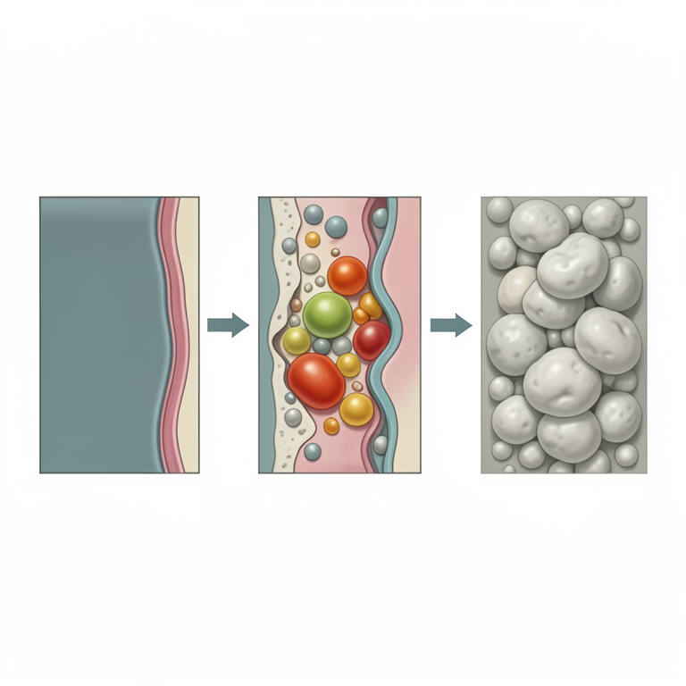
Atherosclerosis Initiation Process
AI-generated conceptual illustration (human coronary artery wall and lipoproteins). It explains the idea and does not show study data.
View source evidence ↗Figures & tables
From the original paper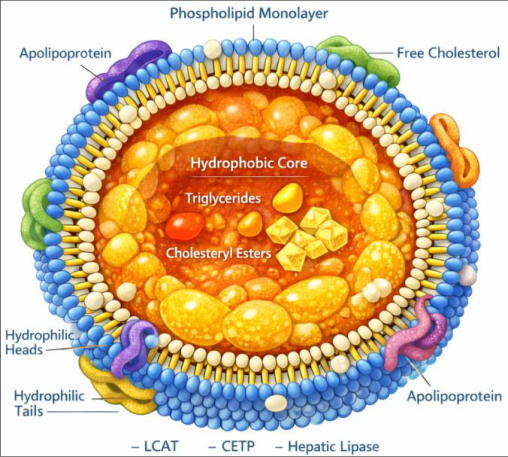 Figure 1
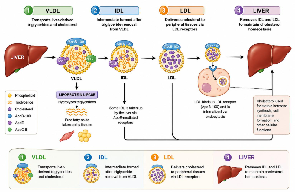 Figure 2
 Table 1
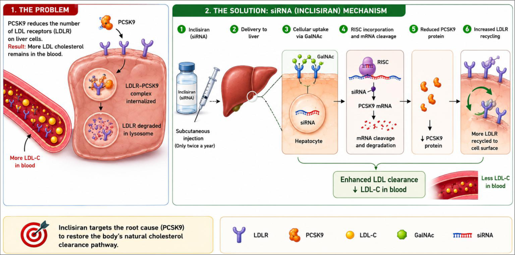 Figure 3
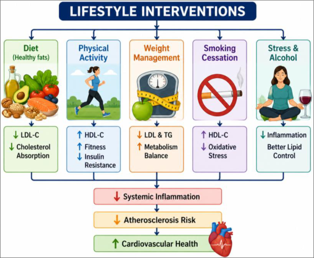 Figure 4
Introduction
Day by Day, worldwide CVDs increases and leading cause of mortality rate, accounting WHO every year 17.9 million deaths. These diseases are largely driven by dysregulation of lipid metabolism, and lipoproteins are vital for the movement and distribution of lipids like triglycerides and cholesterol in the blood [1,2] . Lipoproteins are elobrate macromolecular structures which made up of amphipathic phospholipids, cholesterol, and apolipoproteins that surround by hydrophobic lipid core. Having varying sizes, densities, and functions, they can be categorised as chylomicrons, LDL, HDL and VLDL [3] . Although LDL may penetrate the artery wall and undergo oxidative alteration, which can lead to inflammatory reactions and plaque formation, it is considered as one of the most atherogenic of these [4] . On the other hand, HDL is thought to be beneficial because it stimulates reverse cholesterol transfer and has anti-inflammatory and antioxidant effects; however, new research shows that HDL functionality may be more crucial than its levels in the blood [5] . A chronic inflammatory disease, atherosclerosis, which builds up by lipid accumulation, endothelial failure and immunological activation within the artery wall. Endothelial damage starts the process via allowing LDL particles into the intima, where they are maintained and altered. Fatty streaks and gradual plaque growth result from the stimulation of monocyte influx, macrophage differentiation, and foam cell generation by these defective lipoproteins. Long-term, plaque can result in myocardial infarction and stroke [6] . Lipoprotein metabolism and cardiovascular risk are influenced by genetic, environmental and lifestyle such as smoking and unhealthy food eating; it is greatly influenced by genes linked to familial hypercholesterolemia and regulation mechanisms of DNA methylation and microRNA-mediated gene transcription [5] . Targeted regulation of lipoprotein metabolism has replaced traditional lipid-lowering methods in the cardiovascular disease treatment. Small interfering RNA (siRNA)-based medicines like inclisiran, Proprotein convertase subtilisin or kexin type 9 (PCSK9) inhibitors and gene-editing technologies are examples of novel methods showing great promise in lowering the cardiovascular disease risk [6][7][8] . With an emphasis on operational and structural properties, metabolic pathways, genetic regulation, and novel therapeutic strategies, this study seeks to offer a detailed examination of the role of lipoproteins in the growth and course of atherosclerosis. Furthermore, a combination of improved lipid profiling, customized medicine, and multi-omics approaches is anticipated to improve risk assessment and treatment outcomes.
Overview of lipoproteins 2.1 Structural organization of lipoproteins
The structure of lipoproteins, consist of the core-shell paradigm, amphipathic outer monolayer. Monolayer composed of free cholesterol, phospholipids, and apolipoproteins. Lipoproteins are complex structure, spherical particles that facilitate the transport of hydrophobic lipids in the plasma; this special structure allows lipids to remain soluble in the circulation [9,10,11] . As shown in Figure 1, The hydrophobic tails of the external phospholipids point inwards into the lipid core, while the hydrophilic heads face the plasma. This layer's apolipoproteins serve as ligands for receptors and cofactors for enzymes, which are necessary for stabilizing the molecule and regulating its metabolic relationships [3] . Hepatic lipase, lecithin-cholesterol acyltransferase (LCAT) and Cholesterol Ester Transfer Protein (CETP) is an enzyme that functions to regularly alter lipoproteins, affecting their atherogenicity, composition, and functioning [3,9,10,12] .
Classification of lipoproteins 2.2.1 Major classes Chylomicrons
Chylomicrons are the biggest and less dense lipoproteins and are synthesized in the intestinal mucosa following absorption of dietary fats. They are involved for the exogenous triglycerides transport and cholesterol from the gut to peripheral tissues such as adipose tissue and muscle via the lymphatic and circulatory systems. Fundamentally, chylomicrons are rich in triglycerides and contain apolipoprotein B-48 as the key structural proteins needed for their assembly and secretion. In the circulation their acquire additional apolipoproteins including ApoC-II and ApoE which promote lipoprotein lipase activation and remnant clearance by the liver [6,3] .
Very Low-Density Lipoproteins (VLDL)
VLDL are formed in the liver and are help the transport of endogenous triglycerides and cholesterol to peripheral tissues. They include apolipoprotein B-100 structural component, along with ApoC and ApoE acquired in circulation. VLDL particles deliver triglycerides to tissues by lipoprotein lipase hydrolysis of triglycerides to free fatty acids. VLDL is converted to IDL (intermediate-density lipoproteins) and then to LDL as triglycerides are removed. This metabolic conversion is necessary for lipid homeostasis [3] .
Intermediate-Density Lipoproteins (IDL)
IDL are transient particles and formed during the catabolism of VLDL. They are in an intermediate stage between VLDL and LDL. IDL particles are rich in cholesterol esters and relatively poor in triglycerides. These particles are either removed by the liver by a receptor-mediated process involving apolipoprotein E or further metabolized to LDL in the circulation [3,13] .
Low-Density Lipoproteins (LDL)
LDL are cholesterol-rich particles that act as the dominant carriers cholesterol to peripheral tissues and they are apolipoprotein B-100 and cellular uptake. High levels LDL are associated with atherosclerosis because of the LDL risk. It gathers in the wall of arterial and is oxidatively modified, and plaque formation [3,13] .
High-Density Lipoproteins (HDL)
It has a special feature that the core mainly contains cholesteryl esters and small amount triglycerides. This core have an amphipathic monolayer phospholipids and soluble cholesterol. Phospholipids and soluble cholesterol are apolipoproteins and ApoA-I. ApoA-I work as a structural framework and activating LCAT enzymes. HDL exists in discoidal and spherical modes and undergoes remodeling, which is fundamental to HDL's function in cholesterol transport and cardiovascular defence [13] .
Apolipoproteins
Apolipoproteins are the lipoproteins that are essential for their structural integrity and metabolic regulation, act as enzyme receptor ligands, cofactors, and determinants of lipoprotein fate.
Apolipoprotein A-I (ApoA-I) and B (ApoB)
ApoA-I is the key protein part of HDL and required for restore cholesterol transport. ApoA-I promote lecithincholesterol acyltransferase (LCAT), which promotes cholesterol esterification. Apolipoprotein B (ApoB) is a key structural protein of atherogenic lipoproteins, present as and ApoB-100 and ApoB-48 in chylomicrons in IDL, LDL and VLDL. ApoB-100 interacts with LDL receptors to allow cellular uptake of cholesterol. It is required for lipoprotein assembly and a marker of cardiovascular risk [3] .
Apolipoprotein C (ApoC) and Apolipoprotein E (ApoE)
Apolipoprotein C (ApoC) comes in a number of isoforms. ApoC-II stimulates triglyceride hydrolysis by activating lipoprotein lipase (LPL), but ApoC-III inhibits lipolysis and is linked to hypertriglyceridemia and high plasma triglyceride levels. Apolipoprotein E (ApoE) mediates receptor-dependent hepatic absorption of lipoprotein remnants, which is essential for lipid metabolism. It aids in the redistribution of lipids between tissues and is necessary for the removal of intermediate-density lipoproteins and chylomicron remnants [3,10,12] .
Pathways of lipoprotein metabolism
For the body to transport cholesterol and triglycerides, lipoprotein metabolism is necessary. Lipids have to be transported in the bloodstream as complexes known as lipoproteins because they are hydrophobic. The lipids and apolipoproteins that make up these molecules aid in preserving structural integrity and controlling metabolism. The exogenous and endogenous pathway, and reverse cholesterol transport are the three main pathways that contribute to lipoprotein metabolism; each is essential for preserving lipid balance and cardiovascular health [14] .
Exogenous pathway
Lipids from food sources are transported via this route. The breakdown of lipids from food into cholesterol and monoglycerides by pancreatic enzymes begins in the small intestine. These molecules are absorb by intestinal epithelial cells [13,15] . Chylomicrons are particles of lipoprotein that contain apolipoprotein B-48 (ApoB-48) and rich in triglycerides. After being absorbed in the intestines, chylomicrons travel via the lymphatic system before entering the bloodstream. HDL supply them with ApoC-II and ApoE there, enabling them to efficiently take part in lipid transport and metabolism. LPL enzyme is located on the capillaries endothelial membrane of muscle and and adipose tissue, stimulated by ApoC-II. While glycerol flows to the liver, the released fatty acids are absorbed by tissues for energy generation or stored as fat [5,6,13,15] . ApoE receptors in the liver identify these leftovers, which are then taken up by receptor-mediated endocytosis. Their lipid contents are either stored, recycled, or transformed into bile acids for excretion inside the liver [14] .
Endogenous pathway
The endogenous pathway is the transporter of lipids synthesised. Triglycerides and cholesterol are generated by liver cells and combined with apolipoprotein B-100 (ApoB-100) to form VLDL. VLDL accumulates ApoC-II and ApoE from HDL after being secreted into the circulation. Like chylomicrons, lipoprotein lipase hydrolyses VLDL triglycerides, liberating free fatty acids tissues can absorb. VLDL is converted into IDL as triglycerides are depleted [16] . IDL particles can either undergo more triglyceride removal to create low-density lipoproteins (LDL) or be directly accepted by the liver through ApoE-mediated receptors. Figure 2, Graphical representation of the endogenous lipid transport system. The main carriers of cholesterol in the blood are LDL particles, which are rich in cholesterol [14][15][16][17] . Through receptor-mediated endocytosis activating LDL receptors that identify ApoB-100, LDL transports cholesterol to peripheral organs. Steroid hormone synthesis, cell membrane creation, and other cellular functions depend on cholesterol. On the other hand, high LDL levels can cause cholesterol to accumulate in artery walls, which accelerates the development of atherosclerosis [18] . Excess LDL can be digested by macrophages, which then transform into foam cells and fatty streaks, and it gives early signs of plaque development. As a result, LDL is frequently referred to as "bad cholesterol." The distribution of lipids produced through the liver and the preservation of cholesterol homeostasis depend large on the endogenous route.
Reverse Cholesterol Transport (RCT)
A defence mechanism called RCT, that eliminates extra cholesterol from outer tissues and returns it to the liver for excretion, this process mediator by HDL. In the gut and liver, HDL is synthesised as a young, lipid-poor particle that contains apolipoprotein A-I (ApoA-I) [19] . These particles function as cholesterol scavengers and move throughout the bloodstream. Through ATP-binding cassette transporters (ABCA1 and ABCG1), peripheral cells including macrophages in artery walls transfer extra cholesterol to HDL (Rosenson et al., 2012). After HDL absorbs free cholesterol, ApoA-I promotes the enzyme lecithin-cholesterol acyltransferase, which then process it into cholesterol esters. The HDL particle becomes an experienced structure as a result of these esters migrating to its core [1] . There are two ways that HDL can carry cholesterol esters to the liver. First, HDL allows for the selective uptake of cholesterol by binding directly with the hepatic scavenger receptor class B type I (SR-BI). Second, cholesteryl ester transfer protein (CETP) allows HDL to transfer cholesterol esters to LDL and VLDL, which are then absorbed by the liver (Nelson & Cox, 2021). Cholesterol is either transformed into bile acids in the liver or released straight into bile, which is then removed by the digestive system. This procedure is essential for lowering the risk of cardiovascular disorder and cholesterol development in tissues [19,20] .
Global burden of cardiovascular diseases
Today, the worldwide leading cause of death and major threat to public health is CVDs. CVD included various disorder such as Stroke, Hypertensive heart condition, Coronary and Peripheral artery diseases. According to WHO, every year 17.9 million (around 32%) people die from CVDs. Additionally, 85% people are died by strokes and heart attacks (World Health Organization, 2020). The burden of CVDs is particularly severe in low-and middleincome countries (LMICs), accounting for more than a third of CVD-related deaths [19][20][21] . In urbanisation, CVDs increase due to diabetes, excessive alcohol consumption, low-quality dietary habits and physical inactivity. With a population getting older and increasing life expectancy, more people are living with chronic heart problems. Additionally, high costs of medicinal and healthcare systems increase deaths from CVDs and create loss of job performance, and long-term disability. Despite breakthroughs in diagnosis, treatment and prevention, the incidence of CVDs continues to increase due to rising exposure to modifiable risk variables. Effective public health measures such as healthy diet, smoking cessation, blood pressure control, lipid management, regular exercise, avoiding diabetes and early detection are necessary to reduce the worldwide burden of cardiovascular diseases [19, 20] .
Lipoproteins in atherosclerosis development
The lipids buildup, fibrous components, and immune cells in wall of artery is typical of atherosclerosis, a long-term inflammatory disease. LDL in molecule is a key factor in the development and course of this illness. The endothelial dysfunction, lipid infiltration, plaque progression, and the final development of a necrotic core are some of the stages that atherosclerosis goes through. These mechanisms are intimately associated with anomalies in the metabolism and function of lipoproteins [20,21] .
Initiation: Endothelial dysfunction
Endothelial dysfunction, which affects the inner membrane of blood arteries, is the initial stage of atherosclerosis. By managing blood circulation, preventing clot formation, and regulating the passage of chemicals into and out of the artery wall, the endothelium normally maintains vascular homeostasis. The endothelium can be effected human health such smoking, high cholesterol, diabetes and high blood pressure. In addition to increasing vascular permeability, it stimulates the production of extra immune cells and adhesion molecules (ICAM-1 and VCAM-1) [22] . Endothelial dysfunction, which affects the inner lining of blood arteries, is the initial stage of atherosclerosis. Moreover, it produces prostacyclin and thrombomodulin that maintain smooth blood flow and thrombomodulin. This damage increases vascular permeability and promotes the expression of adhesion molecules [22] . Additionally, selectins (such as E-selectin and P-selectin) are upregulated, facilitating the rolling and adhesion of leukocytes on the endothelial surface, an important step in immune cell recruitment. Lower nitric oxide levels cause oxidative stress and vasoconstriction, which fosters inflammation and fat buildup. Reactive Oxygen Species (ROS), which further deactivate nitric oxide, are produced at the same time. The risk factor of thrombosis is raised due to decreased fibrinolytic property and increased tissue factor expression.
Additionally, LDL and lipoproteins can more easily enter the subendothelial region due to damaged endothelium [22,23] . The switch on of inflammatory nuclear factor-kappa B (NF-κB), which promotes the cytokines production like tumor necrosis factor-alpha (TNF-α) and interleukin-1 (IL-1) is another vital feature. plaque development, foam cell production, and lipid deposition due to Long-term endothelial dysfunction. As a result, endothelial dysfunction is a major factor of atherosclerosis progressing [23] .
LDL infiltration and oxidation
Endothelial dysfunction increases the vascular endothelium permeability. It allowing LDL particles in flow to reach the subendothelial area (intima). Once inside, LDL binds to extracellular matrix elements, capturing it and increasing its stay on the artery wall [21] . ROS synthesized by endothelial and smooth muscle cells, enable LDL to undergo oxidative alteration inside the intima. This mechanism is aided by myeloperoxidase and lipoxygenase enzymes, which change normal LDL into oxidised LDL (oxLDL), a form of atherogenic [23] . OxLDL plays a vital role in initiating and intensifying inflammation by causing endothelial cells to express binding molecules like ICAM-1 and VCAM-1 and that release chemokines (monocyte chemoattractant protein-1); it promote the circulating monocytes transfer. These monocytes go through normal cell division after passage into the intima [22] . Unlike native LDL, oxLDL gets absorbed by macrophages through SR-A and CD36. This uncontrolled intake leads to macrophages accumulating large amounts of fat and becoming foam cells, which is halmark of early atherosclerosis plaques. Also, oxLDL has pro-inflammatory and cytotoxic properties; it stimulates the release of inflammatory cytokines like TNF-α and IL-1 and lowers the availability of nitric oxide (NO), it leads to endothelial dysfunction [21] . Also, modified LDL, including glycated LDL, that commonly appears in diabetes, has a higher potential for atherogenicity and is more vulnerable to oxidation. Because they can easier pass via the artery wall, small, dense LDL particles are at high risk [22] .
Plaque progression
Plaque development is the critical stage of atherosclerosis, early lipid deposits grow into complex, fibrous lesions within the arterial wall. When fatty streaks appear, LDL buildup and storage in the subendothelial area maintain oxidative alteration and inflammatory signaling [2] . Extracellular matrix cover the lipid-rich core with fibrous cap, elastin, and proteoglycans [24,25] . Solidifying the plaque and keeping thrombogenic substances out of the bloodstream, the fibrous cap supports protection, but it is weakened by the ROS and release of Matrix Metalloproteinases (MMPs), which break down collagen.
The necrotic core forms and grows due to the accumulation of cellular debris caused by foam cell death and impaired efferocytosis [25] . When the necrotic core grows and plaque are more inclined to burst. Plaque rupture exposes prothrombotic factors such as tissue factor, that can lead to thrombosis and perhaps acute cardiac events including myocardial infarction and stroke. Plaque formation is influenced by hemodynamic stress, dyslipidemia and diabetes. However, inflammation, proteolysis, and SMCs' collagen synthesis are stabilising and provide plaque stability [2] . Vascular remodeling, Cellular death, lipid accumulation and ongoing inflammation are all essential conditions of plaque formation. Understanding the plaque progression this is necessary to understanding and risk factors of reducing cardiovascular disease.
Fatty streaks and foam cell
The first visible step of atherosclerosis is fatty streak formation. It creates lipid-filled foam cells in the intimal membrane of the arterial wall and this process start Endothelial dysfunction which gradually increases arterial permeability and makes it easier for LDL to enter the subendothelial area [2] . LDL undergoes oxidative processing after preservation, resulting in oxidized LDL (oxLDL), a highly atherogenic and pro-inflammatory molecule that is crucial for the formation of lesions. OxLDL stumulates recruitment and adherence of circulating monocytes to the endothelium by the production of adhesion molecules such VCAM-1 and ICAM-1. Under the influence of growth factors and cytokines, these monocytes go inside the intima and transform into macrophages [21] . Scavenger receptors, such as SR-A and CD36, are produced by macrophages and mediate the uncontrolled uptake of modified lipoproteins, leading to excessive lipid buildup and foam cell transformation [24,25] . Under a microscope, foam cells appear frothy due to internal lipid droplets. Fatty streaks, which can be visible throughout early infancy and may continue without symptoms, are the result of these cells aggregating. They nevertheless serve as a crucial prelude to more severe atherosclerotic lesions [2,21] . Foam cells increase local inflammation and encourage the recruitment of more immune cells by releasing chemokines, pro-inflammatory cytokines, and ROS. Fatty streaks may be reversible in the early stages, but inflammation and persistent lipid buildup propel the development of fibrous plaque. Table 1, Sequential processes that lead to the development of foam cells and fatty streaks in the early phases of atherosclerosis. Thus, fatty streaks and foam cell formation are pivotal early events linking lipid metabolism with chronic vascular inflammation in atherogenesis.
| Key event | Major cell and molecules involved | Outcomes |
|---|---|---|
| Endothelial dysfunction | Endothelial cells, LDL | Increased vascular permeability and LDL entry into the intima |
| Fatty streak formation | Foam cells | Foam cell development producing the first noticeable atherosclerotic lesion |
| Plaque formation | Vascular smooth muscle cells (VSMCs) | Development fibrous plaques and atherosclerosis progression. |
| Foam cell formation | SR-A, CD36 and oxLDL | Uncontrolled uptake of oxLDL leading to lipid-rich foam cells |
| Monocyte recruitment | ICAM-1, monocytes and VCAM-1 | Monocyte movement into the intima and attachment to the vasculature |
Table text extracted from the original PDF; superscript letters are shown inline. The original table image is below.
View original table image
Necrotic core formation
One feature of mature atherosclerotic plaques and a major factor in determining plaque susceptibility is necrotic core development. It results from long-term lipid buildup, persistent inflammation, and increasing artery wall cellular damage. Because of oxidative damage, endoplasmic reticulum stress, and high lipid loading, macrophages experience apoptosis after foam cell production [25] . In general, phagocytes utilize efferocytosis to efficiently remove apoptotic cells; in chronic lesions, this process fails, resulting in the accumulation of necrotic cells and cellular debris inside the plaque [2] . The expanding necrotic core, which is composed of extracellular cholesterol crystals, lipid droplets, and dead cells, increases inflammatory responses by initiating innate immune pathways such inflammasome signaling [2] . Cholesterol crystals cause inflammatory cascades, cytokine production and formation of lesions.
Increasing of inflammatory cells and the generation of proteolytic enzymes promote plaque instability. Inactivated MMPs that break down elastin and other external matrix components, causing the fibrous cap to shrink [26] . When a plaque bursts, its chemicals released into the bloodstream and formation highly thrombogenic, this pathway is reliable for stroke. Thus, necrotic core growth represents a key mechanism linking the impacts of lipid accumulation and chronic inflammation to plaque edema and poor clinical results.
6.
Role of genetics and epigenetics for hypercholesterolemia 6.1 Genetic mutations Hypercholesterolemia is primarily caused by genetic abnormalities, particularly in hereditary disorders like familial hypercholesterolemia (FH). Low-density lipoprotein-cholesterol (LDL-C) levels are noticeably higher in FH, and the risk of early cardiovascular disease is greatly enhanced. Mutations in genes related to LDL metabolism and clearance are the main cause of the condition. The LDL receptor gene (LDLR), which genes for a receptor that eliminates LDL particles from circulation, has one of the most frequent mutations. LDL increases in plasma as a result of decreased hepatic absorption caused by defective or missing LDL receptors. Whether the mutation is homozygous or heterozygous determines the clinical severity; the latter results in more severe hypercholesterolemia from an early age [27] . Hypercholesterolemia caused by mutations in the Apolipoprotein B (APOB) gene. The main ligand for LDL receptor binding is ApoB, and structural changes in ApoB lower its affinity for the receptor, which hinders LDL clearance [13,28] . Impaired cholesterol homeostasis may result from rare autosomal recessive mutations in LDLRAP1 that further interfere with LDL receptor internalization. When taken as a whole, these genetic flaws accelerate atherosclerosis by modifying the usual control of plasma LDL levels and encouraging cholesterol build up in artery walls.
Epigenetic modifications
Epigenetic mechanisms are gradually linked to hypercholesterolemia and control gene expression of the DNA sequence, Diet and lifestyle have an effect on these dynamic shifts. DNA methylation and regulation of microRNAs (non-coding RNAs), are important epigenetic alterations that have substantial effects on the stability of cholesterol and metabolism of lipids.
Methylation of DNA
DNA methylation, the presence of a methyl group to cytosines, predominantly at CpG dinucleotides, leads to the blockage of gene transcription. In the case of lipid metabolism, methylation regulation alterations of vita genes (LDLR, PCSK9 and APOB) may trigger disruption of cholesterol metabolism. Changes in the methylation of genes involved in lipid formation and transport, such as SREBP and ABCA1 may also affect cholesterol synthesis and reversible cholesterol transport. Hypomethylation of lipogenic genes that stimulste lipid synthesis may cause dyslipidemia [29,30] . Lifestyle choices like smoking, obesity, and high-fat diets can cause epigenetic changes that alter the expression of genes related to lipid metabolism. Crucially, DNA methylation alterations are reversible in contrast to genetic abnormalities, providing possible therapeutic options through medications that target epigenetics and lifestyle modifications.
microRNAs regulating lipid metabolism
By binding to target miRNAs and mRNAs which are tiny, non-coding RNA molecules of nearly 20 to 22 nucleotides, control gene expression at the post-transcriptional stage. By controlling genes related to cholesterol synthesis, transport, and efflux, they play a crucial part in preserving lipid homeostasis. Co-expressed with the SREBP genes, miR-33 is one of the most researched miRNAs in lipid metabolism. MiR-33 inhibits the expression of ABCA1 and ABCG1 and the synthesis of HDL. Suppression of these transporters results to increased fat accumulation and decreased cholesterol elimination from outer tissues [31] . Strongly expressed in the liver, miR-122 is crucial for controlling the metabolism of cholesterol and fatty acids. Its therapeutic potential can be seen by the fact that blocking miR-122 has been shown to reduce levels of plasma cholesterol [32] . Additionally, miR-370 and miR-758 have been connected to lipid metabolism via altering genes involving fatty acid oxidation and cholesterol transport. Dysregulation of miRNAs can lead to issues related to lipid metabolism, including atherosclerosis and hypercholesterolemia. Additionally, because of their stability in circulation and simplicity of detection, miRNAs are being studied as biomarkers for cardiovascular diseases.
Modulation of lipoprotein in cardiovascular therapy
7.1 siRNA-based therapies highly specific method is SiRNA-based treatments that provide treating hypercholesterolemia. These drugs use a natural process of RNAi to specifically decrease genes involved in fat metabolism. siRNAs degrade mRNA molecules and reduce the proteins synthesis that increase cholesterol levels [18,33] . The PCSK9 gene is the most essential siRNA therapy target in lipid diseases. PCSK9 plays a part in the regulation of LDLR numbers by promoting its degradation. Increased PCSK9 activity lowers circulating LDLR levels on hepatocytes and minimizes the clearance of LDL-C from the normal circulation. siRNA molecules that are suppress PCSK9, mRNA reduce its protein expression, which promotes LDL receptor renewal and enhances LDL clearance. The molecular mechanism of inclisiran-mediated PCSK9 silencing and its role in lowering LDL cholesterol are display in Figure 3.
The medication inclisiran, which supress the liver's synthesis of PCSK9, is a great illustration of a siRNA-based treatment. With just two doses provide annually, subcutaneous injection of inclisiran has shown long-lasting decreases in LDL-C levels. In patients with inclisiran and hypercholesterolemia has been shown in clinical trials like the ORION series to reduce LDL cholesterol by about 50 percent [18] . SiRNA treatments are superior to traditional drugs. Some of these that can enhance patient adherence include long duration of high precision, effect, and less frequent dosing. Additionally, people with genetic diseases like statin resistance or familial hypercholesterolemia advantage most from siRNA techniques. Despite these advantages, there are still problems with cost, long-term safety, and distribution efficiency. However, lipid nanoparticles like N-acetylgalactosamine (GalNAc) and conjugate transport systems have greatly enhanced liverspecific targeting and reduced off-target impacts [18,33] .
PCSK9 inhibitors
Importance PCSK9 inhibitors are a novel class of potent lipid-lowering agents for the management of hypercholesterolemia and individuals with 3-fold greater risk of experiencing cardiovascular events than those that do not achieve good control on statins. PCSK9 is a liverderived protein that interacts with LDLR on hepatocytes to actively mediate their breakdown. This leads to reduced expression of LDL receptors, which clears circulating LDL-C from the circulation, leading to increased plasma cholesterol levels. Monoclonal antibody therapy targeting PCSK9 inhibits the heart's ability to recycle LDL receptors by binding and neutralizing circulating PCSK9. As a result, the bulk of the LDL receptors eventually enters into a recycling pathway that redistributes these receptors back to the surfaces of hepatocytes where they continue to mediate cellular uptake of LDL-C and subsequently generated up to 50 to 70% drop in plasma LDL-C level. Alirocumab and Evolocumab-two major PCSK9 inhibitors. When combined with statin therapy in currently implemented practice, these agents reduce LDL-C by nearly 50 to 60% [34,35] . The case when there is a homozygous form of Familial Hypercholesterolemia, in which people inherit defective genetic mutations that impact the function and production of the LDL receptor biosynthesis or an atherosclerotic cardiovascular disease (ASCVD) where very potent specific lipid lowering drops are required, where PCSK9 inhibitors have proven to be particularly effective. These are administered by SC injection every 2-4 weeks and reduce CV events such as MI and stroke. PCSK9 inhibitors are well tolerated, relatively specific, and effective. Other patients mostly reported mild injection-site reactions, nasopharyngitis or flu-like symptoms. However, they are costly and administrated by injection, which might also hamper their use [34] . PCSK inhibitors seem to offer a new and exciting class of lipid lowering therapies which we can now add on to our current treatment modalities of statins, newer classes including siRNA-based agents yet particularly promising in the high-risk patients or genetically determined disorders.
CRISPR targeting lipid genes
CRISPR-based genome editing represents a potential new strategy for chronic management of hypercholesterolemia through direct modification of lipid metabolism genes. The most widely used of these systems, CRISPR-Cas9, now makes it possible to target and edit virtually any specific DNA sequence with relative ease-with the possibility to cure root causes in patients with one permanent therapeutic intervention. Inheritable loss-of-function mutations in PCSK9 are inherently associated with lower levels LDL-C and protection from heart disease. Data from experimental studies, by disrupting PCSK9 in hepatocytes using CRISPR-Cas9 approaches, revealed these mechanisms to dramatically reduce circulating LDL-C levels in animal models [36] . CRISPR-based approaches seek to permanently inactivate PCSK9, thereby mimicking the action of these naturally occurring genetic variants that protect against CAD [36][37][38][39] . An additional significant target which we will be covering is the ANGPTL3 (Angiopoietin-like 3) gene, contributing to lipid regulation by inhibiting lipoprotein lipase and endothelial lipase. ANGPTL3 is a beneficial target for treatment mixed dyslipidemia because CRISPR-mediated gene elimination of the protein lowers levels of triglycerides, LDL cholesterol and atherogenic lipoproteins [37] . Recently prime and Base editing methods have been developed making it possible to achieve more correct genetic alterations without breaking double strand of DNA. These methods will reduce off-target influence and improve safety. Lipid nanoparticles have been utilized in a phase I clinical study to deliver CRISPR in vivo for targeted liver delivery. Although CRISPR therapy are still challenges with longterm safety, immunological responses, off-target mutations, and ethical considerations. Furthermore, approaches to effective and organ-specific administration continue to be a barrier to therapeutic applications [38] . In short, CRISPRbased targeting of lipid-related genes represents a paradigm shift in cardiovascular medicine and provides a chance of long-term or perhaps permanent therapy for hypercholesterolemia.
HDL mimetics
An innovative approach to treating CVD is the use of HDL mimetics, that aim to mimic important beneficial properties of native HDL particles. The elimination of excess cholesterol from outer tissues and macrophage foam cells in atherosclerotic plaques, as well as its eventual transport back to the liver for secretion, are among the potent atheroprotective functions of this actively new form of HDL. Approaches to mimicking HDL are designed to potentiate much of these actions, specially in patients with low HDL levels. HDL mimetics generally represent synthetic polypeptides or reconstituted lipoprotein particles which reproduce the structural and functional features of Apolipoprotein A-I (ApoA-I), the main HDL protein constituent. Herein, ApoA-I represents the core-activator of LCAT and its interventions in cholesterol efflux as through ABCA1. HDL mimetics promote cholesterol removal by imitating ApoA-I and restoring lipid homeostasis. In clinical studies, infusion of ApoA-I Milano complexes causes regression of atherosclerotic plaques (Nissen et al., 2003) A different approach is represented by CER-001, a recombinant HDL particle designed to enhance cholesterol efflux and reduce plaque bulk. Likewise, CSL112, a reconstituted ApoA-I formulation exhibited an ability to increase cholesterol efflux capacity and lower cardiovascular risk in early phase clinical trials [39,40] . Apart from enhancing reverse cholesterol transport, HDL mimetics are also anti-inflammatory, antioxidant and endothelial-protective and slow the progress of atherosclerosis and boost lesion controll. Early research study on HDL mimetics has shown prominent, but there are hurdles limiting widespread implementation in clinical application; barriers are IV delivery and inconsistent phase III clinical trial data.
Anti-inflammatory therapies 7.5.1 IL-1β inhibitors
A significant pro-inflammatory cytokine implicated with atherosclerosis is interleukin-1 beta (IL-1β). It forms when NLRP3 inflammasome of macrophage triggers, and it promotes the release of descending mediators such as C-Reactive Protein (CRP) and interleukin-6 (IL-6). These inflammatory signals heighten vascular inflammation, increase endothelial activation, and hasten the formation of plaque. One crucial therapeutic medication that directly targets this pathway is canakinumab, a human monoclonalantibody that neutralizes IL-1β [41] . The notable fact that this benefit occurred without notable changes in LDL cholesterol levels offers strong medical evidence that inflammation is an independent therapy target. IL-1β inhibition represents proof of principle for antiinflammatory therapy in cardiac sickness, but it has constraints, such as higher susceptibility to illnesses due to immune suppression and expensive medication.
Colchicine
Colchicine, an oral anti-inflammatory medication, received a lot of attention since it can lower cardiovascular risk. Colchicine reduces IL-1β production and NLRP3 inflammasome stimulation by blocking microtubule assembly. Leukocyte adhesion is reduced, vascular inflammation is reduced, and atherosclerotic plaques stabilize as a result. Numerous large clinical trials have confirmed the cardiovascular benefits of colchicine. The COLCOT study found that low-dose colchicine significantly reduced the risk of ischemic cardiovascular events in people following a myocardial infarction [42,43] . Unlike molecular medications, colchicine is inexpensive, widely available, and taken orally, making it a good choice for long-term treatment. Furthermore, it affects several pathways of inflammation, offering broader anti-inflammatory effects than specific cytokine inhibitors. GI distress, and, in rare cases, drug interactions may limit its use, particularly in people with kidney disorder.
Lifestyle interventions
The primary approach for controlling and preventing hypercholesterolemia still involves lifestyle modifications in place of medication therapies (Figure 4). These therapies have risk factors that can be changed, such as diet, smoking, physical inactivity, and obesity, which have an impact on lipid metabolism and cardiovascular health. Significantly, lifestyle modifications boost HDL levels, lower triglycerides, and lessen systemic inflammation in addition to lowering the level of LDL-C. LDL-C levels are lowered by diets low in trans and saturated fats, which are frequently present in refined food, red meat, and fried foods. Lipid profiles are improved by substituting saturated fats from foods including nuts, seeds, fish, and vegetable oils. It has been demonstrated that eating a diet high in fruits and vegetables and olive oil, like the Mediterranean diet, lowers the risk of heart disease and lowers cholesterol [44] . Furthermore, consuming more soluble fiber (such as legumes and oats) helps minimize the cholesterol absorption in the intestines. Lipid regulation is significantly affected by regular physical activity. Exercises that boost HDL cholesterol and improve cardiovascular fitness include swimming, biking, and brisk walking. A minimum of 150 minutes of moderate-intensity exercise each week is advised by current guidelines. Additionally, exercise promotes weight control and increases insulin sensitivity, both of which have an indirect positive impact on lipid metabolism [1] . Another significant consideration is controlling one's weight. Reduced HDL and increased LDL and triglyceride levels are linked to obesity. For long-term weight management, behavioral techniques like calorie restriction and increased exercise are crucial. Quitting smoking is equally crucial since it accelerates atherosclerosis by lowering HDL cholesterol and increasing oxidative stress and endothelial dysfunction. Vascular health and HDL levels quickly improve after quitting smoking.
Future directions
The treatment of hypercholesterolemia is changing quickly, with long-term treatments, integrated approaches that target inflammation and lipid metabolism, and precision medicine being key elements of future efforts. It is anticipated that advances in molecular biology and technologies would change treatment from lifelong management to potentially curative interventions. The development of gene-editing tools like CRISPR-Cas9 is one important trend. These methods seek to permanently alter critical lipid metabolism regulators such as PCSK9 and ANGPTL3. The idea of onetime treatments for diseases like familial hypercholesterolemia has been raised by early research showing that in vivo gene editing in the liver can result in lasting reductions in LDL cholesterol levels [18,45] . Clinical translation will demand ongoing advancements in distribution strategies and editing precision. Another interesting treatment RNA-based therapies such as mall interfering RNA (siRNA) and antisense oligonucleotides (ASOs). The efficacy of inclisiran treatment indicates the potential of long-acting treatments that sustain significant LDL reductions with only sporadic administration. For future studies, genes linked to lipoprotein, triglyceride metabolism, and inflammatory pathways may be the focus and [18] . Biomarker, epigenomics and genome examinations will be prominent clinicians to tailor treatments using a patient's genetic and metabolic profile. This new approach improves treatment efficacy and reduces side effects and mortality for a patient's basic illness mechanism. A more common goal of Combination treatments is cholesterol and inflammation. For example, mixing lipid-lowering medications with antiinflammatory treatments like cytokine blockers may minimize residual heart disease risk more than decreasing cholesterol [21] .
Conclusion
Lipoproteins have a critical role in the progression of CVDs as well as in maintaining lipid homeostasis. Even though healthy lipoprotein metabolism is what ensures the transport of cholesterol and triglycerides to meet cellular and metabolic demands, disruptions in these pathways, increased LDL and impaired HDL function, result in endothelial dysfunction, oxidative stress, chronic inflammation, and the development of atherosclerotic plaque. Genetic mutations, epigenetic regulation, and external variables that further affect lipoprotein metabolism interact to cause individual variations in cardiovascular risk. In addition to conventional statin medication, highly targeted biological therapies have recently been included in cardiovascular therapies. PCSK9 inhibitors, siRNA-based therapies like inclisiran, CRISPR-mediated gene editing, HDL mimetics, and anti-inflammatory drugs have demonstrated outstanding potential in lowering cardiovascular events, stabilizing atherosclerosis plaques, and lowering atherogenic lipoproteins. These medications better lower blood pressure when combined with lifestyle modifications such as walking frequently, weight control, quitting smoking and eating heart-healthy diet. Cardiovascular healthcare will use genetic, epigenomic, integration and lipidomic data to advance standardised approaches, identify patients at high risk and personalize treatments using their individual molecular profiling. Ongoing advances in gene-editing technologies, RNA-based therapy, and multi-omics investigation are likely to replace the treatment of dyslipidemia and atherosclerotic cardiovascular disease. Eventually, an improved comprehension of lipoprotein biology and how it interacts with metabolic and inflammatory functions will help develop more personalized, safer and better therapies that will boosts long-term health outcomes for patients and reduce the global burden of heart conditions.
Acknowledgement
AcknowledgementsThe authors are grateful to the Honourable Chancellor Madam of Seacom Skills University and MR Bangur Hospital for completing this review work.
References
45 in the original paper- Ference BA, Ginsberg HN, Graham I, Ray KK, Packard CJ, Bruckert E, et al. Low-density lipoproteins cause atherosclerotic cardiovascular disease. 1. Evidence from genetic, epidemiologic, and clinical studies. A consensus statement from the European Atherosclerosis Society Consensus Panel. Eur Heart J. 2017;38(32):2459-2472.
- Ji E, Lee S. Antibody-based therapeutics for atherosclerosis and cardiovascular diseases. Int J Mol Sci. 2021;22(11):5770.
- Chapman MJ, Ginsberg HN, Amarenco P, Andreotti F, Borén J, Catapano AL, et al. Triglyceride-rich lipoproteins and high-density lipoprotein cholesterol in patients at high risk of cardiovascular disease: evidence and guidance for management. Eur Heart J. 2011;32(11):1345-1361.
- Rohatgi A, Khera A, Berry JD, Givens EG, Ayers CR, Wedin KE, et al. HDL cholesterol efflux capacity and incident cardiovascular events. N Engl J Med. 2014;371(25):2383-2393.
- Martemucci G, Fracchiolla G, Muraglia M, Tardugno R, Dibenedetto RS, D'Alessandro AG. Metabolic syndrome: a narrative review from the oxidative stress to the management of related diseases. Antioxidants. 2023;12(12):2091.
- Fernández-Hernando C, Suárez Y, Rayner KJ, Moore KJ. MicroRNAs in lipid metabolism. Curr Opin Lipidol. 2011;22(2):86-92.
- Paryani M, Gupta N, Jain SK, Butani S. Lowering LDL cholesterol by PCSK9 inhibition: A new era of gene silencing, RNA, and alternative therapies. Naunyn Schmiedebergs Arch Pharmacol. 2025;398(6):65976615.
- Ray KK, Ference BA, Severin T, Blom D, Nicholls SJ, Shiba MH, et al. World Heart Federation cholesterol roadmap 2022. Glob Heart. 2022;17(1):75.
- Wu NQ, Li ZF, Lu MY, Li JJ. Monoclonal antibodies for dyslipidemia in adults: a focus on vulnerable patient groups. Expert Opin Biol Ther. 2024;24(3):157-169.
- Feingold KR, Grunfeld C. Introduction to lipids and lipoproteins. In: Feingold KR, Anawalt B, Blackman MR, et al., editors. Endotext. South Dartmouth (MA): MDText.com, Inc.; 2023.
- Martemucci G, Fracchiolla G, Muraglia M, Tardugno R, Dibenedetto RS, D'Alessandro AG. Metabolic syndrome: A narrative review from the oxidative stress to the management of related diseases. Antioxidants. 2023;12(12):2091.
- World Health Organization. World Health Organization Cardiovascular Diseases (CVDs) fact sheet. World Health Organ. 2020;42(1):207-216.
- Borén J, Taskinen MR. Metabolism of triglyceride-rich lipoproteins. In: Prevention and treatment of atherosclerosis: improving state-of-the-art management and search for novel targets. 2021. p. 133-156.
- Nelson DL, Cox MM. Lehninger principles of biochemistry. 8th ed. New York: W.H. Freeman; 2021.
- Ferraro RA, Fischer NM, Xun H, Michos ED. Nutrition and physical activity recommendations from the United States and European cardiovascular guidelines: A comparative review. Curr Opin Cardiol. 2020;35(5):508-516.
- Shah AS, Tan L, Long JL, Davidson WS. The protean presentations of apolipoprotein A-I and lecithin cholesterol acyltransferase deficiency. Curr Opin Lipidol. 2013;24(3):220-228.
- Glomset JA. The mechanism of the plasma cholesterol esterification reaction: plasma fatty acid transferase. Biochim Biophys Acta. 1962;65:128-135.
- Ray KK, Wright RS, Kallend D, Koenig W, Leiter LA, Raal FJ, et al. Two phase 3 trials of inclisiran in patients with elevated LDL cholesterol. N Engl J Med. 2020;382(16):1507-1519.
- Rosenson RS, Brewer HB Jr, Davidson WS, Fayad ZA, Fuster V, Goldstein J, et al. Cholesterol efflux and atheroprotection: advancing the concept of reverse cholesterol transport. Circulation. 2012;125(15):19051919.
- Ray KK, Raal FJ, Kallend DG, Jaros MJ, Koenig W, Leiter LA, et al. Inclisiran and cardiovascular events: a patient-level analysis of phase III trials. Eur Heart J. 2023;44(2):129-138.
- Libby P. The changing landscape of atherosclerosis. Nature. 2021;592(7855):524-533.
- Ross R. Atherosclerosis -An inflammatory disease. N Engl J Med. 1999;340(2):115-126.
- Hansson GK. Inflammation, atherosclerosis, and coronary artery disease. N Engl J Med. 2005;352(16):1685-1695.
- Bennett MR, Sinha S, Owens GK. Vascular smooth muscle cells in atherosclerosis. Circ Res. 2016;118(4):692-702.
- Tabas I, Lichtman AH. Monocyte-macrophages and T cells in atherosclerosis. Immunity. 2017;47(4):621-634.
- Bennett MR, Sinha S, Owens GK. Vascular smooth muscle cells in atherosclerosis. Circ Res. 2016;118(4):692-702.
- Goldstein JL, Brown MS. A century of cholesterol and coronaries: from plaques to genes to statins. Cell. 2015;161(1):161-172.
- Seidah NG, Awan Z, Chrétien M, Mbikay M. PCSK9: a key modulator of cardiovascular health. Circ Res. 2014;114(6):1022-1036.
- Guay SP, Brisson D, Lamarche B, Gaudet D, Bouchard L. Epipolymorphisms within lipoprotein genes contribute independently to plasma lipid levels in familial hypercholesterolemia. Epigenetics. 2014;9(5):718-729.
- Irvin MR, Zhi D, Joehanes R, Mendelson M, Aslibekyan S, Claas SA, et al. Epigenome-wide association study of fasting blood lipids in the genetics of lipid-lowering drugs and diet network study. Circulation. 2014;130(7):565-572.
- Rayner KJ, Esau CC, Hussain FN, McDaniel AL, Marshall SM, van Gils JM, et al. Inhibition of miR33a/b in nonhuman primates raises plasma HDL and lowers VLDL triglycerides. Nat Med. 2011;17(12):1519-1524.
- Esau C, Davis S, Murray SF, Yu XX, Pandey SK, Pear M, et al. miR-122 regulation of lipid metabolism revealed by in vivo antisense targeting. Cell Metab. 2006;3(2):87-98.
- Nair JK, Willoughby JL, Chan A, Charisse K, Alam MR, Wang Q, et al. Multivalent Nacetylgalactosamine-conjugated siRNA localizes in hepatocytes and elicits robust RNAi-mediated gene silencing. J Am Chem Soc. 2014;136(49):16958-16961.
- Sabatine MS, Giugliano RP, Keech AC, Honarpour N, Wiviott SD, Murphy SA, et al. Evolocumab and clinical outcomes in patients with cardiovascular disease. N Engl J Med. 2017;376(18):1713-1722.
- Schwartz GG, Steg PG, Szarek M, Bhatt DL, Bittner VA, Diaz R, et al. Alirocumab and cardiovascular outcomes after acute coronary syndrome. N Engl J Med. 2018;379(22):2097-2107.
- Ding Q, Strong A, Patel KM, Ng SL, Gosis BS, Regan SN, et al. Permanent alteration of PCSK9 with in vivo CRISPR-Cas9 genome editing. Circ Res. 2014;115(5):488-492.
- Mudla A, Quintana DD, Savoy LR, Atallah CF, Leu AIJ, Dam T, et al. Targeted epigenetic modulation outperforms nuclease-and deaminase-based editing for durable PCSK9 silencing in a clinically relevant delivery system. bioRxiv. 2026 Mar.
- Gillmore JD, Gane E, Taubel J, Kao J, Fontana M, Maitland ML, et al. CRISPR-Cas9 in vivo gene editing for transthyretin amyloidosis. N Engl J Med. 2021;385(6):493-502.
- Nissen SE, Tsunoda T, Tuzcu EM, Schoenhagen P, Cooper CJ, Yasin M, et al. Effect of recombinant ApoA-I Milano on coronary atherosclerosis in patients with acute coronary syndromes: a randomized controlled trial. JAMA. 2003;290(17):2292-2300.
- Gibson CM, Korjian S, Tricoci P, Daaboul Y, Yee M, Jain P, et al. Safety and tolerability of CSL112, a reconstituted, infusible, plasma-derived apolipoprotein AI, after acute myocardial infarction: the AEGIS-I trial (ApoA-I event reducing in ischemic syndromes I). Circulation. 2016;134(24):1918-1930.
- Ridker PM, Everett BM, Thuren T, MacFadyen JG, Chang WH, Ballantyne C, et al. Antiinflammatory therapy with canakinumab for atherosclerotic disease. N Engl J Med. 2017;377(12):1119-1131.
- Tardif JC, Kouz S, Waters DD, Bertrand OF, Diaz R, Maggioni AP, et al. Efficacy and safety of low-dose colchicine after myocardial infarction. N Engl J Med. 2019;381(26):2497-2505.
- Nidorf SM, Fiolet ATL, Mosterd A, Eikelboom JW, Schut A, Opstal TS, et al. Colchicine in patients with chronic coronary disease. N Engl J Med. 2020;383(19):1838-1847.
- Estruch R, Ros E, Salas-Salvadó J, Covas MI, Corella D, Arós F, et al. Primary prevention of cardiovascular disease with a Mediterranean diet. N Engl J Med. 2013;368(14):1279-1290.
- Musunuru K, Chadwick AC, Mizoguchi T, Garcia SP, DeNizio JE, Reiss CW, et al. In vivo CRISPR base editing of PCSK9 durably lowers cholesterol in primates. Nature. 2021;593(7859):429-434.