
Research article · 2026 · Full text
International Journal of Advanced Chemistry ResearchComprehensive Assessment of Electrophoretic Haemoglobin Variants and Enzymological Metabolic Study β-thalassemia Trait Patients in Al-Anbar Governorate, Iraq
Volume 8(8) · Pages 21–27 · DOI: 10.33545/26646781.2026.v8.i8a.728 ↗
Research areasHemoglobinopathies and Related DisordersIron Metabolism and DisordersHemoglobin structure and function
Abstract
Author-writtenThis study aimed at providing a detailed examination of electrophoretic hemoglobin variants and enzymological and metabolic changes in patients with β-thalassemia trait in the Al-Anbar Governorate, Iraq. β-Thalassemia trait is a relatively mild clinical condition with decreased production of β-globin chains that can be associated with subtle biochemical and metabolic changes. The haemoglobin electrophoresis method was used to analyze the distribution and percentage of haemoglobin fractions (HbA, HbA2, and HbF) in affected subjects and healthy controls. Furthermore, certain enzymological parameters and metabolic parameters were examined, to assess the possibility of biochemical disturbances related to the trait condition. The results showed significant differences in haemoglobin pattern mainly in the high level of HbA2, confirming the existence of β-thalassemia trait in the studied population. Additionally, changes in enzyme activity and metabolic status indicated mild oxidative and biochemical imbalance, which indicated that there was underlying oxidative and biochemical imbalance even in the absence of any symptoms. The results herein emphasize the need to use electrophoretic analysis in addition to enzymological and metabolic profiling to gain a better insight into the biological consequences of the β-thalassemia trait. The study also gives important epidemiological data for Al-Anbar Governorate and helps in the development of screening and early detection of carriers which is vital for genetic counseling and prevention programs in the regions with high prevalence of hemoglobinopathies.
Summary
Source-checkedPlain-language reading of the full paper, prepared with AI and checked against the text. Each point links to the authors' own words.
Why it matters
- Thalassemia is a significant public health issue, particularly in the Mediterranean, India, and Southeast Asian regions. View evidence ↗
- The β-thalassemia trait is a mild condition characterized by reduced β-globin chain production, which may lead to subtle metabolic and biochemical alterations. View evidence ↗
- Elevated levels of Malonaldehyde (MDA) serve as a biomarker for oxidative stress in individuals with β-thalassemia. View evidence ↗
The question
- The study aimed to compare electrophoretic hemoglobin variants and oxidative stress biomarkers between Iraqi patients with β-thalassemia trait and healthy controls matched for age and sex. View evidence ↗
What they did
- The study enrolled 132 participants, consisting of 66 patients diagnosed with β-thalassemia trait and 66 healthy controls. View evidence ↗
- Participants in the study ranged in age from 5 to 25 years. View evidence ↗
- Hemoglobin electrophoresis was employed to determine the percentages of HbA, HbA2, and HbF fractions. View evidence ↗
- ELISA assays were used to assess the levels of SOD, GSH, CAT, MDA, and GPX1. View evidence ↗
What they found
- Patients with the trait had significantly lower mean HbA levels (94.05±0.11%) compared to the control group (96.99±0.06%). View evidence ↗
- HbA2 levels were significantly higher in patients (5.52±0.10%) than in controls (2.73±0.05%). View evidence ↗
- HbF levels were significantly increased in the patient group (2.14±0.13%) compared to the control group (0.55±0.02%). View evidence ↗
- MDA levels, a marker of oxidative stress, were significantly higher in patients (7.14±0.19 ng/mL) than in controls (2.21±0.08 ng/mL). View evidence ↗
- Superoxide dismutase (SOD) activity was significantly lower in patients (132.7±2.44 U/mL) than in controls (175.8±3.20 U/mL). View evidence ↗
- Glutathione (GSH) levels were significantly reduced in patients (208.5±6.00 ng/mL) compared to controls (353.5±7.25 ng/mL). View evidence ↗
- Catalase (CAT) activity was significantly lower in patients (35.25±1.18 U/mL) than in controls (67.17±1.49 U/mL). View evidence ↗
- Glutathione peroxidase (GPX1) levels were significantly lower in patients (38.09±1.26 ng/mL) than in controls (72.93±1.17 ng/mL). View evidence ↗
What it means
- The authors conclude that biochemical and oxidative changes are measurable in individuals with β-thalassemia trait, suggesting a need for more extensive studies. View evidence ↗
- The authors recommend combining electrophoretic analysis with enzymological and metabolic profiling to better understand the biological impact of the β-thalassemia trait. View evidence ↗
Visual summary
1 visualsIllustrations explain the paper; they do not show study data.
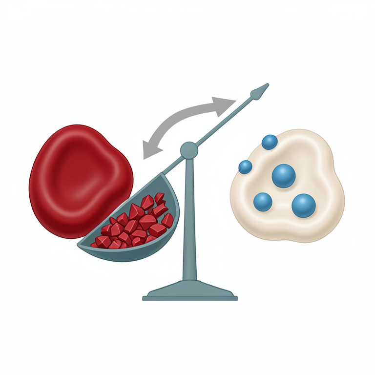
Oxidative Stress Imbalance in β-Thalassemia Trait
AI-generated conceptual illustration (human red blood cells). It explains the idea and does not show study data.
View source evidence ↗Figures & tables
From the original paper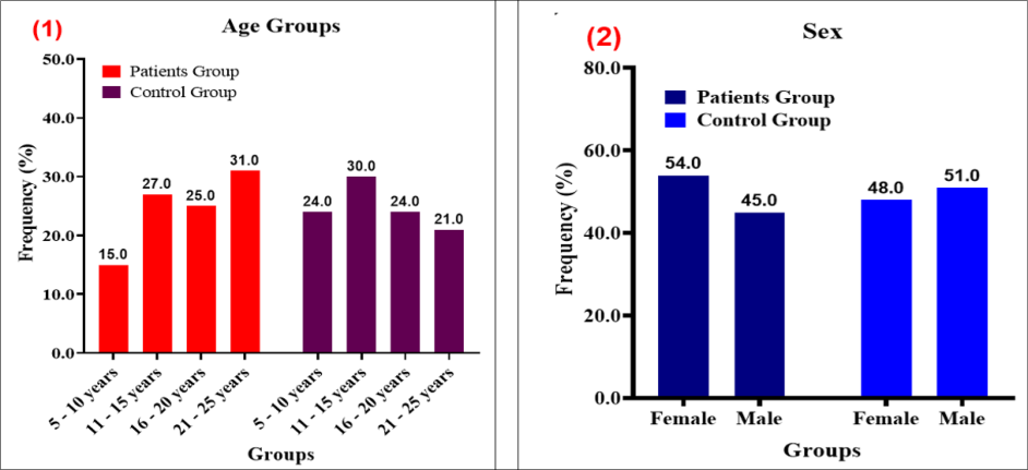 Figure 1
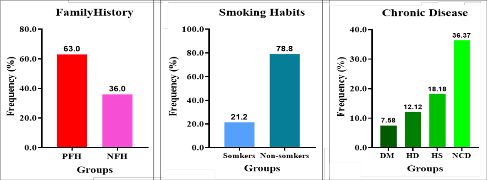 Figure 2
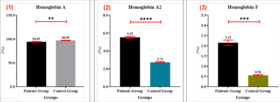 Figure 3
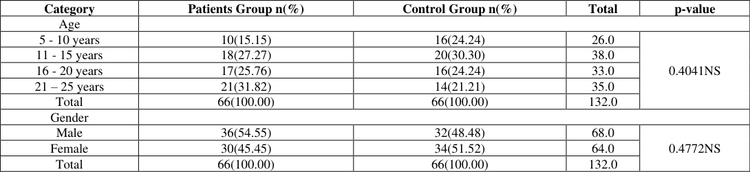 Table 1
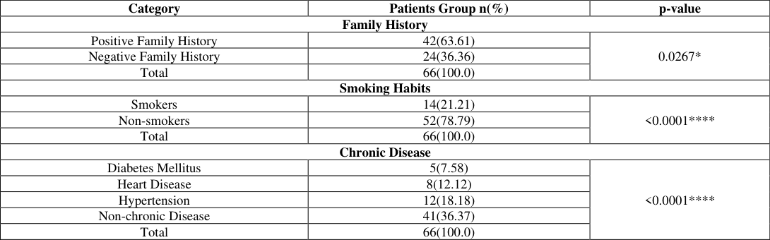 Table 2
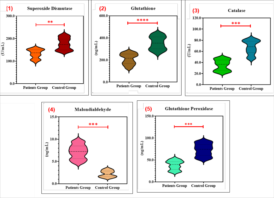 Figure 4
 Table 3
 Table 4
Introduction
Thalassemia is a serious public health problem and is most prevalent in the Southeast Asian region, India and the Mediterranean region. If the patient suffers from β-thalassemia major, he has chronic hemolytic anaemia and requires blood transfusion for his lifetime. [1,2] . There are three types of thalassemia: alpha (α) [1,2] , beta (β) and delta-beta (δβ), with β-thalassemia being the most common. In the world, the 3% of patients are in the category of β-thalassemia of the 5% of patients [3] . Iron chelation therapy and systematic red blood cell (RBC) transfusions are most often used supportive treatments for thalassemia, and these treatments have significantly increased the patient's life span. In any case, endocrine disorders associated with β-thalassemia may occur, and the most frequent symptoms are hypoxia, iron overload and chronic anaemia [4] . It appears that there was a higher prevalence of thalassemia in tropical and subtropical areas where malaria was very common [5,6] . The symptoms observed in patients suffering β-thalassemia intermedia are similar to those of both carriers and significant cases of the disease. Even in moderately anaemic patients, morbidity such as ulcers, pulmonary hypertension and discomfort can be caused by ineffective erythropoiesis and hemolysis [7] . The prevalence of β thalassemia is highest in the Mediterranean, Middle East and Southeast Asian regions, and lowest in Northern Europe and North America [7,8] . The disease has long been endemic in the native population of Middle East, due to high frequency of carrier status and consanguineous marriage, and large migration from other areas, such as Southeast Asia, where there is a high prevalence of the disease [9] . Decreased synthesis of δ globin chains may result in reduced levels of HbA2, which can be caused by transcriptional errors, or, in rare instances, by post-translational modifications that impair the HbA2 tetramer assembly. The dimerization of charged αand non-α subunits is an important step in the formation of Hb tetramer [10] . Oxidative stress is the imbalance between the ability to stabilize or reduce the damage caused by ROS and the production of ROS itself [11] .
Too much ROS can lead to damage of lipids, proteins and DNA [12] . The enzymatic antioxidants (Catalase (CAT) [5,6,8,7] and Superoxide dismutase (SOD) and nonenzymatic antioxidants (GSH, Vitamin C and E) play an important role in protection from oxidative stress [13,14] . It is found that Malonaldehyde (MDA) [13,14] is elevated in numerous people with βthalassemia, which is a biomarker of oxidative stress. This may be due to iron overload, which is known to occur to varying extents in the condition, as reflected by the elevated ferritin (the form of body iron that is stored) levels in affected people [15] . Therefore, the present study aimed to evaluate electrophoretic hemoglobin variants and oxidative stress biomarkers in Iraqi patients with β-thalassemia trait and to compare them with ageand sex-matched healthy controls.
Materials and Methods Study Design:
The study was a cross-sectional study carried out during December, 2025-March, 2026 to assess electrophoretic hemoglobin pattern and its association with serum level of hemoglobin and some biochemical parameters of the patients with beta thalassemia trait and age matched healthy controls. A total of 132 people were enrolled, comprising of 66 patients with diagnosis of βthalassemia trait and 66 apparently normal controls. Blood samples were collected from Fallujah Teaching Hospital, Ramadi Teaching Hospital, and Fallujah Maternity and Children's Hospital, Iraq. The participants were between 5 and 25 years of age.
Inclusion and Exclusion criteria:
Patients were selected from hospital outpatients based on clinical laboratory findings, including hematological and biochemical parameters such as abnormal hemoglobin (Hb), reduced mean corpuscular volume (MCV), reduced mean corpuscular hemoglobin (MCH) and raised hemoglobinA2 (HbA2) level. The severity and classification were based on the hemoglobin electrophoresis distribution. In the study, people suffering from β-thalassemia major, iron deficiency anemia, recent blood transfusion dependency or chronic inflammatory malignancies were excluded. The samples of the health control group were taken within normal haematological values and there were no clinical and genetic data for haemoglobinopathies
Collection of samples:
A total of 5 mL of venous blood was collected from each participant. The blood samples were divided into two portions. The first portion was transferred into an EDTA tube for hematological analysis, while the second portion was transferred into a plain gel tube for serum separation. The blood in the gel tube was allowed to clot at room temperature for 30 minutes, after which the serum was separated by centrifugation at 3000 rpm for 10 minutes and stored at -20 °C until biochemical analyses and ELISA assays were performed.
Statistical analysis:
GraphPad Prism software version 9.5.1 (GraphPad Software, San Diego, CA, USA) was used for statistical analysis. Continuous variables are presented as mean ± standard error (SE). The Mann-Whitney U test was used for non-parametric data and Fisher's exact test was used for categorical data for comparisons between two independent groups. Statistically significant values were pvalues <0.05
Results
Results
Distribution of Demographic Characteristics:
The demographic characteristics of the study participants were comparable between the patient and control groups. There was no statistically significant difference in age distribution between the two groups Φ (p=0.4041) ), indicating successful age matching and minimizing the possibility of selection bias. Similarly, gender distribution did not differ significantly between the patient and control groups (p=0.4772), demonstrating a balanced sex composition across the study population. Male participants constituted the majority in both groups. Overall, these findings confirm that the patient and control groups were demographically comparable, thereby reducing the potential influence of age and gender as confounding variables on the study outcomes. The detailed demographic data are presented in Table (1), while the gender distribution is illustrated in Figure (1). Distribution of Lifestyle and Clinical Characteristics: Analysis of the patients' lifestyle and clinical characteristics revealed that a positive family history was the most frequently reported factor, accounting for 63.61% of the study population, with a statistically significant difference (p=0.0267). Regarding smoking status, the majority of patients were non-smokers (78.79%), and this distribution was also statistically significant (p=0.001) Among the assessed comorbidities, hypertension was more prevalent than both heart disease and diabetes mellitus. Nevertheless, the largest proportion of patients had no associated chronic comorbidities. These findings are summarized in Table (2) and illustrated in Figure (2). Estimation of Hemoglobin Electrophoretic Parameters: A significant alteration in the distribution of hemoglobin electrophoretic fractions was observed between the patient and control groups. The mean level of hemoglobin A (HbA) was significantly lower in the patient group (94.05±0.11) than in the control group (96.99±0.06) (p=0.0043) indicating a reduction in normal adult hemoglobin. In contrast, hemoglobin A2 (HbA2) was markedly elevated in patients (5.52±0.10) compared with controls (2.73±0.05), showing a highly significant difference (p<0.0001). Likewise, fetal hemoglobin (HbF) levels were significantly higher in the patient group (2.14±0.13) than in the control group (0.55±0.02) (p=0.0008) Overall, these findings demonstrate the characteristic hemoglobin electrophoretic pattern associated with β-thalassemia trait, characterized by reduced HbA levels and increased HbA2 and HbF levels. The detailed results are presented in Table (3), while the distribution of hemoglobin fractions is illustrated in Figure (3).
Estimation of Enzymological Findings:
There was clear biochemical evidence of oxidative stress among the patients. The patients showed significantly lower levels of antioxidant enzymes, including (SOD), (GSH), (CAT) and (GPX1), compared to the control group (p≤0.01 to p<0.0001). The (MDA) levels were markedly higher in patients than in controls (p=0.0002). Although all parameters demonstrated highly to extremely significant differences, these findings indicate a pronounced imbalance between oxidant production and antioxidant mechanisms in patients. The reduction in antioxidant capacity alongside elevated MDA reflects increased lipid peroxidation and cellular damage as shown in Table (4) and figure (4). The p>0.05= Non significant difference (NS), the p≤0.05=Significant: (), the p≤0.01=Very significant: (), the p≤0.001=Highly significant: (), and the p≤0.0001=Excellent significant: (). The p>0.05= Non significant difference (NS), the p≤0.05=Significant: (), the p≤0.01=Very significant: (), the p≤0.001=Highly significant: (), and the p≤0.0001=Excellent significant: (). The p>0.05= Non significant difference (NS), the p≤0.05=Significant: (), the p≤0.01=Very significant: (), the p≤0.001=Highly significant: (), and the p≤0.0001=Excellent significant: (). The p>0.05= Non significant difference (NS), the p≤0.05=Significant: (), the p≤0.01=Very significant: (), the p≤0.001=Highly significant: (), and the p≤0.0001=Excellent significant: ().
Discussion
There was no significant difference between age group distribution in the patient group compared to the control group (p=0.4041); likewise, there was no difference between gender distribution (p =0.4772) This suggests that there was no confounding effect caused by demographic parameters as both groups were comparable in respect of age and sex. The importance of considering demographic factors to improve comparability and prevent bias has been emphasized. The accuracy of the correlation between biomarkers and disease outcomes can be improved, and bias reduced, if individuals are well-distributed by age and sex [16] . Hb electrophoretic analysis showed that the study subjects showed highly significant difference (p≤0.0001) from the control group. The increased level of HbA2 was attributed to an increased production of the δ-globin chain as a compensatory mechanism for the decreased production of the β-globin chain, and this finding was confirmed in the present study where most of those who were diagnosed as having βthalassemia trait had increased HbA2 despite having other factors such as iron-deficiency anemia [17] .
The current results are consistent with those published by Passarello et. al, [18] It is important to remember that who demonstrated that even when ID HbA₂ is present it is the best laboratory test to diagnose β-thalassemia trait. Their study demonstrated that the value of HbA₂ measurement in the detection of carriers of β-thalassemia is not significantly affected by iron deficiency. The results are comparable to that of the present work in which the level of HbA₂ was found to be significantly high in the patients as compared to healthy individuals, indicating the diagnostic potential and the stability of the level of HbA₂ as a marker of β-thalassemia trait [18] . The present findings are consistent with those reported by Colaco, et al. [19] who demonstrated that the hallmark of carriers of β - thalassemia is an increase in the amount of HbA2 and sometimes a slight increase in HbF. The hematological changes have been thought to play a significant role in the diagnostic features of the carrier state and the basis for present screening programmes for βthalassemia. Similarly, Thilakarathne et al. [20] reported that most cases of diagnosing βthalassemia carrier state rely on HbA₂ as the main laboratory parameter, however, some genetic and haematological parameters could impact the diagnosis. The results of this study are reinforced by the observation that the most effective carrier detection is by high HbA₂ [20] . These results suggest that chronic oxidative stress is linked with the relatively mild clinical phenotype of βthalassemia trait. If there is an imbalance in α and β chains production, excess of unpaired α chains precipitate and oxidize, resulting more than Reactive Oxygen Species (ROS) [18,19,20] . This is because ROS are constantly generated, and the antioxidant resources in cell are gradually used up and further reduced glutathione and SOD/CAT/GPX1 are decreased. Concurrently, it is observed that increased ROS levels cause lipid peroxidation of erythrocyte membranes that causes increased lipid peroxidation products of the membranes (MDA) that are markers of oxidative damage of membranes and erythrocyte fragility. This imbalance between oxidants and antioxidants leads to chronic cellular damage and less survival of red blood cells. In detail, these mechanisms have been described by Fibach, E. & Dana, M [21] , and they brought up the idea of oxidative stress being one of the mechanisms behind the pathophysiology of β-thalassemia. In the present study, the activity of antioxidant enzymes was found to be reduced in the β-thalassemia trait patients indicating ongoing oxidative stress in these cells. The results concur with the notion that cells have enzymatic antioxidant defense systems such as superoxide dismutase (SOD) [21] , catalase (CAT) and glutathione peroxidase (GPX1) as well as non-enzymatic antioxidants like reduced glutathione (GSH) to counteract the production of (ROS) under normal cellular metabolic activity and during pathological status.However, in β thalassemia disease, this constant production of free radicals in the disease creates imbalance between oxidants and antioxidants leading to oxidative stress and loss of antioxidant reserves. This imbalance is experienced by the reduction in the activity of these antioxidant enzymes and the increased oxidative damage reflected in the increased level of malondialdehyde (MDA) in the present study. The results are similar to those reported by [22,23] .
The present study has corroborated the earlier reports of continuous oxidative stress on the blood cells in β-thalassemia patients as evidenced by elevated generation of ROS, enhanced lipid peroxidation and decreased levels of reduced glutathione (GSH). Such a continuous oxidative imbalance is caused by continuous autoxidation of the excess unpaired α-globin chains and formation of free radicals due to excess iron, which leads to the exhaustion of the cellular antioxidant defense system. This results in the decrease in antioxidant capacity and increase in Lipid Peroxides such as Malondialdehyde (MDA). These are likely to be responsible for the significant decrease in antioxidant activity and the increase in the concentration of MDA observed in the present study [24] . The current study's findings are consistent with those of Fatima et al. [25] , who demonstrated that patients with β-thalassemia had reduced antioxidant defense mechanisms and substantial oxidative stress. Their research showed that antioxidant enzyme expression dropped despite an increase in oxidative stress, indicating that both enzymatic and nonenzymatic antioxidant systems were malfunctioning. This diminished antioxidant capacity may be the cause of the current study's elevated levels of malondialdehyde (MDA) [25] and lower antioxidant activity since it leaves the cells susceptible to cellular and molecular damage due to Reactive Oxygen Species (ROS) [25] .
Conclusion
In this study, β-thalassemia trait patients showed a significant decrease in haemoglobin A and increase in haemoglobin A2 and haemoglobin F levels in electrophoretic patterns. Furthermore, oxidative stress was confirmed as increased MDA and decreased antioxidant biomarkers SOD, GSH, CAT and GPX1 were observed. The results indicate that the biochemical and oxidative changes are measurable in the individuals with βthalassemia trait and more extensive studies are needed.
Acknowledgement
AcknowledgmentsThe author would like to express sincere gratitude to the staff of Fallujah Teaching Hospital, Ramadi Teaching Hospital, Fallujah Maternity and Children Hospital, and Al-Gharbia Specialized Laboratories for their valuable cooperation and assistance in sample collection. The author also extends sincere appreciation to the Department of Pathological Analysis, College of Applied Sciences, University of Fallujah, for providing laboratory facilities and technical support that contributed to the successful completion of this study.
Figures and tables from the paper
| Category | Patients Group n(%) | Control Group n(%) | Total | p-value |
|---|---|---|---|---|
| Age | ||||
| 5 - 10 years | 10(15.15) | 16(24.24) | 26.0 | 0.4041NS |
| 11 - 15 years | 18(27.27) | 20(30.30) | 38.0 | |
| 16 - 20 years | 17(25.76) | 16(24.24) | 33.0 | |
| 21 – 25 years | 21(31.82) | 14(21.21) | 35.0 | |
| Total | 66(100.00) | 66(100.00) | 132.0 | |
| Gender | ||||
| Male | 36(54.55) | 32(48.48) | 68.0 | 0.4772NS |
| Female | 30(45.45) | 34(51.52) | 64.0 | |
| Total | 66(100.00) | 66(100.00) | 132.0 |
Table text extracted from the original PDF; superscript letters are shown inline. The original table image is below.
View original table image
| Category | Patients Group n(%) | p-value |
|---|---|---|
| Family History | ||
| Positive Family History | 42(63.61) | 0.0267* |
| Negative Family History | 24(36.36) | |
| Total | 66(100.0) | |
| Smoking Habits | ||
| Smokers | 14(21.21) | <0.0001**** |
| Non-smokers | 52(78.79) | |
| Total | 66(100.0) | |
| Chronic Disease | ||
| Diabetes Mellitus | 5(7.58) | <0.0001**** |
| Heart Disease | 8(12.12) | |
| Hypertension | 12(18.18) | |
| Non-chronic Disease | 41(36.37) | |
| Total | 66(100.0) |
Table text extracted from the original PDF; superscript letters are shown inline. The original table image is below.
View original table image
| Parameters | Patients Group (Mean±SE) | Control Group (Mean±SE) | p-value |
|---|---|---|---|
| Hemoglobin A (%) | 94.05±0.11 | 96.99±0.06 | 0.0043** |
| Hemoglobin A2 (%) | 5.52±0.10 | 2.73±0.05 | <0.0001**** |
| Hemoglobin F (%) | 2.14±0.13 | 0.55±0.02 | 0.0008*** |
Table text extracted from the original PDF; superscript letters are shown inline. The original table image is below.
View original table image
| Parameters | Patients Group (Mean±SE) | Control Group (Mean±SE) | p-value |
|---|---|---|---|
| Superoxide dismutase (U/mL) | 132.7±2.44 | 175.8±3.20 | 0.0083 ** |
| Glutathione (ng/mL) | 208.5±6.00 | 353.5±7.25 | <0.0001 **** |
| Catalase (U/mL) | 35.25±1.18 | 67.17±1.49 | 0.0009 *** |
| Malondialdehyde (ng/mL) | 7.14±0.19 | 2.21±0.08 | 0.0002 **** |
| Glutathione peroxidase (ng/mL) | 38.09±1.26 | 72.93±1.17 | 0.0004 **** |
Table text extracted from the original PDF; superscript letters are shown inline. The original table image is below.
View original table image
References
23 in the original paper- Rao E, Kumar Chandraker S, Misha Singh M, Kumar R. Global distribution of β-thalassemia mutations: An update. Gene. 2024;896:148022.https://doi.org/10.1016/j.gene.2023.148 022
- U J, K W, T P, P K, S H, S S, al. Classification of betathalassemia major and HbE/beta-thalassemia via deep learning of image structure function image. Biomedical Signal Processing and Control. 2025;107265.https://doi.org/10.1016/j.bspc.2024.107265
- Venou TM, Barmpageorgopoulou F, Peppa M, Vlachaki E. Endocrinopathies in beta thalassemia: A narrative review. Hormones. 2024;23(2):205-
- Qian H, Zhao W, Li W, Wu Y, Lin X, Huang J, et al. Analysis of thalassemia genotypes and HbA test results in pregnant women in Shenzhen, China. Scientific Reports. 2025;15(1):3421.https://doi.org/10.1038/s41598-02586541-2
- Kattamis A, Forni GL, Aydinok Y, Viprakasit V. Changing patterns in the epidemiology of β-thalassemia. European Journal of Haematology. 2020;105(6):692703.https://doi.org/10.1111/ejh.13512
- Sharma SK, Chaudhari U, Ghosh M, Das S, Britt RP, Singh S, et al. Prevalence of β-thalassemia and other haemoglobinopathies in six cities in India: A multicentre study. Journal of Community Genetics. 2013;4(1):3342.https://doi.org/10.1007/s12687-012-0114-0
- El-Beshlawy A, Dewedar H, Hindawi S, Alkindi S, Tantawy AY, Yassin MA, et al. Management of transfusion-dependent β-thalassemia (TDT): Expert insights and practical overview from the Middle East. Blood Reviews. 2024;63:101138.https://doi.org/10.1016/j.blre.2023.10113
- Colaco S, Nadkarni A. Borderline HbA levels: Dilemma in diagnosis of beta-2 thalassemia carriers. Mutation Research/Reviews in Mutation Research. 2021;788:108387.https://doi.org/10.1016/j.mrrev.2021.10 8387
- Amin S, Jalal S, Ali K, Rasool L, Osman T, Ali O, et al. Molecular characterization and disease-related morbidities of β-thalassemia patients from the northeastern part of Iraq. International Journal of General Medicine. 2020;13:14531467.https://doi.org/10.2147/IJGM.S277947
- Jallab HR, Eesa ZM. Prevalence of hepatitis B and hepatitis C viruses in β-thalassemia major patient in AD-Diwanya Province, Iraq. Indian Journal of Forensic Medicine & Toxicology. 2020;14(3):11451150.https://doi.org/10.37506/ijfmt
- Eren F, Koca Yozgat A, Firat ğğuz E, Neşelioğlu S, Firat R, Gürlek Gökçebay D, Yarali HN, Özbek NY, Erel Ö. A new perspective for potential organ damage due to ironmediated oxidation in thalassemia major patients. Journal of Clinical Medicine. 2023;12(6):2422.https://doi.org/10.3390/jcm12062422
- Meloni A, Pistoia L, Spasiano A, Cossu A, Casini T, Massa A, et al. Oxidative stress and antioxidant status in adult patients with transfusion-dependent thalassemia: Correlation with demographic, laboratory, and clinical biomarkers. Antioxidants. 2024;13(4):446.https://doi.org/10.3390/antiox13040446
- Basu D, Adhya DG, Sinha R, Chakravorty N. Role of malonaldehyde as a surrogate biomarker for iron overload in the β-thalassemia patient: A systematic meta-analysis. Advances in Redox Research. 2021;3:100017.https://doi.org/10.1016/j.arres.2021.10001
- Dockès J, Varoquaux G, Poline JB. Preventing dataset shift from breaking machinelearning biomarkers. GigaScience. 2021;10(9):giab055.https://doi.org/10.1093/gigascience/gi ab055
- Shalli AAK, Jalal SD, Mohammed DJ. The impact of iron deficiency on the diagnostic level of HbA in betathalassemia trait from the Sulaimani hemoglobinopathies₂ screening program. Advanced Medical Journal. 2022;6(1):12-19.https://doi.org/10.21172/amj
- Passarello C, Giambona A, Cannata M, Vinciguerra M, Renda D, Maggio A. Iron deficiency does not compromise the diagnosis of high HbA β-thalassemia trait.₂ Haematologica. 2012;97(3):472473.https://doi.org/10.3324/haematol.2011.052357
- Colaco S, Colah R, Nadkarni A. Significance of borderline HbA levels in β-₂ thalassemia carrier screening. Scientific Reports. 2022;12(1):5414.https://doi.org/10.1038/s4159820.
- Thilakarathne S, Jayaweera UP, Premawardhena A. Unresolved laboratory issues of the heterozygous state of β-thalassemia: A literature review. Haematologica. 2024;109(1):2332.https://doi.org/10.3324/haematol.2022.282667
- Fibach E, Dana M. Oxidative stress in β-thalassemia. Molecular Diagnosis & Therapy. 2019;23(2):245261.https://doi.org/10.1007/s40291-018-0373-5
- Abdalla MY, Fawzi M, Al-Maloul SR, El-Banna N, Tayyem RF, Ahmad IM. Increased oxidative stress and iron overload in Jordanian β-thalassemic children. Hemoglobin. 2011;35(1):6779.https://doi.org/10.3109/03630269.2010.544624
- Fibach E, Rachmilewitz E. The role of oxidative stress in hemolytic anemia. Current Molecular Medicine. 2008;8(7):609619.https://doi.org/10.2174/156652408786241384
- Amer J, Dana M, Fibach E. The antioxidant effect of erythropoietin on thalassemic blood cells. Anemia. 2010;2010:978710.https://doi.org/10.1155/2010/978710
- Fatima T, Khan S, Khan MM, Kamran R, Uddin MW, Sohrab S. Oxidative stress in beta-thalassemia patients: Role of enzymatic and non-enzymatic modulators. Protein and Peptide Letters. 2023;30(12):10301037.https://doi.org/10.2174/01092986652462702310200 62048