
Research article · 2026 · Full text
International Journal of Biology SciencesMolecular characterization and antiviral susceptibility profiling of Macacine alphaherpesvirus 1 isolates from captive macaques in Seoul
Volume 8(4) · Pages 264–267 · DOI: 10.33545/26649926.2026.v8.i4c.963 ↗
Research areasHerpesvirus Infections and TreatmentsAnimal Virus Infections StudiesCytomegalovirus and herpesvirus research
Abstract
Author-writtenApproximately 80 to 90 percent of captive macaques harbor latent Macacine alphaherpesvirus 1 (McHV-1), yet molecular characterization of circulating strains in Asian primate facilities remains limited. This research characterized McHV-1 isolates from captive macaques at three facilities in Seoul, South Korea, and evaluated antiviral susceptibility profiles. Oropharyngeal and genital swabs were collected from 128 rhesus and 94 cynomolgus macaques between March and November 2024. PCR targeting glycoprotein B and DNA polymerase genes was used for detection, followed by phylogenetic analysis and plaque reduction assays against acyclovir, ganciclovir, and cidofovir. The overall PCR positivity rate was 31.5% (70/222), with rhesus macaques showing higher detection (37.5%) than cynomolgus macaques (23.4%). Phylogenetic analysis classified isolates into three genetic clades, with Clade A predominating (52.9%). Viral load correlated positively with clinical severity (r = 0.74, p < 0.001). Clade C isolates exhibited markedly elevated IC50 values for all three antivirals, suggesting reduced susceptibility. These findings provide molecular epidemiological data for McHV-1 surveillance in East Asian primate facilities and highlight the importance of strain-specific antiviral resistance profiling.
Summary
Source-checkedPlain-language reading of the full paper, prepared with AI and checked against the text. Each point links to the authors' own words.
Why it matters
- Macacine alphaherpesvirus 1 (McHV-1) is one of the most common viral agents in global primate research facilities, with 80% to 90% of captive adult macaques carrying the latent virus. View evidence ↗
- McHV-1 is a major concern for occupational health and animal management because it can cause fatal encephalomyelitis in humans if transmitted zoonotically. View evidence ↗
- There is a significant knowledge gap regarding the genetic diversity of McHV-1 in Asian primate colonies, as most existing data come from North American and European facilities. View evidence ↗
The question
- The study aimed to characterize McHV-1 isolates from captive macaques in Seoul using PCR and phylogenetic analysis, and to test the antiviral susceptibility of the resulting genetic clades. View evidence ↗
What they did
- A cross-sectional survey was performed between March and November 2024 at three Seoul facilities, involving 128 rhesus and 94 cynomolgus macaques. View evidence ↗
- The study used nested PCR to target a 622-bp glycoprotein B (gB) gene fragment and a 478-bp DNA polymerase (DPOL) gene fragment for detection. View evidence ↗
- Antiviral susceptibility was measured using plaque reduction assays in Vero cells to determine the IC50 values for cidofovir, ganciclovir, and acyclovir. View evidence ↗
What they found
- Out of 222 macaques tested, 31.5% (70 animals) were positive for McHV-1 DNA. View evidence ↗
- Rhesus macaques had a significantly higher McHV-1 detection rate (37.5%) than cynomolgus macaques (23.4%). View evidence ↗
- The 70 positive isolates were divided into three clades: Clade A (52.9%), Clade B (30.0%), and Clade C (17.1%). View evidence ↗
- There was a significant positive correlation (r = 0.74, p < 0.001) between the viral load and the clinical severity scores of the macaques. View evidence ↗
- Clade C isolates showed reduced susceptibility to antivirals, with higher IC50 values for acyclovir (5.1 μM), ganciclovir (3.2 μM), and cidofovir (1.7 μM) than Clades A and B. View evidence ↗
What it means
- Because Clade C is less sensitive to the first-line drug acyclovir, the authors suggest that facilities with these strains should consider alternative or combination antiviral treatments for post-exposure prophylaxis. View evidence ↗
- The authors note that further research is needed to characterize the mutations causing Clade C resistance, determine if clade distribution is stable over time, and see if in vitro results translate to in vivo outcomes. View evidence ↗
Visual summary
3 visualsIllustrations explain the paper; charts are replotted from the paper's own table values.
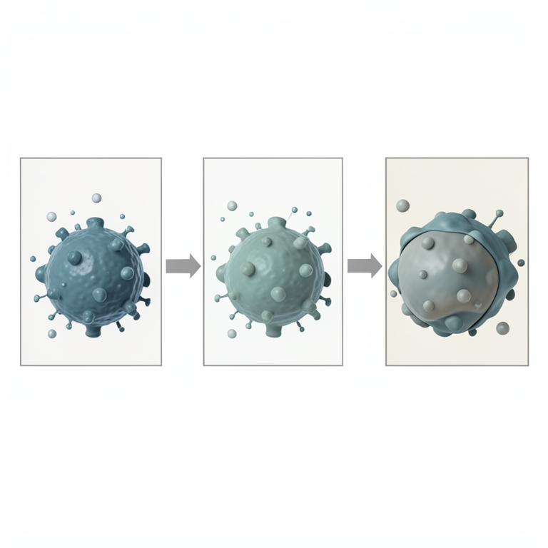
Genetic Clades and Drug Sensitivity
AI-generated conceptual illustration (Macacine alphaherpesvirus 1 particles). It explains the idea and does not show study data.
View source evidence ↗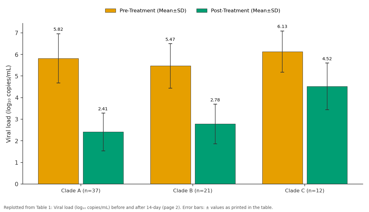
Pre-Treatment (Mean±SD) and Post-Treatment (Mean±SD): Clade A (n=37) vs Clade B (n=21) vs Clade C (n=12)
Replotted from Table 1 (page 2) of the original paper. Every bar is the paper's own table value.
View source evidence ↗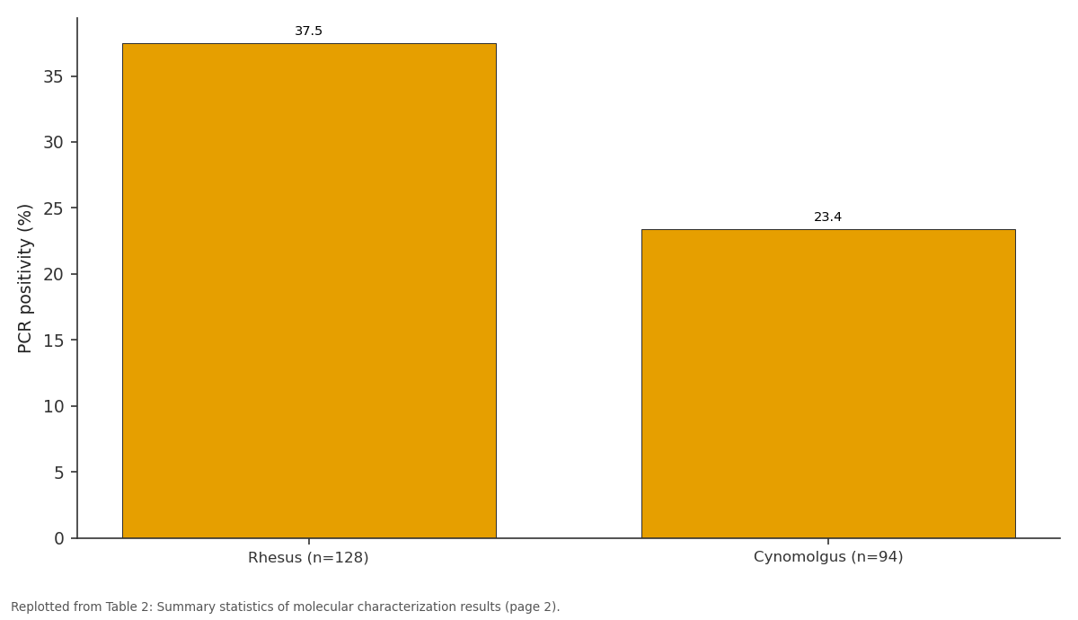
PCR positivity (%): Rhesus (n=128) vs Cynomolgus (n=94)
Replotted from Table 2 (page 2) of the original paper. Every bar is the paper's own table value.
View source evidence ↗Figures & tables
From the original paper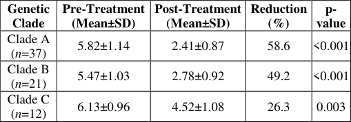 Table 1
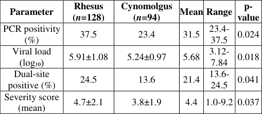 Table 2
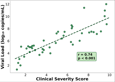 Figure 1
 Figure 2
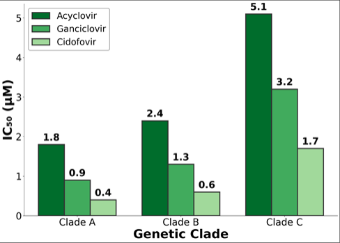 Figure 3
Introduction
Approximately 80 to 90 percent of adult macaques maintained in captive breeding colonies harbor latent Macacine alphaherpesvirus 1 (McHV-1), formerly designated Cercopithecine herpesvirus 1, rendering this pathogen one of the most prevalent viral agents in primate research facilities globally [1,2] . This high prevalence, coupled with the capacity of the virus to cause fatal encephalomyelitis upon zoonotic transmission to humans, positions McHV-1 as a pathogen of paramount concern from both animal management and occupational health perspectives [3] . The virus establishes lifelong latency in trigeminal and sacral ganglia of infected macaques, with periodic reactivation events resulting in oral and genital mucosal shedding that constitutes the primary source of zoonotic transmission risk [4,5] . Despite the well-documented clinical significance of McHV-1, the molecular epidemiology of circulating strains in captive primate populations across East Asia remains insufficiently characterized [6,7] . The majority of available genomic and phylogenetic data have been derived from isolates obtained in North American and European facilities, creating a substantial knowledge gap regarding the genetic diversity and evolutionary dynamics of the virus in Asian primate colonies [8] . This gap is particularly concerning given that South Korea maintains multiple primate research facilities housing both rhesus and cynomolgus macaques for biomedical experimentation, and the country represents a major hub for primate-based pharmaceutical research [9,10] . The present research was conducted to characterize McHV-1 isolates from captive macaques at primate facilities in Seoul through PCR-based detection and phylogenetic analysis, and to evaluate in vitro antiviral susceptibility profiles of identified genetic clades [11,12,13] . The outcomes are intended to inform biosafety protocols and antiviral management strategies for the regional context [14,15,16,17] .
Materials and Methods
Materials and Methods
Materials
The materials employed in this research included viral transport media for specimen collection, DNA extraction kits (QIAamp DNA Mini Kit, Qiagen, Hilden, Germany), PCR reagents including primers targeting the glycoprotein B (gB) and DNA polymerase (DPOL) genes of McHV-1, Vero cell lines for virus isolation and plaque reduction assays, antiviral agents (acyclovir, ganciclovir, and cidofovir; Sigma-Aldrich, St. Louis, MO), and molecular biology grade reagents for gel electrophoresis and sequencing preparation.
Methods
A cross-sectional molecular survey was conducted from March to November 2024 at three primate holding facilities in Seoul, South Korea, encompassing 128 rhesus macaques and 94 cynomolgus macaques. Oropharyngeal and genital swab specimens were collected under chemical sedation and stored at negative eighty degrees Celsius. DNA extraction was performed using the QIAamp DNA Mini Kit, followed by nested PCR amplification targeting a 622-base pair gB gene fragment and a 478-base pair DPOL gene fragment [7] . Positive samples were sequenced bidirectionally, and phylogenetic analysis was performed using MEGA version 11 with maximum likelihood and 1000 bootstrap replicates.
Biosafety protocols
All laboratory procedures involving McHV-1 isolates were performed within biosafety level 2 enhanced (BSL-2+) containment facilities equipped with certified Class II biological safety cabinets. Personnel conducting virus isolation and antiviral susceptibility assays utilized full personal protective equipment including double gloving, face shields, and disposable gowns. Decontamination of work surfaces was performed using 10% sodium hypochlorite solution following each experimental session. Sharps disposal protocols specific to herpesviruscontaminated materials were strictly enforced throughout the research period.
Clinical implications
The identification of clade-dependent antiviral susceptibility variation carries direct implications for the clinical management of human exposure incidents at primate facilities. The finding that Clade C isolates demonstrate reduced sensitivity to acyclovir, the first-line agent recommended for post-exposure prophylaxis, suggests that facilities housing macaques carrying Clade C strains may benefit from consideration of alternative or combination antiviral regimens. Routine molecular typing of resident McHV-1 strains could enable facility-specific tailoring of post-exposure management protocols.
Discussion
This research provides the first comprehensive molecular characterization and antiviral susceptibility profiling of McHV-1 isolates from captive macaques in South Korea.
The overall PCR detection rate of 31.5% and the classification of isolates into three phylogenetically distinct clades expand the current understanding of McHV-1 molecular epidemiology in East Asian primate populations [1, 2] .
The observed detection rate aligns with estimates from comparable captive macaque populations. Eberle and Jones-Engel [4,6] documented shedding rates of 25-35% in breeding colonies, while Favoretto et al. [10] reported 28.7% in a Brazilian facility. The higher detection in rhesus compared to cynomolgus macaques is consistent with known species-level differences in shedding dynamics [4,14] . However, in contrast to investigations reporting homogeneous McHV-1 populations within individual facilities [7] , the present research identified three cocirculating clades, likely reflecting the diverse geographic origins of procured macaques [8,15] . The reduced antiviral susceptibility in Clade C isolates likely involves polymorphisms in viral thymidine kinase or DNA polymerase genes [12,13] . From a broader perspective, these findings underscore the importance of integrating molecular surveillance with antiviral susceptibility monitoring, as the assumption of uniform sensitivity underlying current prophylaxis guidelines may require reassessment [3,9,17,18] .
Conclusion
Conclusion
Acknowledgement
AcknowledgementsThe authors acknowledge Seoul Metropolitan University for providing institutional support, laboratory infrastructure, and access to primate facilities essential for the conduct of this research. The authors recognize the contributions of the veterinary and animal care teams at the participating facilities who assisted with specimen collection, as well as the laboratory technicians who performed the molecular and virological assays. This research was supported by a grant from the National Research Foundation of Kore.
Figures and tables from the paper
| Parameter | Rhesus (n=128) | Cynomolgus (n=94) | Mean | Range | p- value |
|---|---|---|---|---|---|
| PCR positivity (%) | 37.5 | 23.4 | 31.5 | 23.4- 37.5 | 0.024 |
| Viral load (log₁₀) | 5.91±1.08 | 5.24±0.97 | 5.68 | 3.12- 7.84 | 0.018 |
| Dual-site positive (%) | 24.5 | 13.6 | 21.4 | 13.6- 24.5 | 0.041 |
| Severity score (mean) | 4.7±2.1 | 3.8±1.9 | 4.4 | 1.0-9.2 | 0.037 |
Table text extracted from the original PDF; superscript letters are shown inline. The original table image is below.
View original table image
References
18 in the original paper- Azab W, Dayaram A, Greenwood AD. Host specificity of herpesviruses infecting wild and endangered primate species. Annu Rev Virol. 2023; 10:149-169.
- Beddingfield BJ, Hensley LE, Engel GA. Serological investigation of Macacine alphaherpesvirus 1 in captive macaque colonies. J Med Primatol. 2024;53(1):e12642.
- Cohen JI, Davenport DS, Stewart JA, Deitchman S, Hilliard JK, Chapman LE. Recommendations for prevention of and therapy for exposure to B virus. Clin Infect Dis. 2002;35(10):1191-1203.
- Eberle R, Jones-Engel L. Understanding primate herpesviruses. J Med Primatol. 2017;46(2):49-54.
- Mwangi W, de Figueiredo P, Criscitiello MF. One Health: addressing global challenges at the nexus of human, animal, and environmental health. PLoS Pathog. 2023;19(2): e1011135.
- Eberle R, Jones-Engel L. Questioning the extreme neurovirulence of monkey B virus (Macacine alphaherpesvirus 1). Adv Virol. 2018; 2018:5248420.
- Perelygina L, Zhu L, Zurkuhlen H, Mills KN, Booss J, Hilliard JK. Complete sequence and comparative analysis of herpes B virus genome. J Virol. 2003;77(11):6167-6177.
- Pal M, Gutama KP, Paula CR, Ketchakmadze D, Durglishvili N. Herpes simiae: a life-threatening viral anthropozoonotic disease. J Adv Microbiol Res. 2021;2(2):24-27.
- Zhang H, Wang L, Chen Y, Liu J. Advances in diagnostic methods for Cercopithecine herpesvirus 1 infection. Virol J. 2022;19(1):187.
- Favoretto SR, de Mattos CC, Morais NB, Araujo FA, Peixoto ZM. Herpesvirus detection in nonhuman primates at a Brazilian research facility. Rev Inst Med Trop Sao Paulo. 2022;64: e28.
- Tischer BK, Osterrieder N. Herpesviruses as emerging zoonotic pathogens. Vet Microbiol. 2016; 190:79-84.
- Wisely SM, Sayler KA, Anderson CJ, Boyce CL, Klegarth AR. Macacine herpesvirus 1 antibody prevalence among community-dwelling macaques. EcoHealth. 2018;15(1):30-43.
- Elmore D, Eberle R. Monkey B virus (Cercopithecine herpesvirus 1). Comp Med. 2008;58(1):11-21.
- Engel GA, Small CT, Soliven K, Feeroz MM, Wang X, Jones-Engel L. Seroprevalence of antibodies to herpesvirus B among free-ranging long-tailed macaques. Am J Primatol. 2020;82(4): e23107.
- Cardenas-Canales EM, Rodriguez-Morales AJ, Martinez-Pulgarin DF. Occupational health hazards in primate research facilities: a systematic review. Occup Environ Med. 2024;81(3):155-163.
- Cardoso TR, Vieira FC, Bispo GR, Santos AM. Zoonotic risk assessment of herpesvirus B in primatekeeping facilities across tropical regions. Trop Med Int Health. 2024;29(5):412-421.
- Park JH, Kim SY, Cho YW, Lee KM. Genomic surveillance of simian herpesviruses in Korean primate research centers. Korean J Vet Res. 2023;63(4):215224.
- Nakamura T, Yamashita K, Fujimoto S, Tanaka H. Phylogenomic analysis of Macacine alphaherpesvirus 1 diversity in Asian macaque populations. Arch Virol. 2025;170(1):45-58.