
Research article · 2026 · Full text
International Journal of Biology SciencesHaematic fetal mummification as a cause of dystocia in a non-descript doe: A case report
Volume 8(7) · Pages 282–284 · DOI: 10.33545/26649926.2026.v8.i7c.961 ↗
Research areasReproductive Physiology in LivestockVeterinary Medicine and SurgeryRabbits: Nutrition, Reproduction, Health
Abstract
Author-writtenA 3-year-old pluriparous non-descript doe in advanced gestation was presented with prolonged straining and failure to deliver. Ultrasonography confirmed a non-viable singleton fetus with oligohydramnios. Following induction of parturition with cloprostenol (PGF₂α), the cervix dilated completely, and a haematic mummified fetus was successfully delivered by obstetrical correction and manual traction. The doe recovered uneventfully after supportive therapy.
Summary
Source-checkedPlain-language reading of the full paper, prepared with AI and checked against the text. Each point links to the authors' own words.
Why it matters
- Fetal mummification is a rare reproductive disorder where a fetus dies aseptically and is retained in the uterus, leading to economic losses and longer intervals between kiddings. View evidence ↗
- There are two types of mummification: papyraceous (dry, parchment-like membranes) and haematic (chocolate-brown membranes due to blood pigment), the latter of which is common in ruminants like goats. View evidence ↗
- The causes of fetal mummification are diverse, ranging from infectious agents like Toxoplasma gondii to non-infectious factors such as fetal trauma, umbilical cord compression, and nutritional deficiencies. View evidence ↗
The question
- The study aims to report the successful management of dystocia caused by a single mummified fetus in a goat, emphasizing the importance of early diagnosis and intervention. View evidence ↗
What they did
- The subject was a 3-year-old, 20 kg pluriparous non-descript doe in advanced pregnancy presenting with 12 hours of non-progressive straining and vaginal discharge. View evidence ↗
- Diagnosis was aided by trans-abdominal ultrasonography, which showed a single fetal skeleton with no heartbeat and low amniotic fluid (oligohydramnios). View evidence ↗
- The fetus was recovered via manual traction and correction after the perineal area was cleaned with 1% Potassium permanganate and lubricated with liquid paraffin. View evidence ↗
What they found
- The delivered fetus exhibited deep brown pigmentation and shriveled membranes, characteristic of haematic mummification, and lacked eyeballs and a subcutaneous layer. View evidence ↗
- The mummified fetus alone weighed 78.0 gm. View evidence ↗
- The crown-rump length of the mummified fetus was 21 cm. View evidence ↗
- The approximate age of the mummified fetus was determined to be 20-21 weeks. View evidence ↗
- The total length of the placenta associated with the mummified fetus was 77.0 cm. View evidence ↗
- Following the delivery and supportive therapy, the doe recovered without further complications. View evidence ↗
What it means
- The authors conclude that manual per-vaginal delivery of a mummified fetus is feasible provided there is adequate cervical dilation. View evidence ↗
- Because no damage to the reproductive tract was found, the authors state the prognosis for the doe's future fertility is favorable. View evidence ↗
Figures & tables
From the original paper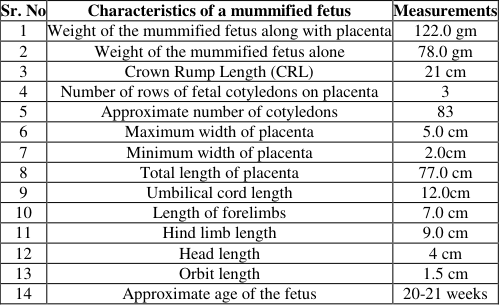 Table 1
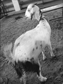 Figure 1
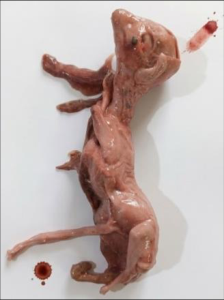 Figure 2
Introduction
Not aborted, not resorbed, but preserved fetal mummification is an uncommon reproductive disorder characterized by aseptic intrauterine fetal death, retention of the fetus, and resorption of fetal fluids (Noakes et al., 2009) [16] resulting in fetal loss, prolongs the intercalving or kidding interval, and causes significant economic losses to livestock farms (Sutradhar et al., 2010) [18] . Fetal mummification is classified into papyraceous which is characterized by dry, shriveled, parchment-like fetal membranes and is commonly reported in dogs, cats, pigs, and horses, whereas hematic mummification is characterized by chocolate-brown fetal membranes due to blood pigment deposition and is predominantly observed in cattle, buffaloes, sheep, and goats (Noakes et al., 2009) [16] . It is a rare obstetrical disorder in small ruminants, typically occurring during the middle to the last third of gestation (Roberts, 1971) [17] . Although an incidence of approximately 4% has been reported among gestational disorders, its occurrence in does and ewes remains uncommon (Vikram et al., 2020) [20] . The etiology of fetal mummification is multifactorial, with major infectious causes in caprine including Toxoplasma gondii, Chlamydophila spp., Border disease virus (Pestivirus), and Coxiella burnetii (Edmondson et al., 2012; Lefebvre, 2015) [10,14] . Noninfectious causes include fetal trauma (Broaddus et al., 2009; Dubey, 2009) [8,9] , umbilical cord compression (Mahajan and Sharma, 2002) [15] , uterine torsion, defective placentation (Irons, 1999) [12] , genetic and chromosomal abnormalities, hormonal imbalances, and protein or energy deficiencies during advanced gestation, with affected animals often appearing weak and anemic (Lefebvre, 2015) [14] . The present case report describes the successful management of dystocia due to a single mummified fetus in a non-descript doe, highlighting early diagnosis and prompt obstetrical intervention.
Anamnesis and Clinical observations
An adult 3 year old pluriparous non-descript doe weighing around 20 kg and in advanced pregnancy was presented to TVCC of MJF Veterinary college, Jaipur with a history of intermittent straining, with a string of clear to whitish vaginal discharge since 12 h without any progress along with inappetence and isolation from the flock. On clinical examination, doe was dull with normal rectal temperature and respiration rate. Per-vaginal examination revealed cervical dilation to the extent of about two finger and detailed trans-abdominal ultra-sonographic examination revealed single fetal skeletons devoid of any fetal heartbeat and with oligohydramniosis. Hence, it was decided to terminate the pregnancy by administering 125 µg of Vetmate® Inj. (Cloprostenol-Synthetic Prostaglandin F2α 250 µg / mL) IM and was advised to observe for fetal expulsion. On the same day evening, the doe was brought back with complaint of non-progressive straining upon gynaecoclinical examination, cervix was completely dilated and a dry rubber like compact structure in pelvic brim with malposition was palpated. The case was tentatively diagnosed as dystocia due to fetal mummifications.
Treatment and discussion
The perineal portion was thoroughly washed with 1% Potassium permanganate and liquid paraffin for lubrication, Then, well lubricated gloved hand was passed per vaginally and a mummified fetus (Fig. 1) was recovered per vaginally via manual traction after correction. The foetal consistency and deep brown colour pigmentation from caruncular hemorrhage was typical of haematic type of mummification. The fetal membranes were shriveled and dried with fetus lacking eye ball and SC layer. The biometry of the mummified fetus delivered was also recorded in (Table.1). (Melonex®) IM for five consecutive days.As a supportive therapy Inj. Milk fever formula®-60 mL slow IV to compensate for the loss of calcium ions due to prolonged straining, along with these, uterine cleanser, Liq Utrevive® 40 mL per oral twice a day for five days was prescribed to evacuate and cleanse the uterus from any remnant.The doe was discharged post 2 h of delivery and owner was advised to follow up the prescribed treatment. Fetal mummification as a cause of dystocia in does has been documented in the literature in association with a single mummified fetus (Bisla et al., 2018) [6] , a mummified cotwin with a normal dead fetus (Bawaskar et al., 2018) [4] , and a mummified fetus accompanied by two normal fetuses (Bisla et al., 2019) [7] . In polytocous species such as goats, the condition is generally associated with prolonged gestation and is best diagnosed using ultrasonography, with Doppler ultrasonography serving as a valuable adjunct for assessing fetal viability. Mummification involving one or two fetuses has also been reported by Bisla et al. (2019) [7] , Vinayak et al. (2020) [21] , Arun et al. (2023) [3] . [5,2,1,11] . However, the present case involved a single mummified fetus, suggesting that factors other than multiple gestation, such as placental insufficiency, fetal trauma, compromised fetal blood supply, defective placentation, umbilical cord abnormalities, or genetic defects, may have contributed to the condition (Jainudeen, 1992) [13] . Although spontaneous expulsion of mummified fetuses may occasionally occur, they are more commonly retained in utero well beyond the normal gestation period (Jainudeen, 1992) [13] . The successful outcome in the present case demonstrates that manual pervaginal delivery is feasible when adequate cervical dilation has occurred. In cases with an incompletely dilated cervix, administration of luteolytic agents such as PGF₂α, along with cervical dilators, has been recommended to facilitate fetal expulsion (Thangamani, 2023) [19] . In the present doe, PGF₂α administration resulted in complete cervical dilation and enhanced uterine contractions, allowing the mummified fetus to descend into the birth canal. However, fetal maldisposition led to dystocia, which was successfully corrected by obstetrical manipulation, adequate lubrication, and gentle manual traction. As no apparent damage to the reproductive tract was observed, the prognosis for subsequent fertility and breeding remains favourable
Acknowledgement
AcknowledgmentThe authors acknowledge the clinical and support staff involved in the management of the case.
Figures and tables from the paper
| Sr. No | Characteristics of a mummified fetus | Measurements |
|---|---|---|
| 1 | Weight of the mummified fetus along with placenta | 122.0 gm |
| 2 | Weight of the mummified fetus alone | 78.0 gm |
| 3 | Crown Rump Length (CRL) | 21 cm |
| 4 | Number of rows of fetal cotyledons on placenta | 3 |
| 5 | Approximate number of cotyledons | 83 |
| 6 | Maximum width of placenta | 5.0 cm |
| 7 | Minimum width of placenta | 2.0cm |
| 8 | Total length of placenta | 77.0 cm |
| 9 | Umbilical cord length | 12.0cm |
| 10 | Length of forelimbs | 7.0 cm |
| 11 | Hind limb length | 9.0 cm |
| 12 | Head length | 4 cm |
| 13 | Orbit length | 1.5 cm |
| 14 | Approximate age of the fetus | 20-21 weeks |
Table text extracted from the original PDF; superscript letters are shown inline. The original table image is below.
View original table image
References
21 in the original paper- Alagar S, Prakash S, Selvaraj M, Ravikumar K, Manokaran S. Papyraceous mummification leading to dystocia of a normal fetus in a Mecheri ewe. Indian Journal of Animal Reproduction. 2017;38:62-63.
- Anil M, Rajashri M, Raju G, Solmon Raju KG, Reddy KC. Fetal mummification in non-descript doe: A case International Journal of Biology Sciences https://www.biologyjournal.net report. International Journal of Environmental Science and Technology. 2017;6:2335-2338.
- Arun R, Sowbharenya C, Uma Rani R, Arulanandam K. Dystocia due to fetal mummification in non-descriptive goat: An investigation of live fetus challenges. International Journal of Veterinary Science and Animal Husbandry. 2023;8(5):341-342.
- Bawaskar MS, Sahatpure SK, Sheetal SK, Takle VU, Bhardwaj A. Mummified Schistosomus reflexus cotwinned with normal goat fetus: A rare case report. International Journal of Science Environment and Technology. 2018;7(4):1479-1482.
- Bhardwaj RK, Kumar S. Management of goat with one mummified and one live fetus. Indian Veterinary Journal. 2014;91:64-65.
- Bisla A, Kumar B, Kurhe R, Behra H, Ngou AA, Shah I, et al. Dystocia due to fetal mummification in a nondescript goat: A case study. Journal of Experimental Biology and Agricultural Sciences. 2018;6(3):613-616.
- Bisla A, Singh SK, Kumar A, Ngou AA, Kurhe RS, Rutela R, et al. Fetal mummification in one of the triplet kids: A cause of dystocia in a non-descript doe: A case report. Exploratory Animal and Medical Research. 2019;9(1):90-92.
- Broaddus CC, Lamm CG, Kapil S, Dawson L, Holyoak GR. Bovine viral diarrhea virus abortion in goats housed with persistently infected cattle. Veterinary Pathology. 2009;46(1):45-53.
- Dubey JP. Toxoplasmosis in sheep: The last 20 years. Veterinary Parasitology. 2009;163(1-2):1-14.
- Edmondson MA, Roberts JF, Baird AN, Bychawski S, Pugh DG. Theriogenology of sheep and goats. In: Pugh DG, Baird AN, editors. Sheep and Goat Medicine. 2nd ed. Maryland Heights (MO): Elsevier Saunders; 2012. p.150-230.
- Hemalatha H, Murugavel K, Kantharaj S, Antoine D, Raju MS. Mummified fetus co-twin to a live kid in a goat. Indian Journal of Animal Reproduction. 2018;39(2):70-71.
- Irons PC. Hysterotomy by a colpotomy approach for treatment of fetal mummification in a cow. Journal of the South African Veterinary Association. 1999;70:127129.
- Jainudeen MR. Reproductive failure in females. In: Hafez ESE, editor. Reproduction in Farm Animals. 6th ed. Baltimore: Williams & Wilkins; 1992. p.277-278.
- Lefebvre RC. Fetal mummification in the major domestic species: Current perspectives on causes and management. Veterinary Medicine: Research and Reports. 2015;6:233-244.
- Mahajan M, Sharma A. Haematic mummification due to umbilical cord torsion in a cow: A case report. Indian Veterinary Journal. 2002;79:1186-1187.
- Noakes DE, Parkinson TJ, England GCW. Veterinary Reproduction and Obstetrics. 9th ed. London: W.B. Saunders Company Ltd.; 2009. p.139.
- Roberts SJ. Veterinary Obstetrics and Genital Diseases. 2nd ed. New Delhi: CBS Publishers and Distributors; 1971. p.308.
- Sutradhar ABC, Das BC, Hossain MF, Faruk MO. [Title not provided]. University Journal of Zoology. 2010;28:61-63.
- Thangamani A, Rajkumar R, Reshma A, Manokaran S, Sarath T, Elango A. Spontaneous expulsion of mummified fetus in crossbred Jersey cow. International Journal of Veterinary Science and Animal Husbandry. 2023;8(4):111-113.
- Vikram R, Joshi V, Khatti A, Babu M, Biam KP, Barman D. Fetal mummification in domestic animals: A critical review. International Journal of Livestock Research. 2020;10(11):16-22.
- Vinayak B, Magnus Paul K, Arun HD, Raghavendra A, Bindu S. Dystocia due to foetal mummification in four goats carrying twin pregnancies. International Journal of Current Microbiology and Applied Sciences. 2020;9(6):1733-1738.