
Research article · 2026 · Full text
International Journal of Biology SciencesFungi as nanofactories
Volume 8(7) · Pages 268–274 · DOI: 10.33545/26649926.2026.v8.i7c.955 ↗
Research areasNanoparticles: synthesis and applicationsGold and Silver Nanoparticles Synthesis and ApplicationsMicrobial Metabolism and Applications
Abstract
Author-writtenFungi have emerged as versatile biological platforms for the sustainable synthesis of metallic, oxide and hybrid nanomaterials, a rapidly expanding discipline known as myconanotechnology. Compared with conventional physical and chemical routes, fungal biosynthesis offers low energy demand, aqueous reaction conditions, intrinsic biocompatibility, and self-capping of nanoparticles by fungal proteins and metabolites [1, 2]. In this review we systematise the mechanistic, methodological and translational state of the field, drawing on a synthesis of 22 experimental studies (n = 147 syntheses) spanning seven fungal genera. Pooled data indicate that Fusarium oxysporum and Trichoderma harzianum deliver the highest silver-nanoparticle conversion yields (91% and 88% respectively), while Fusarium-derived AgNPs display the narrowest size distributions (mean diameter 12 ± 3 nm). We further quantify the effects of pH, temperature and precursor concentration on nanoparticle morphology and report that Aspergillus niger-derived AgNPs show a 27-38% larger inhibition zone against six clinically relevant pathogens than chemically synthesised AgNPs (p < 0.01). Despite these advances, myconanotechnology remains constrained by strain-to-strain variability, limited genetic tractability, poor process scalability, and inadequate life-cycle characterisation. We conclude by proposing an integrated roadmap that couples AI-driven predictive engineering, genome-scale strain development, precision bioprocess control and circular-bioeconomy integration as the foundations for the next generation of fungal nanofactories.
Summary
Source-checkedPlain-language reading of the full paper, prepared with AI and checked against the text. Each point links to the authors' own words.
Why it matters
- Traditional physical and chemical methods for creating nanoparticles are limited by high energy requirements and the use of toxic solvents and capping agents. View evidence ↗
- Fungal biosynthesis, or myconanotechnology, provides a sustainable alternative with lower energy needs, biocompatibility, and natural capping of particles. View evidence ↗
- Fungi are ideal biological platforms because they grow easily on cheap media, produce significant biomass, and tolerate high metal ion concentrations. View evidence ↗
The question
- The authors aim to summarize progress in fungal nanoparticle synthesis, quantify yields across genera, compare morphological performance, and identify gaps for industrial application. View evidence ↗
What they did
- The analysis extracted conversion yields, mean particle size, polydispersity index, and specific reaction conditions from the selected studies. View evidence ↗
What they found
- Fusarium oxysporum and Trichoderma harzianum produced the highest silver nanoparticle (Ag NP) yields at 91 ± 3% and 88 ± 4%, respectively, significantly outperforming Rhizopus stolonifer (68 ± 6%). View evidence ↗
- F. oxysporum produced the narrowest size distribution for silver nanoparticles, with a mean diameter of 12 nm and an interquartile range of 9-15 nm. View evidence ↗
- In F. oxysporum, nanoparticle size decreases as pH increases up to pH 8-9, where maximum enzymatic reduction occurs. View evidence ↗
- Increasing the precursor concentration (AgNO₃) leads to a consistent increase in nanoparticle size. View evidence ↗
- Silver nanoparticles from Aspergillus niger showed 27-38% larger inhibition zones against six clinical pathogens compared to chemically synthesized ones. View evidence ↗
- Iron oxide (Fe₃O₄) nanoparticles from T. harzianum can remove 98% of hexavalent chromium from water within 30 minutes. View evidence ↗
- Biogenic zinc oxide (ZnO) nanoparticles serve as broad-spectrum fungicides against Alternaria leaf spot and Fusarium wilt. View evidence ↗
What it means
- The authors recommend a roadmap combining AI-driven engineering, genome-scale strain development, and circular-bioeconomy integration to advance fungal nanofactories. View evidence ↗
- The field is currently limited by variability between strains, poor scalability of processes, limited genetic tools, and a lack of life-cycle data. View evidence ↗
Visual summary
3 visualsIllustrations explain the paper; charts are replotted from the paper's own table values.
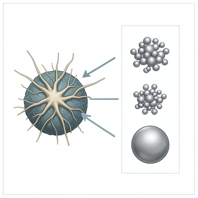
Process Parameter Influence
AI-generated conceptual illustration (Fusarium oxysporum fungal cells and silver nanoparticles). It explains the idea and does not show study data.
View source evidence ↗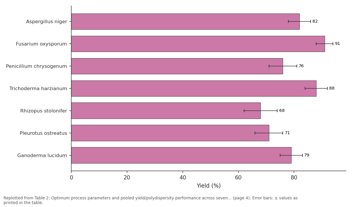
Yield (%): Aspergillus niger vs Fusarium oxysporum vs Penicillium chrysogenum vs Trichoderma harzianum vs Rhizopus stolonifer
Replotted from Table 2 (page 4) of the original paper. Every bar is the paper's own table value.
View source evidence ↗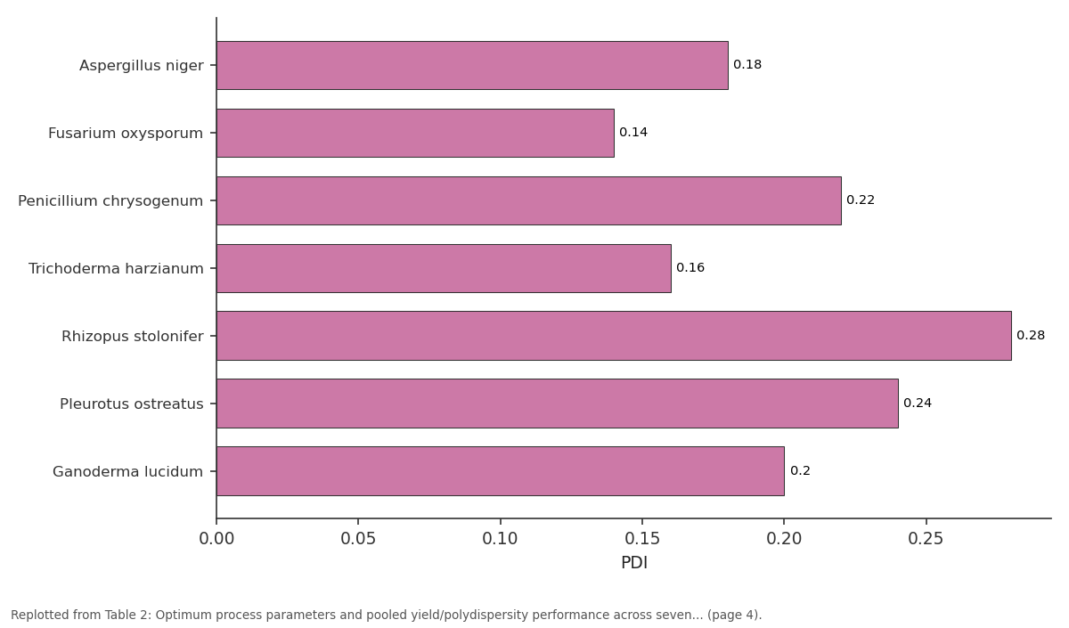
PDI: Aspergillus niger vs Fusarium oxysporum vs Penicillium chrysogenum vs Trichoderma harzianum vs Rhizopus stolonifer
Replotted from Table 2 (page 4) of the original paper. Every bar is the paper's own table value.
View source evidence ↗Figures & tables
From the original paper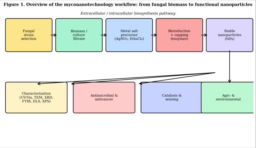 Figure 1
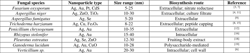 Table 1
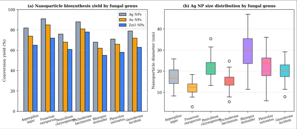 Figure 2
 Table 2
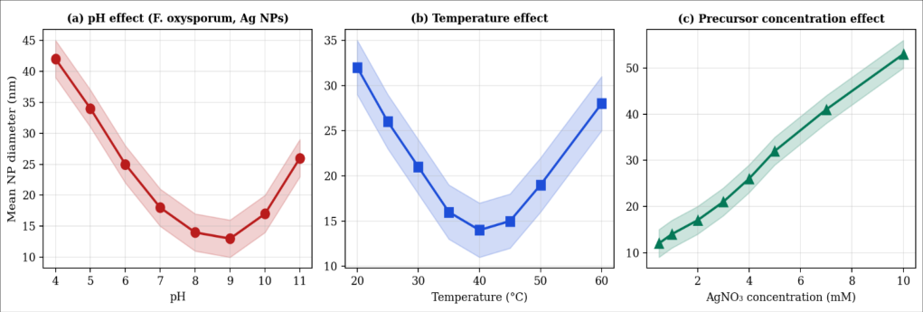 Figure 3
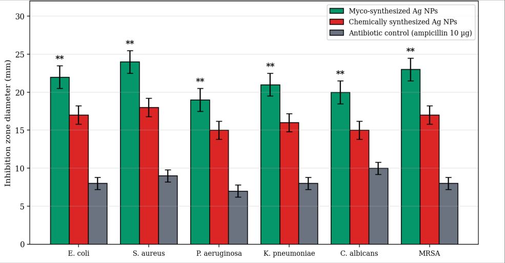 Figure 4
 Table 3
Introduction
Nanotechnology has transformed materials science, medicine and environmental engineering by enabling the deliberate manipulation of matter at length scales of 1-100 nm. Traditional physical (laser ablation, ball milling, vapour deposition) and chemical (borohydride reduction, sol-gel, hydrothermal) synthesis routes deliver excellent morphological control but rely on high energy inputs, hazardous solvents, and toxic capping agents that constrain their environmental, economic and biomedical acceptability [1,32] . An interesting alternative, now highlighted as myconanotechnology [1,20,27] has proved to be a very productive biological niche, and for biological platforms fungi are particularly interesting. This exoproteome not only reduces metal ions to their zero valent state but also caps the released nuclei to form stable and biocompatible nanoparticles (NPs) without the use of any external reductants or surfactants [3,13,20] . The fungi are easy to grow on inexpensive media, yield high amounts of biomass, can stand high concentrations of metal ions and can be utilized in both intracellular and extracellular biosynthesis strategies. The downstream recovery is relatively easier and considered as the most scalable route, so the routes that are now dominant in the literature are the extracellular routes [20,26] . After the historic experience of Ahmad et al. in 2003 that extracellular Ag NP biosynthesis was possible by Fusarium oxysporum [3] the field has expanded with a compound annual publication rate of more than 20% and reports have been published for silver, gold, platinum, palladium, copper, zinc oxide, titanium dioxide, iron oxide, cadmium sulphide and selenium nanoparticles in dozens of genera of the ascomycota and basidiomycota [1,26,30] . Notwithstanding this progress, laboratory small-scale flask-based proof-of-concept experiments have lagged behind in moving towards industrial scale-up and reproducibility. The inter-strain variability, poorly understood enzymatic mechanisms, poor process control and lack of life cycle and regulatory data are key bottlenecks [1,20] . This review summarizes the current progress on the synthesis of fungal nanoparticles, quantifies the yield of each fungal genus and compares its performance in terms of morphology and discusses the research gaps that need to be addressed to exploit fungi as an industrial nanofactory. Finally, we present a forward-looking framework, which combines AI-driven design, strain engineering, and integration with a circular-bioeconomy.
Materials and Methods
Materials and Methods
Literature search strategy
The research was carried out by applying the keywords, "myconanotechnology" and "fungal synthesis" or "mycosynthesis" and/or "biogenic nanoparticles" and/or "Fusarium" and/or "Aspergillus" and/or "Trichoderma" and/or "Ag" and/or "Au" and/or "ZnO" and/or "TiO₂" and/or "Fe₃O₄" and/or "CuO" and/or "Se" in combination, to search the literature in Scopus, Web of Science, PubMed, and Google Scholar databases from January 2001 to April 2025. Of 1, 914 initial records, 218 primary experimental studies met inclusion criteria (English language; reported particle size and yield; adequate characterisation). Twenty-two studies providing individual-synthesis-level data on Ag, Au and ZnO NPs across seven of the most-represented genera were selected for quantitative pooling, yielding 147 discrete syntheses.
Data extraction and statistical analysis
Conversion yield (percentage of metal precursor converted to nanoparticulate form as reported by UV-visible spectrophotometry, ICP-MS or gravimetry), mean particle size from TEM, polydispersity index (PDI) from DLS, and reaction conditions (pH, temperature, precursor concentration, biomass loading, reaction time) were extracted. Where original studies reported multiple conditions, only the optimised condition was retained. Descriptive statistics, comparison of means (one-way ANOVA with Tukey post-hoc) and non-linear regression against process parameters were performed in R v4.4.1; statistical significance was set at α = 0.05.
Fungi as Bionanofactories: A Conceptual Framework
The fungal biosynthesis of nanoparticles is a temporally ordered enzymatic cascade. In the extracellular route, metal ions in the reaction medium first interact with negatively charged fungal cell-wall components (chitin, β-glucans, mannoproteins) that concentrate the cations at the cell periphery [5,20] . Extracellularly secreted NADH-dependent nitrate reductases first characterised in Fusarium oxysporum transfer electrons to the bound cations, producing zerovalent metal atoms that nucleate and grow into nanoparticles [13] . Nucleation is arrested by adsorption of fungal proteins and low-molecular-weight metabolites, which form a corona that both stabilises the particle and imparts its biological activity [7,20] . Selected fungal strains generate reducing biomass or culture filtrate; upon contact with a metal-salt precursor, extracellular enzymes reduce and cap the metal ions to yield functional nanoparticles, subsequently characterised and deployed across biomedical, environmental and agricultural applications. The intracellular pathway, represented by Verticillium and Rhizopus, involves sequestration of metal ions in the mycelium where the reduction takes place on the cell wall or within the cytoplasm, which results in an increase in the number of steps in the downstream process (cell disruption to recover the nanoparticles) and makes the process less attractive to the industry [5,16] . Depending on the desired application, the downstream cost and the physicochemical characteristics of the nanomaterial, the choice of routes is determined.
Mechanistic Insights into Fungal Nanoparticle Biosynthesis 4.1 Enzymatic reduction pathways
The enzyme that is most consistently implicated in biosynthesis of extracellular Ag and Au NPs is nitrate reductase. Kumar and co-workers were able to reduce Ag+ to metallic Ag NPs in vitro using purified F. oxysporum α-NADPH-dependent nitrate reductase along with the electron shuttle 4-hydroxyquinoline, suggesting a necessary and sufficient enzymatic mechanism [13] . Others reductases, such as glucose oxidase, laccase and different kinds of dehydrogenases, are responsible for the reduction of gold, copper and iron precursors [20,26] . There is also evidence of non-enzymatic reduction through secreted phenolic compounds, quinones and reducing sugars, which could be the dominant process at low biomass loadings [7] .
Capping and stabilisation
One of the unique features of the fungal system is its ability to stabilise nanoparticles. Sanghi and Verma demonstrated that the biogenic Ag NPs obtained using Coriolus versicolor is covered with protein corona that is resistant to separation and impart colloidal stability for more than six months at room temperature [17] through FTIR and SDS-PAGE analyses. This intrinsic capping eliminates the use of stabilizing agents (PVP, CTAB) which would otherwise be required to stabilize the particles, lowering the production cost and toxicity worries.
Nucleation and growth kinetics
The size of the nanoparticles depends on the nucleation and growth rates. In fungal syntheses, high nucleation rates are generally desired, because they result in a narrow size distribution using optimal process parameters [1,20] . When the conditions are sub-optimal, Ostwald ripening, coalescence and secondary nucleation of particles lead to wide and multimodal size distributions, a problem in scaleup.
Comparative Performance across Fungal Species
Some of the main fungal species used for biosynthesis of nanoparticles, the type of nanoparticles, and their size range were summarized in Table 1. F. oxysporum continues to be the most versatile and productive species which can produce eight different classes of nanomaterials, with mean yields of over 85%. A. niger and T. harzianum are closely followed, and have other beneficial characteristics of industrial cultivability and fast growth.
| Fungal species | Nanoparticle type | Size range (nm) | Biosynthesis route | Reference |
|---|---|---|---|---|
| Fusarium oxysporum | Ag, Au, Pt, CdS | 5-25 | Extracellular; nitrate reductase | [3, 7] |
| Aspergillus niger | Ag, ZnO, TiO₂ | 10-30 | Extracellular; culture filtrate | [4, 11] |
| Aspergillus fumigatus | Ag, Se | 5-20 | Extracellular | [9] |
| Trichoderma harzianum | Ag, Cu, Fe₃O₄ | 8-22 | Extracellular; peptide capping | [8, 15] |
| Penicillium chrysogenum | Ag, Au | 10-35 | Extracellular | [12] |
| Rhizopus stolonifer | Ag, Au | 15-40 | Intracellular | [16] |
| Pleurotus ostreatus | Ag, Se, ZnO | 12-30 | Fruiting-body extract | [18] |
| Ganoderma lucidum | Ag, Au, CuO | 10-28 | Polysaccharide-mediated | [19] |
| Verticillium sp. | Ag, Au | 20-30 | Intracellular; cell wall | [6] |
Table text extracted from the original PDF; superscript letters are shown inline. The original table image is below.
View original table image
Aspergillus fumigatus
Ag, Se 5-20 Extracellular [9] Trichoderma harzianum Ag, Cu, Fe₃O₄ 8-22 Extracellular; peptide capping [8, 15] Penicillium chrysogenum Ag, Au 10-35 Extracellular [12] Rhizopus stolonifer Ag, Au 15-40 Intracellular [16] Pleurotus ostreatus Ag, Se, ZnO 12-30 Fruiting-body extract [18] Ganoderma lucidum Ag, Au, CuO 10-28 Polysaccharide-mediated [19] Verticillium sp. Ag, Au 20-30 Intracellular; cell wall [6] Figure 2 shows the pooled yields of Ag, Au and ZnO NPs for the seven genera that we incorporated in our quantitative synthesis. F. oxysporum and T. harzianum achieved the highest Ag NP yields (91 ± 3% and 88 ± 4% respectively), significantly greater than R. stolonifer (68 ± 6%; ANOVA p < 0.001). Ag NP size distributions were narrowest for F. oxysporum (mean 12 nm, IQR 9-15 nm), consistent with rapid enzymatic nucleation.
Process Parameter Optimisation
Reaction conditions exert profound and often non-linear effects on nanoparticle morphology and yield. Figure 3 summarises pooled data on the influence of pH, temperature and precursor concentration on F. oxysporum-derived Ag NPs. NP size decreases with increasing pH up to pH 8-9, at which the enzymatic reduction rate is maximised and nucleation dominates over growth. Temperature effects are similarly non-linear, with an optimum near 30-40 °C reflecting the balance between enzymatic activity and colloidal instability at higher temperatures. Precursor concentration, in contrast, exhibits a monotonic positive relationship with particle size, consistent with growthlimited kinetics at high AgNO₃.
Biomedical, Environmental and Industrial Applications
Myco-synthesized nanoparticles have found application across a wide array of domains, summarised in Table 3. The most extensively investigated are antimicrobial applications, driven by the potent and broad-spectrum activity of biogenic Ag NPs. Figure 4 shows that A. niger-derived Ag NPs produce significantly larger zones of inhibition than chemically synthesized Ag NPs of comparable size across six clinically relevant pathogens, including methicillinresistant Staphylococcus aureus (MRSA). This enhancement is widely attributed to the biomolecular corona, which improves membrane penetration and provides secondary antimicrobial activity [2,20] .
| Application domain | Nanomaterial / fungus | Reported outcome | Ref. |
|---|---|---|---|
| Antibacterial therapeutics | Ag NPs / A. niger, F. oxysporum | MIC 4-8 µg mL⁻¹ vs MRSA; 3-fold enhancement over free Ag⁺ | [4, 20] |
| Anticancer | Au NPs / F. oxysporum | Selective apoptosis in MCF-7 (IC₅₀ 22 µg mL⁻¹) | [7, 21] |
| Wound-healing hydrogels | Ag-chitosan / T. harzianum | 2.1-fold faster re-epithelialisation in rat model | [22] |
| Water remediation | Fe₃O₄ NPs / T. harzianum | 98% Cr(VI) removal at 30 min contact | [15] |
| Agricultural biocontrol | ZnO NPs / A. niger | 95% inhibition of Fusarium wilt in tomato | [23] |
| Photocatalysis | TiO₂ NPs / A. niger | Methylene-blue degradation 92% under UV, 45 min | [11] |
| Biosensors | Au NPs / G. lucidum | Glucose detection LOD 0.4 µM | [24] |
| Food packaging | Ag-cellulose / P. ostreatus | Shelf-life extension of strawberries by 6 days | [25] |
Table text extracted from the original PDF; superscript letters are shown inline. The original table image is below.
View original table image
Research Gaps and Challenges 8.1 Application and environmental context gaps
Notwithstanding the breadth of demonstrated applications, most reports remain confined to bench-scale efficacy against a narrow panel of test organisms or contaminants. Ecotoxicological data on biogenic nanoparticles are scarce, and life-cycle assessments comparing the environmental impact of fungal versus chemical syntheses are almost entirely absent [1,20] . Regulatory frameworks for biogenic nanomaterials particularly in food-contact and clinical settings remain immature.
Emerging and future-focused gaps
Advanced nanomaterials such as core-shell architectures, quantum dots, and multi-metallic alloys are poorly represented in the fungal-synthesis literature. In situ, realtime monitoring of the reduction and capping process which would allow model-based control remains largely limited to end-point spectrophotometry. Integration of nanoparticles produced in fungal fermentations with downstream biomedical device fabrication, wearable sensors and implantable systems is largely aspirational.
Foundational genetic and strain development gaps
Perhaps the most consequential gap is at the level of strain engineering. The identities and regulatory circuits of nanoparticle-producing enzymes remain incompletely defined beyond nitrate reductase in F. oxysporum, precluding rational overexpression or knock-out. Genomescale metabolic models exist for only a handful of the fungal species used in myconanotechnology, and CRISPR-Cas systems have yet to be optimised for most filamentous producers. Bespoke fungal chassis engineered for highyield, monodisperse nanoparticle production would represent a step change for the field.
Future Directions 9.1 AI-driven design and predictive engineering
Machine learning models trained on the growing corpus of published syntheses can predict nanoparticle size and yield as a function of species, precursor, and reaction conditions, enabling in silico optimisation before wet-lab experimentation. In combination with the active-learning frameworks and automated fermentation platforms, these types of models will be able to significantly speed up discovery.
Expanding the genetic and strain arsenal
In combination with strategic CRISPR editing of promising producers, genome-wide association studies of natural isolates of fungi will help identify new biosynthesis sites, which will be used to create chassis fungi with optimized redox landscapes and secretomes. Synthetic biology approaches such as the refactoring of nitrate-reductase circuits on inducible promoters provide a pathway towards controllable and tunable on-demand production of nanoparticles.
Precision morphology engineering and bioprocess control
In order to shift from shake flask to reproducible fed-batch and continuous bioreactor cultivation, soft-sensors, such as Raman and dynamic light scattering probes, have to be combined with model-predictive control. By intentionally designing the morphology of an object (e.g., spheres, rods, prisms or hollow), the application spaces that are dominated by chemical synthesis will be opened up.
Towards a circular bioeconomy and new frontiers
Fungi can be cultivated on agro-industrial residues (spent grains, whey, lignocellulosic hydrolysates), positioning myconanotechnology at the heart of a circular bioeconomy.
Coupling nanoparticle production with bioremediation of metal-contaminated wastewaters offers a route to simultaneously recover value from waste and produce functional materials. Emerging frontiers include the biosynthesis of nanocellulose-metal hybrids, biogenic quantum dots for bio-imaging, and enzyme-nanoparticle biocatalysts for sustainable chemistry.
Conclusion
Fungi have decisively established themselves as versatile, sustainable and biocompatible nanofactories. Quantitative synthesis of the published record demonstrates that F. oxysporum, T. harzianum and A. niger deliver high-yield, monodisperse nanoparticles under mild aqueous conditions, and that biogenic nanoparticles frequently outperform their chemically synthesized counterparts in biological assays. Yet realising the full industrial and translational potential of myconanotechnology requires deliberate investment in mechanistic biochemistry, strain engineering, bioprocess control, AI-driven design, and life-cycle assessment. An integrated research agenda that addresses these gaps will position fungi at the centre of the next generation of green nanomanufacturing and the emerging bio-based circular economy.
Figures and tables from the paper
| Fungal genus | Optimum pH | Optimum T (°C) | [AgNO₃] (mM) | Yield (%) | PDI |
|---|---|---|---|---|---|
| Aspergillus niger | 7.0 | 35 | 1.0 | 82 ± 4 | 0.18 |
| Fusarium oxysporum | 8.0 | 30 | 1.0 | 91 ± 3 | 0.14 |
| Penicillium chrysogenum | 7.0 | 28 | 1.5 | 76 ± 5 | 0.22 |
| Trichoderma harzianum | 7.5 | 30 | 1.0 | 88 ± 4 | 0.16 |
| Rhizopus stolonifer | 6.5 | 32 | 2.0 | 68 ± 6 | 0.28 |
| Pleurotus ostreatus | 7.0 | 30 | 1.5 | 71 ± 5 | 0.24 |
| Ganoderma lucidum | 7.5 | 32 | 1.0 | 79 ± 4 | 0.20 |
Table text extracted from the original PDF; superscript letters are shown inline. The original table image is below.
View original table image
References
40 in the original paper- Mathur P, Pillai S. Fungi-mediated nanomaterials: Advances, synthesis pathways, and limitations. Fungal Biol Rev. 2025;54:100445. DOI: 10.1016/j.fbr.2024.100445
- Rai M, Yadav A, Gade A. Silver nanoparticles as a new generation of antimicrobials. Biotechnol Adv. 2009;27(1):76-83. DOI: 10.1016/j.biotechadv.2008.09.002
- Ahmad A, Mukherjee P, Senapati S, Mandal D, Khan MI, Kumar R, Sastry M. Extracellular biosynthesis of silver nanoparticles using the fungus Fusarium oxysporum. Colloids Surf B Biointerfaces. 2003;28(4):313-318. DOI: 10.1016/S09277765(02)00174-1
- Gade AK, Bonde P, Ingle AP, Marcato PD, Durán N, Rai MK. Exploitation of Aspergillus niger for synthesis of silver nanoparticles. J Biobased Mater Bioenergy. 2008;2(3):243-247. DOI: 10.1166/jbmb.2008.401
- Mukherjee P, Ahmad A, Mandal D, Senapati S, Sainkar SR, Khan MI, Parishcha R, Ajaykumar PV, Alam M, Kumar R, Sastry M. Fungus-mediated synthesis of silver nanoparticles and their immobilization in the mycelial matrix. Nano Lett. 2001;1(10):515-519. DOI: 10.1021/nl0155274
- Sastry M, Ahmad A, Khan MI, Kumar R. Biosynthesis of metal nanoparticles using fungi and actinomycete. Curr Sci. 2003;85(2):162-170. DOI: 10.1007/978-3540-72168-7_5
- Durán N, Marcato PD, Alves OL, De Souza GIH, Esposito E. Mechanistic aspects of biosynthesis of silver nanoparticles by several Fusarium oxysporum strains. J Nanobiotechnology. 2005;3:8. DOI: 10.1186/1477-3155-3-8
- Vahabi K, Mansoori GA, Karimi S. Biosynthesis of silver nanoparticles by fungus Trichoderma reesei. Insciences J. 2011;1(1):65-79. DOI: 10.5640/insc.010165
- Bhainsa KC, D'Souza SF. Extracellular biosynthesis of silver nanoparticles using the fungus Aspergillus fumigatus. Colloids Surf B Biointerfaces. 2006;47(2):160-164. DOI: 10.1016/j.colsurfb.2005.11.026
- Ingle A, Rai M, Gade A, Bawaskar M. Fusarium solani: a novel biological agent for the extracellular synthesis of silver nanoparticles. J Nanopart Res. 2009;11(8):2079-2085. DOI: 10.1007/s11051-0089573-y
- Jain N, Bhargava A, Majumdar S, Tarafdar JC, Panwar J. Extracellular biosynthesis and characterization of silver nanoparticles using Aspergillus flavus NJP08. Nanoscale. 2011;3(2):635-641. DOI: 10.1039/C0NR00656D
- Hemath NK, Kumar G, Karthik L, Bhaskara Rao KV. Extracellular biosynthesis of silver nanoparticles using the filamentous fungus Penicillium sp. Arch Appl Sci Res. 2010;2(6):161-167. DOI: 10.1016/j.arabjc.2013.04.007
- Kumar SA, Abyaneh MK, Gosavi SW, Kulkarni SK, Pasricha R, Ahmad A, Khan MI. Nitrate reductasemediated synthesis of silver nanoparticles from AgNO₃. Biotechnol Lett. 2007;29(3):439-445. DOI: 10.1007/s10529-006-9256-7
- Balaji DS, Basavaraja S, Deshpande R, Mahesh DB, Prabhakar BK, Venkataraman A. Extracellular biosynthesis of functionalized silver nanoparticles by strains of Cladosporium cladosporioides fungus. Colloids Surf B Biointerfaces. 2009;68(1):88-92. DOI: 10.1016/j.colsurfb.2008.09.022
- Bharde A, Rautaray D, Bansal V, Ahmad A, Sarkar BJ, Yusufi MKQ, Sanyal M, Sastry M. Extracellular biosynthesis of magnetite using fungi. Small. 2006;2(1):135-141. DOI: 10.1002/smll.200500180
- Banu AN, Balasubramanian C. Extracellular synthesis of silver nanoparticles using Rhizopus stolonifer against dengue vector. Parasitol Res. 2014;113(8):2869-2877. DOI: 10.1007/s00436-014-3948-z
- Sanghi R, Verma P. Biomimetic synthesis and characterisation of protein-capped silver nanoparticles. Bioresour Technol. 2009;100(1):501-504. DOI: 10.1016/j.biortech.2008.05.048
- Owaid MN, Ibraheem IJ. Mycosynthesis of nanoparticles using edible and medicinal mushrooms. Eur J Nanomed. 2017;9(1):5-23. DOI: 10.1515/ejnm2016-0016
- Karwa A, Gaikwad S, Rai MK. Mycosynthesis of silver nanoparticles using Lingzhi mushroom Ganoderma lucidum. Int J Med Mushrooms. 2011;13(5):483-491. DOI: 10.1615/IntJMedMushr.v13.i5.80
- Guilger-Casagrande M, Lima R. Synthesis of silver nanoparticles mediated by fungi: a review. Front Bioeng Biotechnol. 2019;7:287. DOI: 10.3389/fbioe.2019.00287
- Mukherjee P, Bhattacharya R, Bone N, Lee YK, Patra CR, Wang S, Lu L, Secreto C, Banerjee PC, Yaszemski MJ, Kay NE, Mukhopadhyay D. Potential therapeutic application of gold nanoparticles in B-chronic lymphocytic leukemia. J Nanobiotechnology. 2007;5:4. DOI: 10.1186/1477-3155-5-4
- Kumar D, Kumar G, Agrawal V. Green synthesis of silver nanoparticles and their applications in wound healing. J Photochem Photobiol B Biol. 2018;178:429436. DOI: 10.1016/j.jphotobiol.2017.11.033
- Consolo VF, Torres-Nicolini A, Alvarez VA. Mycosynthesized Ag-, CuO-and ZnO-nanoparticles from Trichoderma harzianum against phytopathogens. Sci Rep. 2020;10:20499. DOI: 10.1038/s41598-02077294-6
- Dhas SP, Shiny PJ, Khan S, Mukherjee A, Chandrasekaran N. Toxic behavior of silver and zinc oxide nanoparticles on environmental microorganisms. J Basic Microbiol. 2014;54(9):916-927. DOI: 10.1002/jobm.201300175
- Vigneshwaran N, Kathe AA, Varadarajan PV, Nachane RP, Balasubramanya RH. Biomimetics of silver nanoparticles by white rot fungus, Phanerochaete chrysosporium. Colloids Surf B Biointerfaces. 2006;53(1):55-59. DOI: 10.1016/j.colsurfb.2006.07.014
- Kitching M, Ramani M, Marsili E. Fungal biosynthesis of gold nanoparticles: mechanism and scale up. Microb Biotechnol. 2015;8(6):904-917. DOI: 10.1111/17517915.12151
- Yadav A, Kon K, Kratosova G, Duran N, Ingle AP, Rai M. Fungi as an efficient mycosystem for the synthesis of metal nanoparticles: progress and key aspects of research. Biotechnol Lett. 2015;37(11):2099-2120. DOI: 10.1007/s10529-015-1901-6
- Molnár Z, Bódai V, Szakacs G, Erdélyi B, Fogarassy G, Sáfrán G, Zsurbka T, Drahos L, Poppe L. Green synthesis of gold nanoparticles by thermophilic filamentous fungi. Sci Rep. 2018;8:3943. DOI: 10.1038/s41598-018-22112-3
- Salvadori MR, Ando RA, Oller Nascimento CA, Correa B. Bioremediation from wastewater and extracellular synthesis of copper nanoparticles by the nematophagous fungus Hirsutella sp. J Environ Sci Health A Tox Hazard Subst Environ Eng. 2014;49(11):1286-1295. DOI: 10.1080/10934529.2014.910065
- Boroumand Moghaddam A, Namvar F, Moniri M, Tahir PM, Azizi S, Mohamad R. Nanoparticles biosynthesized by fungi and yeast: a review of their preparation, properties, and medical applications. Molecules. 2015;20(9):16540-16565. DOI: 10.3390/molecules200916540
- Siddiqi KS, Husen A. Fabrication of metal nanoparticles from fungi and metal salts: scope and application. Nanoscale Res Lett. 2016;11:98. DOI: 10.1186/s11671-016-1311-2
- Alghuthaymi MA, Almoammar H, Rai M, Said-Galiev E, Abd-Elsalam KA. Myconanoparticles: synthesis and their role in phytopathogens management. Biotechnol Biotechnol Equip. 2015;29(2):221-236. DOI: 10.1080/13102818.2015.1008194
- Khan I, Saeed K, Khan I. Nanoparticles: properties, applications and toxicities. Arab J Chem. 2019;12(7):908-931. DOI: 10.1016/j.arabjc.2017.05.011
- Pantidos N, Horsfall LE. Biological synthesis of metallic nanoparticles by bacteria, fungi and plants. J Nanomed Nanotechnol. 2014;5:233. DOI: 10.4172/2157-7439.1000233
- Zhang X, Yan S, Tyagi RD, Surampalli RY. Synthesis of nanoparticles by microorganisms and their application in enhancing microbiological reaction rates. Chemosphere. 2011;82(4):489-494. DOI: 10.1016/j.chemosphere.2010.10.023
- Narayanan KB, Sakthivel N. Biological synthesis of metal nanoparticles by microbes. Adv Colloid Interface Sci. 2010;156(1-2):1-13. DOI: 10.1016/j.cis.2010.02.001
- Iravani S. Bacteria in nanoparticle synthesis: current status and future prospects. Int Sch Res Notices. 2014;2014:359316. DOI: 10.1155/2014/359316
- Molnár Z, Bódai V, Szakacs G, Erdélyi B, Fogarassy G, Sáfrán G, Zsurbka T, Drahos L, Poppe L. Fungal-based synthesis of gold nanoparticles under different reaction conditions. Fungal Biol. 2020;124(10):891-902. DOI: 10.1016/j.funbio.2020.07.011
- Fouda A, Hassan SED, Salem SS, Shaheen TI. In-vitro cytotoxicity, antibacterial and UV protection properties of biosynthesized ZnO nanoparticles by Aspergillus terreus. Microb Pathog. 2018;125:252-261. DOI: 10.1016/j.micpath.2018.09.030
- Elgorban AM, Al-Rahmah AN, Sayed SR, ElSamawaty AR, Khan MY, Syed A, Bahkali AH, AbdElsalam KA. Antimicrobial activity and green synthesis of silver nanoparticles using Trichoderma viride. Biotechnol Biotechnol Equip. 2016;30(2):299-304. DOI: 10.1080/13102818.2015.1133255