
Research article · 2026 · Full text
International Journal of Biology SciencesMolecular characterization and pathogenicity assessment of Colletotrichum lindemuthianum isolates from major cowpea-growing regions
Volume 8(5) · Pages 237–240 · DOI: 10.33545/26649926.2026.v8.i5c.960 ↗
Research areasPlant Pathogens and Fungal DiseasesPlant Disease Resistance and GeneticsFungal Plant Pathogen Control
Abstract
Author-writtenThe genetic diversity and virulence heterogeneity within Colletotrichum lindemuthianum populations remain the foremost obstacles to durable anthracnose resistance breeding in cowpea across West Africa. This research characterized 15 C. lindemuthianum isolates collected from cowpea fields across three major growing regions of Ghana (Ashanti, Eastern, Northern) using morphological (colony colour, conidial dimensions, appressorium shape) and molecular (ITS-rDNA sequencing, RAPD markers) approaches. Pathogenicity testing was conducted on ten cowpea varieties under controlled glasshouse conditions. Conidial length ranged from 12.4 to 21.8 µm and width from 3.6 to 6.2 µm across isolates. ITS-rDNA phylogenetic analysis revealed two distinct clades with 94-99% sequence similarity to GenBank reference accessions. RAPD profiling with eight primers generated 86 polymorphic bands, yielding genetic similarity coefficients of 0.42-0.91 among isolates. Pathogenicity testing identified three virulence groups: highly virulent (score 4.0-4.2), moderately virulent (2.6-3.6), and weakly virulent (1.4-1.8). Northern region isolates exhibited the highest genetic diversity and virulence variability.
Summary
Source-checkedPlain-language reading of the full paper, prepared with AI and checked against the text. Each point links to the authors' own words.
Why it matters
- Genetic diversity and varying virulence in Colletotrichum lindemuthianum populations are the primary challenges for breeding cowpea with durable resistance to anthracnose in West Africa. View evidence ↗
- The pathogen's high genetic plasticity allows it to create new virulence phenotypes that can bypass existing resistance genes in as little as three to five growing seasons. View evidence ↗
- Colletotrichum lindemuthianum is a hemibiotrophic ascomycete fungus that causes anthracnose in common bean and cowpea in subtropical and tropical areas. View evidence ↗
The question
- The study aimed to characterize 15 C. lindemuthianum isolates from three major Ghanaian growing regions using morphological and molecular methods and to evaluate their virulence on ten cowpea varieties. View evidence ↗
What they did
- Fifteen isolates were collected during the 2023 growing season from five districts in each of the Northern, Eastern, and Ashanti regions of Ghana. View evidence ↗
- Morphological analysis involved measuring conidial dimensions (50 per isolate), classifying appressorium shape, and assessing colony color on PDA after 10 days at 25 °C. View evidence ↗
- Molecular characterization used ITS-rDNA sequencing and RAPD analysis with eight specific decamer primers. View evidence ↗
- Pathogenicity was tested by spray-inoculating 21-day-old seedlings with a 1 × 10⁶/mL conidial suspension and scoring severity on a 1-5 scale after 14 days. View evidence ↗
What they found
- Across the isolates, conidial length ranged from 12.4 to 21.8 µm (mean 16.8 ± 2.7 µm) and width ranged from 3.6 to 6.2 µm (mean 4.8 ± 0.7 µm). View evidence ↗
- RAPD analysis showed that Northern region isolates had the highest genetic diversity, with similarity coefficients of 0.42-0.78, compared to Eastern (0.61-0.86) and Ashanti (0.68-0.91) isolates. View evidence ↗
- Phylogenetic analysis identified two clades: Clade I (9 isolates, mostly Ashanti/Eastern) with 97-99% similarity to the reference, and Clade II (6 isolates, mostly Northern) with 94-96% similarity. View evidence ↗
- The cowpea varieties Kirkhouse and Padi-Tuya showed low disease scores across all virulence groups, making them promising sources for broad-spectrum resistance breeding. View evidence ↗
What it means
- The authors recommend that breeding programs prioritize screening against highly virulent isolates from the Northern region and deploy varieties like Padi-Tuya and Kirkhouse. View evidence ↗
- The authors state that future work should use pathogenomic approaches and multi-locus sequencing to better understand genetic structure and identify genomic regions associated with virulence. View evidence ↗
Visual summary
3 visualsIllustrations explain the paper; charts are replotted from the paper's own table values.
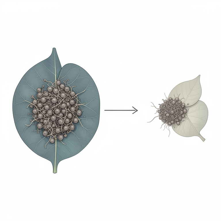
Phylogenetic Clade Structure
AI-generated conceptual illustration (Colletotrichum lindemuthianum fungus on cowpea plants). It explains the idea and does not show study data.
View source evidence ↗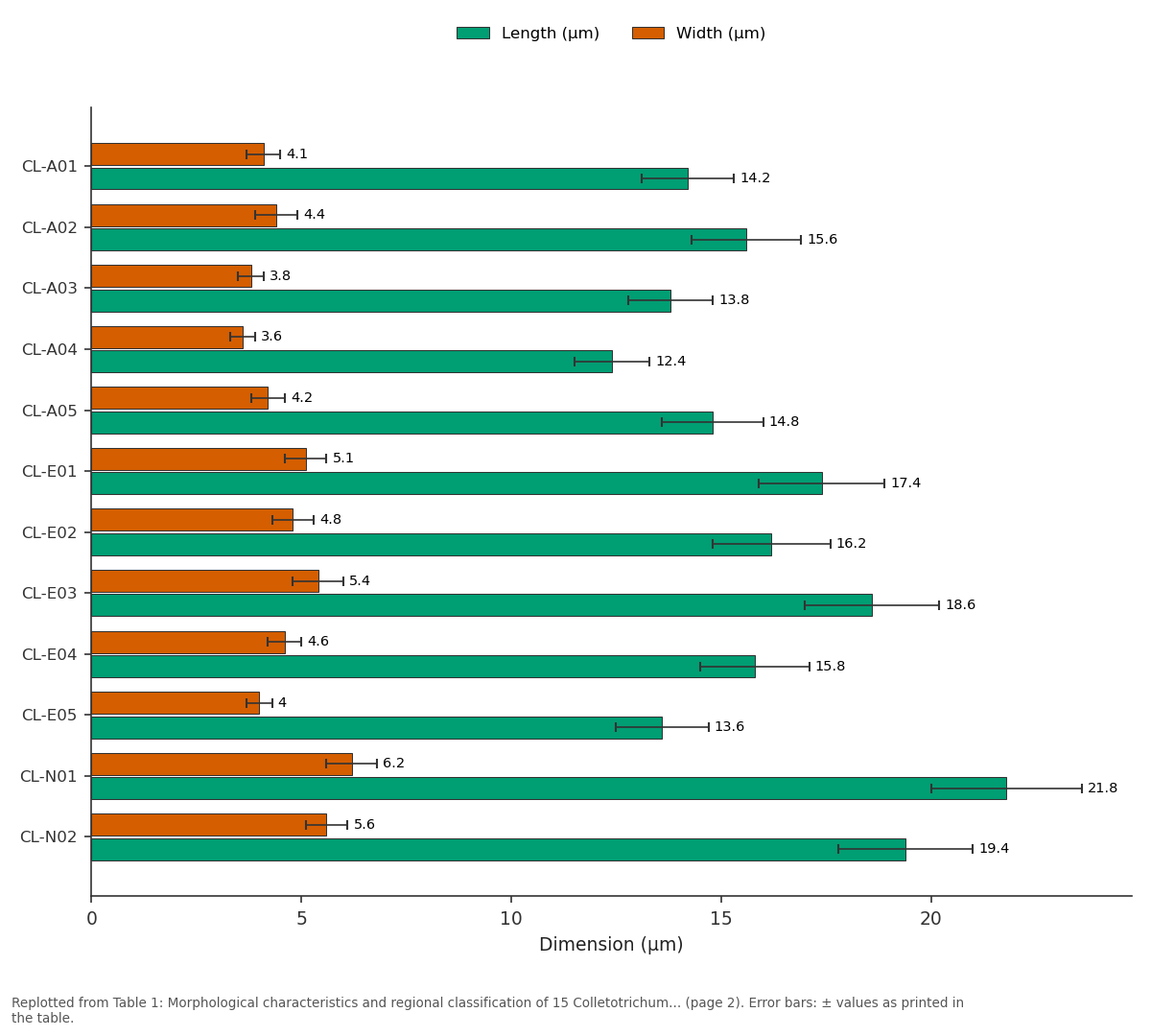
Length (µm) and Width (µm): CL-A01 vs CL-A02 vs CL-A03 vs CL-A04 vs CL-A05
Replotted from Table 1 (page 2) of the original paper. Every bar is the paper's own table value.
View source evidence ↗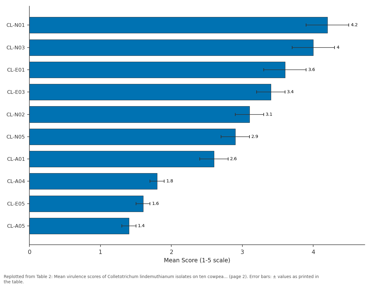
Mean Score: CL-N01 vs CL-N03 vs CL-E01 vs CL-E03 vs CL-N02
Replotted from Table 2 (page 2) of the original paper. Every bar is the paper's own table value.
View source evidence ↗Figures & tables
From the original paper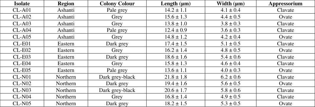 Table 1
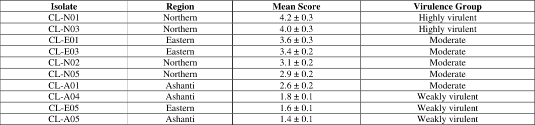 Table 2
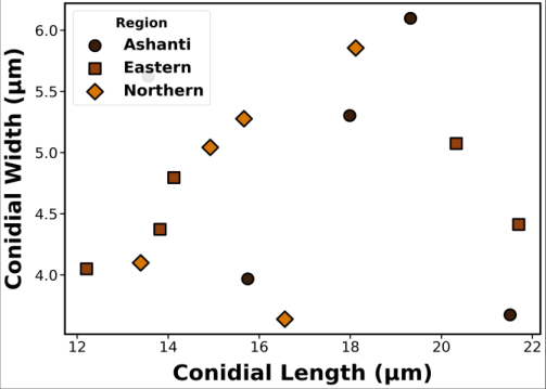 Figure 1
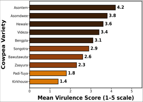 Figure 2
Introduction
The sustained deployment of disease-resistant cowpea cultivars has been repeatedly undermined by the extraordinary genetic plasticity of Colletotrichum lindemuthianum populations, which generate new virulence phenotypes capable of overcoming previously effective resistance genes within as few as three to five growing seasons [1,2] . Understanding the genetic structure, diversity, and virulence distribution within pathogen populations is therefore essential for designing durable resistance breeding strategies and predicting the breakdown risk associated with specific resistance gene deployments [3] . Colletotrichum lindemuthianum (Sacc. & Magnus) Briosi & Cavara is a hemibiotrophic ascomycete responsible for anthracnose disease in cowpea and common bean across tropical and subtropical regions [4,5] . Morphological characterization of isolates based on colony colour, growth rate, conidial dimensions, and appressorium morphology provides preliminary differentiation but is insufficient for reliable phylogenetic classification due to phenotypic plasticity under variable cultural conditions [6] . Molecular approaches including internal transcribed spacer (ITS) rDNA sequencing and random amplified polymorphic DNA (RAPD) markers enable more robust assessment of genetic relationships and population structure [7,8] . Despite the economic importance of cowpea anthracnose in West Africa, systematic molecular characterization of C. lindemuthianum populations from Ghanaian cowpeagrowing regions has not been undertaken [3,9] . This research characterized 15 isolates from three major growing regions using integrated morphological and molecular approaches and assessed their virulence profiles on ten cowpea varieties to inform resistance breeding priorities.
Materials and Methods
Materials and Methods
Materials
Fifteen isolates of C. lindemuthianum were collected from infected cowpea plants exhibiting typical anthracnose symptoms across five districts each in Ashanti, Eastern, and Northern regions of Ghana during the 2023 growing season (five isolates per region). Isolates were purified by singlespore isolation on potato dextrose agar (PDA). Seeds of ten cowpea varieties (Asontem, Asomdwee, Hewale, Videza, Bengpla, Songotra, Bawutawuta, Zaayura, Padi-Tuya, Kirkhouse) were obtained from CSIR-Savanna Agricultural Research Institute, Tamale. All laboratory work was conducted at the Molecular Biology Laboratory, Kumasi Research Centre for Plant Sciences, Kumasi, Ghana, between August 2023 and April 2024.
Methods
Morphological characterization included colony colour assessment on PDA after 10 days at 25 °C, measurement of conidial length and width (n = 50 per isolate) using a calibrated ocular micrometer, and appressorium shape classification [6] . Genomic DNA was extracted using the CTAB method. ITS-rDNA regions were amplified with primers ITS1/ITS4 and sequenced (Macrogen, South Korea). Sequences were aligned using ClustalW and phylogenetic analysis was performed using MEGA 11 with the neighbor-joining method (1000 bootstrap replicates) [7] . RAPD analysis employed eight decamer primers (OPA-02, OPA-04, OPA-09, OPA-13, OPB-06, OPB-10, OPC-04, OPC-08) with PCR conditions: 94 °C/5 min; 40 cycles of 94 °C/1 min, 36 °C/1 min, 72 °C/2 min; 72 °C/10 min [8] . Amplified products were resolved on 1.5% agarose gels. Band scoring (1/0) was used to calculate Jaccard similarity coefficients and construct UPGMA dendrograms using NTSYSpc v2.2. Pathogenicity was assessed by sprayinoculating 21-day-old seedlings with conidial suspensions (1 × 10⁶/mL) and scoring disease severity on a 1-5 scale at 14 days post-inoculation [5,10] . Data were analyzed by ANOVA with Duncan's test at p < 0.05.
Results
The morphological characteristics and regional origin of 15 C. lindemuthianum isolates are classified in Table 1. Colony colour ranged from pale grey to dark grey-black, with Northern region isolates exhibiting the darkest pigmentation. Conidial length varied from 12.4 to 21.8 µm (mean 16.8 ± 2.7 µm) and width from 3.6 to 6.2 µm (mean 4.8 ± 0.7 µm). Appressorium morphology was predominantly clavate (60%) or ovate (40%). The pathogenicity assessment results, summarizing mean virulence scores of isolates on ten cowpea varieties, are presented in Table 2. Isolates were classified into three virulence groups: highly virulent (CL-N01, CL-N03 with scores of 4.2 and 4.0), moderately virulent (8 isolates, scores 2.6-3.6), and weakly virulent (CL-A04, CL-E05, CL-A05 with scores 1.4-1.8). Varieties Asontem and Asomdwee were the most susceptible, while Kirkhouse and Padi-Tuya showed the lowest disease scores. Selected isolates shown. Scores are mean ± SD across 10 varieties with 5 replications. Scale: 1 = no symptoms; 5 = plant death. The distribution of conidial dimensions across regional populations is illustrated in Figure 1. Northern region isolates showed the widest morphometric variation, while Ashanti isolates clustered more tightly. This pattern was confirmed by RAPD analysis, which revealed that genetic similarity coefficients among Northern isolates (0.42-0.78) were lower than those among Ashanti (0.68-0.91) or Eastern (0.61-0.86) isolates, indicating greater genetic diversity in the Northern population. Clade II contained 6 isolates (predominantly from Northern region) with 94-96% similarity, suggesting possible incipient genetic divergence within the Northern population that correlates with the elevated virulence observed in this group [4,7] .
| Isolate | Region | Mean Score | Virulence Group |
|---|---|---|---|
| CL-N01 | Northern | 4.2 ± 0.3 | Highly virulent |
| CL-N03 | Northern | 4.0 ± 0.3 | Highly virulent |
| CL-E01 | Eastern | 3.6 ± 0.3 | Moderate |
| CL-E03 | Eastern | 3.4 ± 0.2 | Moderate |
| CL-N02 | Northern | 3.1 ± 0.2 | Moderate |
| CL-N05 | Northern | 2.9 ± 0.2 | Moderate |
| CL-A01 | Ashanti | 2.6 ± 0.2 | Moderate |
| CL-A04 | Ashanti | 1.8 ± 0.1 | Weakly virulent |
| CL-E05 | Eastern | 1.6 ± 0.1 | Weakly virulent |
| CL-A05 | Ashanti | 1.4 ± 0.1 | Weakly virulent |
Table text extracted from the original PDF; superscript letters are shown inline. The original table image is below.
View original table image
| Isolate | Region | Colony Colour | Length (µm) | Width (µm) | Appressorium |
|---|---|---|---|---|---|
| CL-A01 | Ashanti | Pale grey | 14.2 ± 1.1 | 4.1 ± 0.4 | Clavate |
| CL-A02 | Ashanti | Grey | 15.6 ± 1.3 | 4.4 ± 0.5 | Ovate |
| CL-A03 | Ashanti | Grey | 13.8 ± 1.0 | 3.8 ± 0.3 | Clavate |
| CL-A04 | Ashanti | Pale grey | 12.4 ± 0.9 | 3.6 ± 0.3 | Clavate |
| CL-A05 | Ashanti | Grey | 14.8 ± 1.2 | 4.2 ± 0.4 | Ovate |
| CL-E01 | Eastern | Dark grey | 17.4 ± 1.5 | 5.1 ± 0.5 | Clavate |
| CL-E02 | Eastern | Grey | 16.2 ± 1.4 | 4.8 ± 0.5 | Ovate |
| CL-E03 | Eastern | Dark grey | 18.6 ± 1.6 | 5.4 ± 0.6 | Clavate |
| CL-E04 | Eastern | Grey | 15.8 ± 1.3 | 4.6 ± 0.4 | Clavate |
| CL-E05 | Eastern | Pale grey | 13.6 ± 1.1 | 4.0 ± 0.3 | Ovate |
| CL-N01 | Northern | Dark grey-black | 21.8 ± 1.8 | 6.2 ± 0.6 | Clavate |
| CL-N02 | Northern | Dark grey | 19.4 ± 1.6 | 5.6 ± 0.5 | Ovate |
| CL-N03 | Northern | Dark grey-black | 20.6 ± 1.7 | 5.8 ± 0.6 | Clavate |
| CL-N04 | Northern | Grey | 16.8 ± 1.4 | 4.9 ± 0.5 | Clavate |
| CL-N05 | Northern | Dark grey | 18.2 ± 1.5 | 5.3 ± 0.5 | Ovate |
Table text extracted from the original PDF; superscript letters are shown inline. The original table image is below.
View original table image
Discussion
This research provides the first comprehensive molecular and pathogenicity characterization of C. lindemuthianum populations from Ghanaian cowpea-growing regions. The identification of three distinct virulence groups among 15 isolates confirms substantial pathogenic heterogeneity within the Ghanaian population, consistent with the high race diversity reported for C. lindemuthianum in common bean across Latin America and East Africa [1,2] . The predominance of highly virulent isolates from the Northern region, which represents Ghana's primary cowpea production zone, has direct implications for variety deployment decisions and resistance breeding target prioritization [3,9] . The RAPD-based genetic similarity coefficients (0.42-0.91) indicate moderate to high genetic diversity within the Ghanaian C. lindemuthianum population, comparable to the 0.38-0.89 range reported by Sharma et al. [4] for isolates from East African bean-growing regions. The correlation between geographic origin and genetic clustering observed in the UPGMA dendrogram suggests that regional populations experience limited gene flow, potentially driven by the spatial isolation of cowpea production areas and the seed-borne transmission ecology of the pathogen [6,8] . The differential susceptibility patterns observed across ten cowpea varieties provide actionable information for resistance deployment strategies. Varieties Kirkhouse and Padi-Tuya, which maintained low disease scores across all virulence groups, represent promising resistance sources for breeding programs targeting broad-spectrum anthracnose resistance [5,10] . Conversely, the high susceptibility of Asontem and Asomdwee to Northern region isolates warrants reconsideration of their cultivation in the primary production zones where highly virulent pathogen populations predominate [3,11] .
Phylogenetic Implications
The two-clade phylogenetic structure revealed by ITS-rDNA analysis suggests that Ghanaian C. lindemuthianum populations may harbor cryptic genetic lineages with distinct evolutionary trajectories. The lower sequence similarity (94-96%) of Clade II isolates to reference accessions raises the possibility that these Northern region isolates represent a divergent genetic group with enhanced virulence attributes, warranting further investigation through multi-locus sequence typing approaches [7,8] .
Resistance Breeding Recommendations
Cowpea breeding programs operating in Ghana should incorporate pathogenicity testing against representative isolates from all three virulence groups identified in this research to ensure that candidate varieties possess resistance effective against the full spectrum of virulence present in the target environment [3,5] . Deployment of varieties with narrow-spectrum resistance effective only against weakly virulent populations would create strong directional selection pressure favoring the proliferation of virulent genotypes.
International Journal of Biology Sciences https://www.biologyjournal.net
Conclusion
Within the context of developing durable anthracnose management strategies for cowpea production in West Africa, this research characterized the morphological, molecular, and pathogenic diversity of 15 C. lindemuthianum isolates from three major cowpea-growing regions of Ghana. The findings highlight that Northern region isolates exhibited the greatest genetic diversity (similarity coefficients 0.42-0.78) and virulence variability, with two isolates classified as highly virulent (scores ≥4.0). ITS-rDNA phylogenetic analysis resolved two distinct clades, while RAPD profiling confirmed regional genetic structuring within the pathogen population.
Resistance breeding programs should prioritize screening against highly virulent Northern region isolates and deploy varieties such as Kirkhouse and Padi-Tuya that demonstrated broad-spectrum resistance across all virulence groups. Future investigations should employ multi-locus sequencing and pathogenomic approaches to further resolve population genetic structure and identify virulence-associated genomic regions.
Acknowledgement
AcknowledgementsThis research was funded by the Ghana National Science Foundation (Grant No. GNSF/2023/BIO-0518). Kumasi Research Centre for Plant Sciences is acknowledged for molecular biology laboratory facilities. Contributions: KOM designed the research and performed molecular analyses; DAA conducted morphological characterization; FNB managed glasshouse pathogenicity trials and drafted the manuscript.
Figures and tables from the paper
References
15 in the original paper- Padder BA, Sharma PN, Awale HE, Kelly JD. Colletotrichum lindemuthianum, the causal agent of bean anthracnose. J Plant Pathol. 2017;99(2):317-330.
- Sharma KD, Winter P, Kahl G, Muehlbauer FJ. Molecular mapping of Fusarium oxysporum f. sp. ciceris race 3 resistance gene in chickpea. Theor Appl Genet. 2004;108(7):1243-1248.
- Satpathy MR, Parida D, Khillar AK. Assessment of fungitoxic effects of botanicals on the management of growth in Colletotrichum lindemuthianum, the incitant of cowpea anthracnose. J Curr Res Food Sci. 2022;3(1):21-24.
- Sharma M, Ghosh R, Telangre R, Rathore A, Saifulla M, Mahalinga DM, et al. Environmental influences on pathogenic variability and molecular diversity of Colletotrichum truncatum causing anthracnose of lentil. Phytopathology. 2016;106(5):469-477.
- Boukar O, Belko N, Chamarthi S, Togola A, Batieno J, Owusu E, et al. Cowpea (Vigna unguiculata): Genetics, genomics and breeding. Plant Breed. 2019;138(4):415424.
- Cai L, Hyde KD, Taylor PWJ, Weir BS, Waller JM, Abang MM, et al. A polyphasic approach for studying Colletotrichum. Fungal Divers. 2009;39:183-204.
- Schoch CL, Seifert KA, Huhndorf S, Robert V, Spouge JL, Levesque CA, et al. Nuclear ribosomal internal transcribed spacer (ITS) region as a universal DNA barcode marker for fungi. Proc Natl Acad Sci U S A. 2012;109(16):6241-6246.
- Williams JGK, Kubelik AR, Livak KJ, Rafalski JA, Tingey SV. DNA polymorphisms amplified by arbitrary primers are useful as genetic markers. Nucleic Acids Res. 1990;18(22):6531-6535.
- Ogunsola KE, Ogunfunmilayo AO. Survey and identification of anthracnose disease of cowpea in southwestern Nigeria. Afr J Agric Res. 2022;18(3):182191.
- Nene YL, Thapliyal PN. Evaluation of fungicides. In: Fungicides in plant disease control. 3rd ed. New Delhi: Oxford and IBH Publishing; 2018. p. 531-542.
- Adomako-Bonsu AG, Owusu-Ansah F, Asante SK. Antifungal properties of neem-based preparations against seed-borne pathogens. Crop Prot. 2023;168:106218.
- Dean R, Van Kan JAL, Pretorius ZA, HammondKosack KE, Di Pietro A, Spanu PD, et al. The top 10 fungal pathogens in molecular plant pathology. Mol Plant Pathol. 2012;13(4):414-430.
- Pande S, Sharma M, Ghosh R. Role of host resistance and fungicides in management of foliar diseases of food legumes. Indian Phytopathol. 2019;72(1):1-18.
- Agbenin ON, Marley PS. In vitro assay of some plant extracts against Fusarium oxysporum f. sp. lycopersici. J Plant Prot Res. 2024;46(3):215-220.
- Gurjar MS, Ali S, Akhtar M, Singh KS. Efficacy of plant extracts in plant disease management. Agric Sci. 2012;3(3):425-433.