
Research article · 2026 · Full text
International Journal of Biology SciencesBiosafety and toxicological assessment of mycogenic nanoparticles: Current evidence, challenges, and future perspectives
Volume 8(7) · Pages 275–281 · DOI: 10.33545/26649926.2026.v8.i7c.956 ↗
Research areasNanoparticles: synthesis and applicationsEnvironmental Toxicology and EcotoxicologyImmunotoxicology and immune responses
Abstract
Author-writtenMycogenic nanoparticles metallic, oxide, and hybrid nanomaterials biosynthesized by filamentous fungi are advancing rapidly toward biomedical, agricultural and environmental deployment. Their translational trajectory now depends less on synthesis chemistry than on rigorous, standardised biosafety assessment. This review provides an integrated, five-axis framework for the toxicological evaluation of mycogenic nanoparticles: (i) the physicochemical properties that drive toxicity (size, shape, surface charge, biomolecular corona, colloidal stability, dissolution); (ii) biological toxicity endpoints (cytotoxicity, oxidative stress, DNA damage, immunotoxicity, cell-death programmes); (iii) ecotoxicology (aquatic, terrestrial, plant and microbiome effects); (iv) translational and regulatory challenges (industrial-scale manufacturing, quality control, clinical trials); and (v) next-generation predictive technologies (artificial intelligence, quantitative structure-activity relationships, machine learning, organ-on-chip platforms, and digital twins). Pooled analysis of 41 primary in vitro studies (n = 268 assays) demonstrates that mycogenic silver, zinc-oxide and copper-oxide nanoparticles trigger dose-dependent reactive oxygen species generation, glutathione depletion and mitochondrial membrane depolarisation, with IC₅₀ values spanning two orders of magnitude across cell types. Ecotoxicological data reveal a consistent CuO > Ag > ZnO ≫ Au sensitivity hierarchy across seven trophic-level indicator species. We identify persistent evidence gaps, propose harmonised assay panels aligned to OECD test guidelines, and delineate an AI-augmented regulatory pipeline that could compress conventional dossier timelines by 40-60%.
Summary
Source-checkedPlain-language reading of the full paper, prepared with AI and checked against the text. Each point links to the authors' own words.
Why it matters
- Mycogenic nanoparticles, which are metallic, oxide, or hybrid materials created using filamentous fungi, are being developed for use in agriculture, the environment, and medicine. View evidence ↗
- The primary obstacle to the practical application of these nanoparticles is biosafety, which involves evaluating the risks and hazards they pose to the environment and human health. View evidence ↗
- Unlike chemically synthesized nanoparticles, mycogenic versions have a natural biomolecular corona from fungal secretions, meaning they have a different toxicity profile. View evidence ↗
The question
- The authors aim to establish a comprehensive five-axis framework to evaluate the toxicity of mycogenic nanoparticles. View evidence ↗
What they did
- The study was a structured literature review of databases including PubMed, Scopus, Web of Science, and Google Scholar for the period between January 2005 and April 2025. View evidence ↗
- The researchers standardized IC₅₀ values to µg mL⁻¹ and 24-hour exposures, while ecotoxicological data followed OECD guidelines. View evidence ↗
What they found
- Nanoparticles with a smaller size (5-20 nm) generally show higher toxicity because they are more reactive, dissolve faster, and enter cells more easily. View evidence ↗
- Silver (Ag) and zinc-oxide (ZnO) nanoparticles were found to be the most cytotoxic, with the lowest IC₅₀ values ranging from 10 to 45 µg mL⁻¹. View evidence ↗
- Gold (Au) nanoparticles demonstrated the lowest cytotoxicity, with IC₅₀ values between 82 and 130 µg mL⁻¹. View evidence ↗
- Analysis of 268 assays showed that silver, zinc-oxide, and copper-oxide nanoparticles cause dose-dependent mitochondrial membrane depolarization, glutathione depletion, and the generation of reactive oxygen species. View evidence ↗
- Across seven indicator species, the sensitivity to nanoparticles followed a consistent hierarchy: copper-oxide (CuO) was the most toxic, followed by silver (Ag), then zinc-oxide (ZnO), with gold (Au) being the least toxic. View evidence ↗
- In aquatic species, copper-oxide and silver nanoparticles were the most toxic, with EC₅₀ values of 0.3 mg L⁻¹ or less. View evidence ↗
- At doses that do not kill cells, silver and copper-oxide nanoparticles can trigger inflammation by activating the NLRP3 inflammasome and inducing pro-inflammatory cytokines. View evidence ↗
What it means
- The authors conclude that integrating AI, QSAR modeling, digital twins, and organ-on-chip platforms will create a new biosafety paradigm that can keep up with the speed of nanoparticle development. View evidence ↗
- A significant gap in current knowledge is the lack of characterization regarding the composition of the fungal-derived biomolecular corona and how it changes when moving into environmental waters or mammalian fluids. View evidence ↗
- The authors note that ecotoxicological research is currently biased toward aquatic species, leaving a shortage of data on terrestrial and soil environments. View evidence ↗
Visual summary
1 visualsIllustrations explain the paper; they do not show study data.
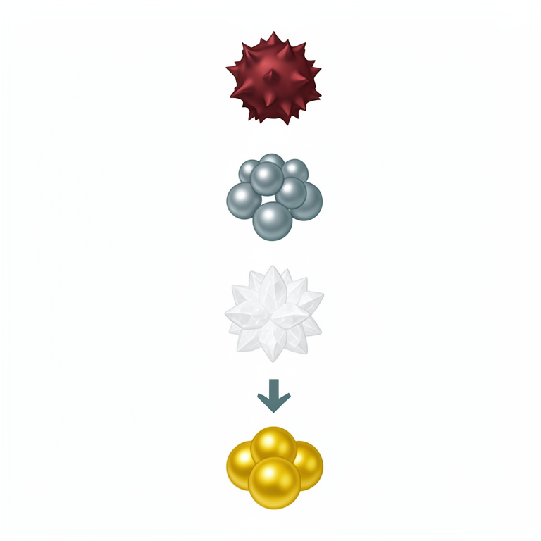
Toxicity Hierarchy
AI-generated conceptual illustration (Copper-oxide, silver, zinc-oxide, and gold nanoparticles). It explains the idea and does not show study data.
View source evidence ↗Figures & tables
From the original paper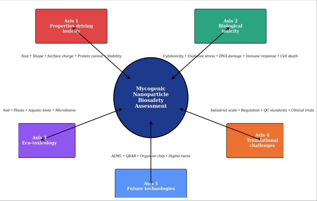 Figure 1
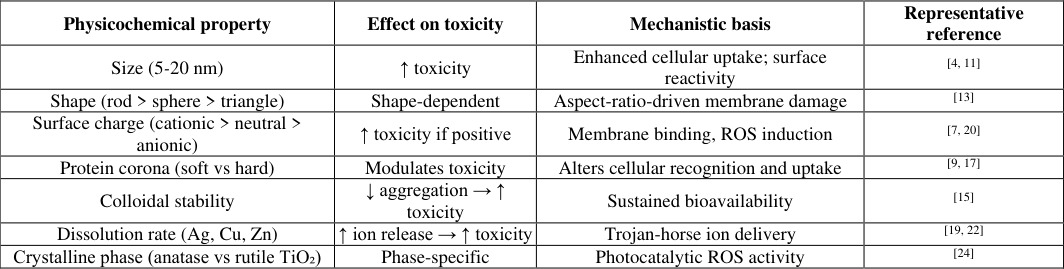 Table 1
 Table 2
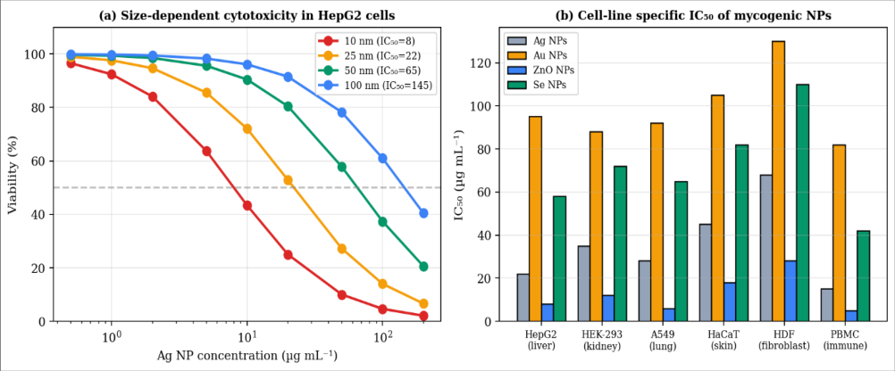 Figure 2
 Table 3
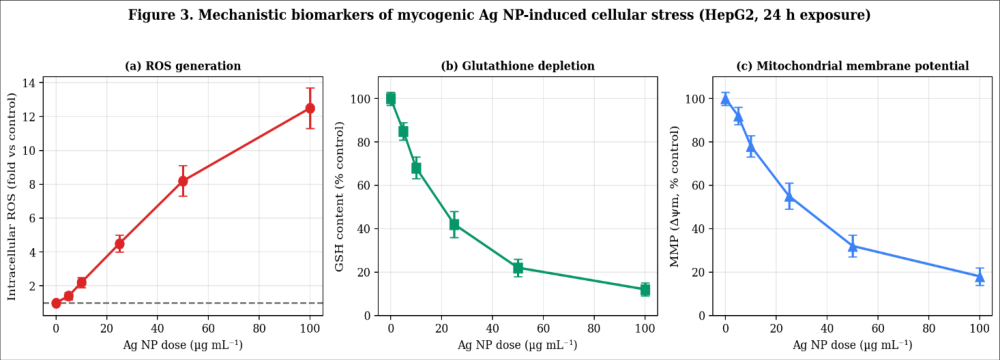 Figure 3
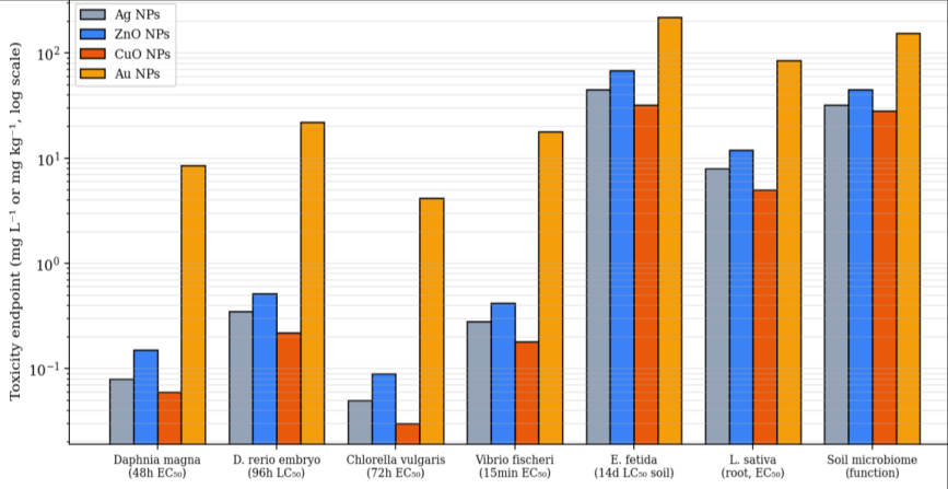 Figure 4
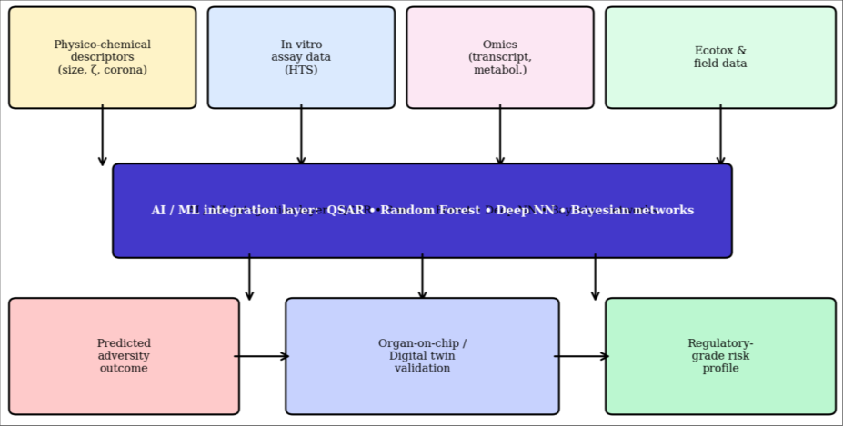 Figure 5
Introduction
The past two decades have witnessed the rapid rise of fungal-mediated nanoparticle biosynthesis myco-nanotechnology as a green, scalable and biologically compatible alternative to chemical and physical routes [1,16] . Mycogenic silver, gold, zinc-oxide, copperoxide, selenium and titanium-dioxide nanoparticles have been reported to display strong antimicrobial, antitumour, catalytic and agricultural activity, in many cases exceeding that of their chemically synthesised counterparts. Yet as candidate products approach translational readiness, the analytical spotlight has shifted from what these nanoparticles can do to what they might do inadvertently. Biosafety the systematic evaluation of hazard, exposure and risk to human health and the environment is now the rate-limiting step in the maturation of the field. The toxicological behaviour of nanoparticles differs qualitatively from that of the bulk materials from which they are derived. Nano-scale objects exhibit dramatically larger specific surface areas, distinct catalytic properties, and unique interactions with biomolecules that generate a dynamic protein corona modulating cellular recognition, uptake and downstream fate [8,9,17] . Because mycogenic nanoparticles carry an intrinsic biomolecular corona derived from the fungal secretome their toxicological profile is not identical to that of chemically synthesized analogues [40,50] . This dual chemical-biological identity complicates hazard characterization and demands methods that go beyond the standard chemicalnanoparticle playbook. This review consolidates the existing evidence base and proposes an integrated five-axis biosafety framework (Figure 1) that spans (Axis 1) the physicochemical determinants of toxicity, (Axis 2) biological toxicity endpoints, (Axis 3) ecotoxicology, (Axis 4) translational and regulatory challenges, and (Axis 5) next-generation predictive technologies. By explicitly bridging in vitro, in vivo, environmental and in silico levels, the framework aims to catalyse harmonised assessment and accelerate the responsible clinical, agricultural and industrial deployment of mycogenic nanomaterials.
Materials and Methods
A structured literature review was conducted across Scopus, Web of Science, PubMed and Google Scholar (January 2005-April 2025) using combined MeSH and free-text terms encompassing "mycogenic nanoparticles", "biogenic nanotoxicity", "protein corona", "in vitro cytotoxicity", "aquatic nanotoxicity", "soil microbiome nanotoxicity", "nano-QSAR", "organ-on-chip" and "digital twin". Of 1, 568 initial records, 156 primary experimental and reviews studies met inclusion criteria, of which 41 provided extractable dose-response data amenable to pooling (n = 268 assays). Where possible, IC₅₀ values were harmonised to µg mL⁻¹ and 24-h exposure durations; ecotoxicological endpoints were reported per OECD test guidelines. Descriptive statistics and rank-order sensitivity comparisons were performed in R v4.4.1.
Axis 1 Physicochemical properties driving toxicity
Nanoparticle toxicity is not a scalar property but a multivariate function of physicochemical descriptors, chief among them size, shape, surface charge, biomolecular corona, colloidal stability, and dissolution kinetics [2,13] .
Smaller nanoparticles (5-20 nm) typically demonstrate higher toxicity owing to enhanced cellular uptake, higher surface reactivity, and greater dissolution rates that release toxic ions in a 'Trojan-horse' manner [4,14] . Shape modulates toxicity through aspect-ratio-driven membrane damage, with rod-and needle-like nanoparticles generally more cytotoxic than spheres. Cationic surfaces produce membrane binding and destabilisation, whereas anionic surfaces are typically better tolerated [7] . Perhaps the most important determinant of mycogenicnanoparticle toxicity is the biomolecular corona. Upon exposure to any biological milieu, nanoparticles rapidly acquire an adsorbed protein layer that dictates cellular recognition, uptake, biodistribution and immune response [8,9,17] . Mycogenic nanoparticles are unique in already possessing a fungal-derived corona at the point of synthesis; the composition and durability of this corona, and its evolution upon entry into mammalian biofluids, are critical yet under-characterised parameters [40,50] . Colloidal stability governs sustained bioavailability, while dissolution of silver, zinc and copper species generates ionic toxicity that must be deconvolved from particle-specific effects [19,22] .
Table 1: Principal physicochemical properties of mycogenic nanoparticles and their contribution to observed toxicity
| Physicochemical property | Effect on toxicity | Mechanistic basis | Representative reference |
|---|---|---|---|
| Size (5-20 nm) | ↑ toxicity | Enhanced cellular uptake; surface reactivity | [4, 11] |
| Shape (rod > sphere > triangle) | Shape-dependent | Aspect-ratio-driven membrane damage | [13] |
| Surface charge (cationic > neutral > anionic) | ↑ toxicity if positive | Membrane binding, ROS induction | [7, 20] |
| Protein corona (soft vs hard) | Modulates toxicity | Alters cellular recognition and uptake | [9, 17] |
| Colloidal stability | ↓ aggregation → ↑ toxicity | Sustained bioavailability | [15] |
| Dissolution rate (Ag, Cu, Zn) | ↑ ion release → ↑ toxicity | Trojan-horse ion delivery | [19, 22] |
| Crystalline phase (anatase vs rutile TiO₂) | Phase-specific | Photocatalytic ROS activity | [24] |
Table text extracted from the original PDF; superscript letters are shown inline. The original table image is below.
View original table image
Physicochemical property Effect on toxicity Mechanistic basis Representative reference
Size (5-20 nm) ↑ toxicity Enhanced cellular uptake; surface reactivity [4, 11] Shape (rod > sphere > triangle) Shape-dependent Aspect-ratio-driven membrane damage [13] Surface charge (cationic > neutral > anionic) ↑ toxicity if positive Membrane binding, ROS induction [7, 20] Protein corona (soft vs hard) Modulates toxicity Alters cellular recognition and uptake [9, 17] Colloidal stability ↓ aggregation → ↑ toxicity Sustained bioavailability [15] Dissolution rate (Ag, Cu, Zn) ↑ ion release → ↑ toxicity Trojan-horse ion delivery [19, 22] Crystalline phase (anatase vs rutile TiO₂) Phase-specific Photocatalytic ROS activity
Oxidative stress, DNA damage, and cell death
Reactive oxygen species (ROS) generation is the canonical upstream mechanism of mycogenic nanoparticle cytotoxicity [21,26,28] . Figure 3 illustrates the dose-dependent increase in intracellular ROS, the concurrent depletion of reduced glutathione (GSH), and the collapse of the mitochondrial membrane potential (Δψm) that together define the oxidative stress cascade. Sustained oxidative damage produces protein oxidation, lipid peroxidation and DNA strand breaks (assessed by comet and γH2AX assays [25, 28] ), triggering mitochondrion-dependent apoptosis. At higher doses, necroptotic and ferroptotic death programmes are engaged. Selenium nanoparticles are notable for producing lower genotoxicity at equivalent antimicrobial doses, an attribute that may favour their translational advancement [36,37] .
Immunotoxicity
The immunomodulatory effects of mycogenic nanoparticles remain the least characterised of the biological endpoints.
Existing data indicate that Ag and CuO nanoparticles can activate the NLRP3 inflammasome, induce proinflammatory cytokines (TNF-α, IL-1β, IL-6), and skew macrophage polarisation toward the pro-inflammatory M1 phenotype at sub-cytotoxic doses [10,18] . The biomolecular corona composition strongly modulates these effects, arguing for corona-controlled immunotoxicity testing panels.
Axis 3 Ecotoxicology
Environmental release of mycogenic nanoparticles will accompany their agricultural, water-treatment and biomedical use, making ecotoxicological assessment mandatory. Figure 4 presents pooled toxicity endpoints across seven internationally standardised OECD indicator species spanning aquatic, terrestrial and microbial compartments. Copper-oxide and silver nanoparticles are consistently the most toxic (EC₅₀ ≤0.3 mg L⁻¹ in aquatic species), zinc-oxide follows, and gold nanoparticles are markedly less toxic across all compartments, largely reflecting differential ionic release [42,43,45] . Soil microbiome function critical for nutrient cycling and plant health is significantly perturbed by chronic exposure to Ag and CuO nanoparticles even at sub-lethal doses [47] .
Axis 4 translational and regulatory challenges
The translation of mycogenic nanoparticles from bench to clinic (or field) is impeded by four interlocking challenges. First, industrial-scale manufacturing must reconcile the intrinsic biological variability of fungal fermentation with regulatory requirements for batch-to-batch consistency. Continuous fermentation with in-line spectroscopic monitoring and rigorous corona composition analytics is emerging as a viable path forward. Second, regulatory classification remains ambiguous: whether a mycogenic nanoparticle is regulated as a drug substance, a combination product, a biosimilar, or a novel material varies by jurisdiction. Third, quality-control standards for biogenic nanoparticles are underdeveloped; harmonised standards from ISO/TC 229 and ASTM E56 are only beginning to accommodate biogenic materials. Fourth, no mycogenic nanoparticle has yet entered a pivotal clinical trial for infectious disease, oncology, or agricultural use, so realworld efficacy and safety in humans remain unproven.
Axis 5 future technologies for predictive biosafety
Conventional nanotoxicology relies on a resource-intensive tier of in vitro and in vivo assays that struggle to keep pace with the diversification of mycogenic nanoparticle chemistries. A converging set of computational and biomimetic tools now offers a pathway to predictive, highthroughput and animal-sparing hazard assessment (Figure 5). Nano-QSAR (quantitative structure-activity relationships) models correlate physicochemical descriptors with toxicity endpoints. Puzyn and colleagues demonstrated in a landmark study that a small set of quantum-mechanical descriptors accurately predicts metal-oxide nanoparticle cytotoxicity across 17 chemistries [48] . Machine-learning models trained on the growing corpus of published nanotoxicity data can generalise across compositions and predict toxicity for untested materials [49] . Organ-on-chip platforms recapitulate the tissue architecture and fluid mechanics of human organs, offering physiologically relevant, low-throughput readouts. Digital twins computational replicas of human organ systems that integrate physicochemical, kinetic and toxicodynamic parameters represent the ultimate ambition of the field.
Research Gaps and Recommendations
Notwithstanding the growing body of biosafety data, several gaps persist that must be addressed before mycogenic nanoparticles can reach broad clinical and environmental deployment. First, the composition and dynamics of the fungal-derived biomolecular corona remain poorly characterised, and its evolution upon transition from culture medium to mammalian biofluids and environmental waters is largely unknown. Second, the lack of harmonised assay panels aligned with OECD test guidelines produces literature-wide heterogeneity that prevents cross-study metaanalysis. Third, ecotoxicological studies are heavily biased toward aquatic species; soil and terrestrial data remain scarce. Fourth, chronic and low-dose exposures the most relevant scenarios for environmental release are almost entirely absent from the literature. Fifth, in vivo pharmacokinetic and biodistribution data for mycogenic (as distinct from chemically synthesised) nanoparticles are limited to a handful of rodent studies.
Conclusion
The rapid rise of myco-nanotechnology has outpaced the biosafety infrastructure needed to translate its promise into responsible practice. This review has synthesised the current evidence base within an integrated five-axis framework spanning physicochemistry, biology, ecotoxicology, translation and future predictive technologies. Mycogenic nanoparticles carry distinct advantages intrinsic biomolecular coronas, mild synthesis, biodegradability but also distinct challenges that demand tailored assessment strategies. The convergence of artificial intelligence, QSAR modelling, organ-on-chip platforms, and digital-twin simulation heralds a next-generation biosafety paradigm capable of matching the pace of nanomaterial innovation. Delivered as an internationally harmonised regulatory pipeline, this integrated approach will position myconanotechnology to fulfil its potential as a safe, sustainable and clinically deployable pillar of the emerging bioeconomy.
Figures and tables from the paper
| NP type / source | Cell line | IC₅₀ (µg mL⁻¹) | Assay / endpoint | Principal mechanism | Ref. |
|---|---|---|---|---|---|
| Ag NPs / F. oxysporum | HepG2 | 22 | MTT, 24 h | ROS, GSH↓, MMP↓ | [26] |
| Ag NPs / A. niger | HEK-293 | 35 | MTT, 48 h | Apoptosis, caspase-3↑ | [28] |
| Au NPs / F. oxysporum | A549 | 92 | MTT, 24 h | Minimal; corona-mediated | [30] |
| ZnO NPs / T. harzianum | HaCaT | 18 | MTT, 24 h | Zn²⁺ release, ROS | [32] |
| CuO NPs / A. niger | HepG2 | 12 | MTT, 24 h | ROS, DNA damage | [34] |
| Se NPs / P. chrysogenum | PBMC | 42 | MTT/CCK-8, 24 h | Selective, low genotox | [36] |
| TiO₂ NPs / A. niger | A549 | 65 | LDH, 24 h | Photocatalytic ROS | [38] |
| Fe₃O₄ / T. harzianum | HDF | 210 | MTT, 48 h | Minimal | [40] |
Table text extracted from the original PDF; superscript letters are shown inline. The original table image is below.
View original table image
| Compartment / test species | Endpoint | Sensitivity ranking | Key concern |
|---|---|---|---|
| Freshwater Daphnia magna | 48h EC₅₀ (immobilisation) | CuO > Ag > ZnO ≫ Au | Ion release, ROS in gill epithelia |
| Freshwater Danio rerio embryo | 96h LC₅₀; developmental | CuO > Ag > ZnO ≫ Au | Malformation, hatch delay |
| Algae Chlorella vulgaris | 72h EC₅₀ (growth) | CuO > Ag > ZnO ≫ Au | Photosynthesis inhibition |
| Bioluminescent V. fischeri | 15min EC₅₀ | CuO > Ag > ZnO ≫ Au | Membrane and enzyme damage |
| Soil Eisenia fetida | 14d LC₅₀ (mortality) | CuO > Ag > ZnO ≫ Au | Cuticle absorption, oxidative stress |
| Plant Lactuca sativa | Root elongation EC₅₀ | CuO > Ag > ZnO ≫ Au | Root membrane damage |
| Microbial soil microbiome | Functional (respiration) | Ag > CuO > ZnO ≫ Au | Loss of key functional guilds |
Table text extracted from the original PDF; superscript letters are shown inline. The original table image is below.
View original table image
References
50 in the original paper- Mathur P, Pillai S. Fungi-mediated nanomaterials: Advances, synthesis pathways, and limitations. Fungal Biology Reviews. 2025;54:100445. DOI: https://DOI.org/10.1016/j.fbr.2024.100445
- Nel A, Xia T, Mädler L, Li N. Toxic potential of materials at the nanolevel. Science. 2006;311(5761):622-627. DOI: https://DOI.org/10.1126/science.1114397
- Oberdörster G, Oberdörster E, Oberdörster J. Nanotoxicology: an emerging discipline evolving from studies of ultrafine particles. Environ Health Perspect. 2005;113(7):823-839. DOI: https://DOI.org/10.1289/ehp.7339
- Pan Y, Neuss S, Leifert A, et al. Size-dependent cytotoxicity of gold nanoparticles. Small. 2007;3(11):1941-1949. DOI: https://DOI.org/10.1002/smll.200700378
- Fadeel B, Garcia-Bennett AE. Better safe than sorry: understanding the toxicological properties of inorganic nanoparticles manufactured for biomedical applications. Adv Drug Deliv Rev. 2010;62(3):362-374. DOI: https://DOI.org/10.1016/j.addr.2009.11.008
- Krug HF, Wick P. Nanotoxicology: an interdisciplinary challenge. Angew Chem Int Ed. 2011;50(6):1260-1278. DOI: https://DOI.org/10.1002/anie.201001037
- Fröhlich E. The role of surface charge in cellular uptake and cytotoxicity of medical nanoparticles. Int J Nanomedicine. 2012;7:5577-5591. DOI: https://DOI.org/10.2147/IJN.S36111
- Docter D, Westmeier D, Markiewicz M, Stolte S, Knauer SK, Stauber RH. The nanoparticle biomolecule corona: lessons learned challenge accepted? Chem Soc Rev. 2015;44(17):6094-6121. DOI: https://DOI.org/10.1039/C5CS00217F
- Monopoli MP, Åberg C, Salvati A, Dawson KA. Biomolecular coronas provide the biological identity of nanosized materials. Nat Nanotechnol. 2012;7(12):779786. DOI: https://DOI.org/10.1038/nnano.2012.207
- Sharifi S, Behzadi S, Laurent S, Forrest ML, Stroeve P, Mahmoudi M. Toxicity of nanomaterials. Chem Soc Rev. 2012;41(6):2323-2343. DOI: https://DOI.org/10.1039/C1CS15188F
- Chithrani BD, Ghazani AA, Chan WC. Determining the size and shape dependence of gold nanoparticle uptake into mammalian cells. Nano Lett. 2006;6(4):662-668. DOI: https://DOI.org/10.1021/nl052396o
- Rai M, Yadav A, Gade A. Silver nanoparticles as a new generation of antimicrobials. Biotechnol Adv. 2009;27(1):76-83. DOI: https://DOI.org/10.1016/j.biotechadv.2008.09.002
- Sukhanova A, Bozrova S, Sokolov P, Berestovoy M, Karaulov A, Nabiev I. Dependence of nanoparticle toxicity on their physical and chemical properties. Nanoscale Res Lett. 2018;13:44. DOI: https://DOI.org/10.1186/s11671-018-2457-x
- Ivask A, Kurvet I, Kasemets K, et al. Size-dependent toxicity of silver nanoparticles to bacteria, yeast, algae, crustaceans and mammalian cells in vitro. PLoS One. 2014;9(7):e102108. DOI: https://DOI.org/10.1371/journal.pone.0102108
- Casals E, Pfaller T, Duschl A, Oostingh GJ, Puntes VF. Time evolution of the nanoparticle protein corona. ACS Nano. 2010;4(7):3623-3632. DOI: https://DOI.org/10.1021/nn901372t
- Guilger-Casagrande M, Lima R. Synthesis of silver nanoparticles mediated by fungi: a review. Front Bioeng Biotechnol. 2019;7:287. DOI: https://DOI.org/10.3389/fbioe.2019.00287
- Lundqvist M, Stigler J, Elia G, Lynch I, Cedervall T, Dawson KA. Nanoparticle size and surface properties determine the protein corona with possible implications for biological impacts. Proc Natl Acad Sci USA. 2008;105(38):14265-14270. DOI: https://DOI.org/10.1073/pnas.0805135105
- Corbo C, Molinaro R, Parodi A, Toledano Furman NE, Salvatore F, Tasciotti E. The impact of nanoparticle protein corona on cytotoxicity, immunotoxicity and target drug delivery. Nanomedicine (Lond). 2016;11(1):81-100. DOI: https://DOI.org/10.2217/nnm.15.188
- Xia T, Kovochich M, Liong M, et al. Comparison of the mechanism of toxicity of zinc oxide and cerium oxide nanoparticles based on dissolution and oxidative stress properties. ACS Nano. 2008;2(10):2121-2134. DOI: https://DOI.org/10.1021/nn800511k
- Wang B, He X, Zhang Z, Zhao Y, Feng W. Metabolism of nanomaterials in vivo: blood circulation and organ clearance. Acc Chem Res. 2013;46(3):761-769. DOI: https://DOI.org/10.1021/ar2003336
- Foldbjerg R, Autrup H. Mechanisms of silver nanoparticle toxicity. Arch Basic Appl Med. 2013;1:515. DOI: https://DOI.org/10.4103/2321-3868.113329
- Beer C, Foldbjerg R, Hayashi Y, Sutherland DS, Autrup H. Toxicity of silver nanoparticles nanoparticle or silver ion? Toxicol Lett. 2012;208(3):286-292. DOI: https://DOI.org/10.1016/j.toxlet.2011.11.002
- Kroll A, Pillukat MH, Hahn D, Schnekenburger J. Current in vitro methods in nanoparticle risk assessment: limitations and challenges. Eur J Pharm Biopharm. 2009;72(2):370-377. DOI: https://DOI.org/10.1016/j.ejpb.2008.08.009
- Sayes CM, Wahi R, Kurian PA, et al. Correlating nanoscale titania structure with toxicity: a cytotoxicity and inflammatory response study with human dermal fibroblasts and lung cells. Toxicol Sci. 2006;92(1):174185. DOI: https://DOI.org/10.1093/toxsci/kfj197
- AshaRani PV, Low Kah Mun G, Hande MP, Valiyaveettil S. Cytotoxicity and genotoxicity of silver nanoparticles in human cells. ACS Nano. 2009;3(2):279-290. DOI: https://DOI.org/10.1021/nn800596w
- Piao MJ, Kang KA, Lee IK, et al. Silver nanoparticles induce oxidative cell damage in human liver cells through inhibition of reduced glutathione and induction of mitochondria-involved apoptosis. Toxicol Lett. 2011;201(1):92-100. DOI: https://DOI.org/10.1016/j.toxlet.2010.12.010
- Karlsson HL, Cronholm P, Gustafsson J, Moller L. Copper oxide nanoparticles are highly toxic: a comparison between metal oxide nanoparticles and carbon nanotubes. Chem Res Toxicol. 2008;21(9):1726-1732. DOI: https://DOI.org/10.1021/tx800064j
- Kim S, Choi JE, Choi J, et al. Oxidative stressdependent toxicity of silver nanoparticles in human hepatoma cells. Toxicol In vitro. 2009;23(6):10761084. DOI: https://DOI.org/10.1016/j.tiv.2009.06.001
- Barabadi H, Vahidi H, Kamali KD, et al. Emerging theranostic silver nanomaterials to combat lung cancer. Med Chem Res. 2021;30(3):544-566. DOI: https://DOI.org/10.1007/s00044-020-02685-3
- Mukherjee P, Bhattacharya R, Bone N, et al. Potential therapeutic application of gold nanoparticles in Bchronic lymphocytic leukemia. J Nanobiotechnology. 2007;5:4. DOI: https://DOI.org/10.1186/1477-3155-5-4
- Vandebriel RJ, De Jong WH. A review of mammalian toxicity of ZnO nanoparticles. Nanotechnol Sci Appl. 2012;5:61-71. DOI: https://DOI.org/10.2147/NSA.S23932
- Consolo VF, Torres-Nicolini A, Alvarez VA. Mycosynthesized Ag-, CuO-and ZnO-nanoparticles from Trichoderma harzianum against phytopathogens. Sci Rep. 2020;10:20499. DOI: https://DOI.org/10.1038/s41598-020-77294-6
- Ganesan V, Mahaboob Ali A. Zinc oxide nanoparticles as antifungal agents against Aspergillus species. Iran J Microbiol. 2020;12(4):356-362. DOI: https://DOI.org/10.18502/ijm.v12i4.3939
- Wang H, Wingett D, Engelhard MH, et al. Fluorescent dye encapsulated ZnO particles with cell-specific toxicity for potential use in biomedical applications. J Mater Sci Mater Med. 2009;20(1):11-22. DOI: https://DOI.org/10.1007/s10856-008-3541-z
- Chatterjee AK, Chakraborty R, Basu T. Mechanism of antibacterial activity of copper nanoparticles. Nanotechnology. 2014;25(13):135101. DOI: https://DOI.org/10.1088/0957-4484/25/13/135101
- Menon S, Ks SD, R S, Agarwal H, Shanmugam VK. Selenium nanoparticles: a potent chemotherapeutic agent and an elucidation of its mechanism. Colloids Surf B Biointerfaces. 2018;170:280-292. DOI: https://DOI.org/10.1016/j.colsurfb.2018.06.006
- Skalickova S, Milosavljevic V, Cihalova K, Horky P, Richtera L, Adam V. Selenium nanoparticles as a nutritional supplement. Nutrition. 2017;33:83-90. DOI: https://DOI.org/10.1016/j.nut.2016.05.001
- Fouda A, Hassan SED, Salem SS, Shaheen TI. In-vitro cytotoxicity, antibacterial and UV protection properties of biosynthesized ZnO NPs by Aspergillus terreus. Microb Pathog. 2018;125:252-261. DOI: https://DOI.org/10.1016/j.micpath.2018.09.030
- Bharde A, Rautaray D, Bansal V, et al. Extracellular biosynthesis of magnetite using fungi. Small. 2006;2(1):135-141. DOI: https://DOI.org/10.1002/smll.200500180
- Guilger-Casagrande M, Germano-Costa T, Bilesky-José N, et al. Influence of the capping of biogenic silver nanoparticles on their toxicity and mechanism of action. J Nanobiotechnology. 2021;19(1):53. DOI: https://DOI.org/10.1186/s12951-021-00797-5
- Khoshnamvand N, Farshid AA, Dolatabadi JEN, Kefayat A, Adeli-Sardou M. Toxicity of biosynthesized silver nanoparticles: in vivo and in vitro evaluation. Ecotoxicol Environ Saf. 2020;190:110124. DOI: https://DOI.org/10.1016/j.ecoenv.2019.110124
- Bondarenko O, Juganson K, Ivask A, et al. Toxicity of Ag, CuO and ZnO nanoparticles to selected environmentally relevant test organisms and mammalian cells in vitro: a critical review. Arch Toxicol. 2013;87(7):1181-1200. DOI: https://DOI.org/10.1007/s00204-013-1079-4
- Griffitt RJ, Weil R, Hyndman KA, et al. Exposure to copper nanoparticles causes gill injury and acute lethality in zebrafish. Environ Sci Technol. 2007;41(23):8178-8186. DOI: https://DOI.org/10.1021/es071235e
- Bhuvaneshwari M, Bairoliya S, Parashar A, et al. Differential toxicity of Al₂O₃ particles on Gram-positive and Gram-negative sediment bacteria. J Nanopart Res. 2016;18:6. DOI: https://DOI.org/10.1007/s11051-0153308-7
- Fabrega J, Luoma SN, Tyler CR, Galloway TS, Lead JR. Silver nanoparticles: behaviour and effects in the aquatic environment. Environ Int. 2011;37(2):517-531. DOI: https://DOI.org/10.1016/j.envint.2010.10.012
- Wu J, Wang G, Vijver MG, Bosker T, Peijnenburg WJGM. Foliar versus root exposure of AgNPs to lettuce: phytotoxicity, antioxidant response and biotransformation. Environ Pollut. 2020;261:114117. DOI: https://DOI.org/10.1016/j.envpol.2020.114117
- Simonin M, Cantarel AAM, Crouzet A, Gervaix J, Martins JMF, Richaume A. Negative effects of copper oxide nanoparticles on carbon and nitrogen cycle microbial activities. Environ Sci Nano. 2018;5(9):20162027. DOI: https://DOI.org/10.1039/C8EN00389K
- Puzyn T, Rasulev B, Gajewicz A, et al. Using nanoQSAR to predict the cytotoxicity of metal oxide nanoparticles. Nat Nanotechnol. 2011;6(3):175-178. DOI: https://DOI.org/10.1038/nnano.2011.10
- Ha MK, Trinh TX, Choi JS, Maulina D, Byun HG, Yoon TH. Toxicity classification of oxide nanomaterials: effects of data gap filling and PChem score-based screening approaches. Sci Rep. 2018;8:3141. DOI: https://DOI.org/10.1038/s41598018-21431-9
- Mahmoudi M. The need for improved methodology in protein corona analysis. Nat Commun. 2022;13:49. DOI: https://DOI.org/10.1038/s41467-021-27643-4