
Research article · 2026 · Full text
International Journal of Biology SciencesIris prolapse in mare: A case report
Volume 8(7) · Pages 105–107 · DOI: 10.33545/26649926.2026.v8.i7b.936 ↗
Research areasTraumatic Ocular and Foreign Body InjuriesVeterinary Equine Medical ResearchOcular Disorders and Treatments
Abstract
Author-writtenIris prolapse is a frequent complication of deep corneal injuries or ulcers, often leading to vision loss. An eight-year-old mare presented with left eye trauma was diagnosed for iris prolapse by visual examination and surgically repaired by rotational bulbar conjunctival graft under general anesthesia. This case report highlights the importance of prompt diagnosis and treatment of iris prolapse with appropriate graft selection in ensuring tectonic stability and functional recovery of the eye. Timely intervention is critical, as any delay greatly diminishes the chances of preserving the globe and, consequently, maintaining vision.
Summary
Source-checkedPlain-language reading of the full paper, prepared with AI and checked against the text. Each point links to the authors' own words.
Why it matters
- Horses are more prone to corneal ulcers and other ocular disorders due to their large globes, pronounced corneal prominence, and the lateral orientation of their eyeballs. View evidence ↗
- Corneal injuries in horses often heal slowly and can lead to serious complications, including iris prolapse. View evidence ↗
- Iris prolapse occurs when a full-thickness loss of the cornea allows dark brown or black iris tissue to protrude through the defect. View evidence ↗
The question
- The study aims to demonstrate how prompt diagnosis and the selection of an appropriate graft can ensure the tectonic stability and functional recovery of an eye affected by iris prolapse. View evidence ↗
What they did
- The subject was an eight-year-old mare weighing 500 kg with a 14-day history of trauma to her left eye. View evidence ↗
- Examination identified a central corneal defect of approximately 3 mm through which the brown iris had prolapsed. View evidence ↗
- Surgery was performed under general anesthesia, induced with intravenous xylazine (1 mg/kg) and ketamine (5 mg/kg), and maintained with 2.5% isoflurane. View evidence ↗
- A rotational conjunctival graft, consisting of a 4 × 8 mm section of bulbar conjunctiva, was sutured to the corneal defect using PDS 4-0 in a simple interrupted pattern. View evidence ↗
- Postoperative care included intravenous systemic antibiotics (Ampicillin and Cloxacillin at 10-50 mg/kg) twice daily for five days and analgesics (meloxicam at 0.2-0.5 mg/kg) once daily for three days. View evidence ↗
What they found
- Two weeks after surgery, the graft remained intact, episcleral congestion was gone, and there was moderate corneal opacification and neovascularization around the graft. View evidence ↗
- By six weeks, the conjunctival graft had successfully healed the corneal defect, which preserved the globe. View evidence ↗
- Although severe corneal opacity developed, the mare retained partial peripheral vision. View evidence ↗
What it means
- The authors conclude that timely surgical intervention is critical to preserve the globe and maintain vision in cases of iris prolapse. View evidence ↗
- A limitation of using conjunctival pedicle grafts is that the remaining graft tissue causes permanent corneal opacity, which can impair the animal's vision. View evidence ↗
Visual summary
1 visualsIllustrations explain the paper; they do not show study data.
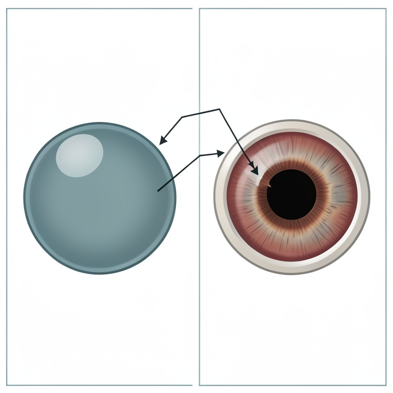
Mechanism of Iris Prolapse
AI-generated conceptual illustration (mare eye (equine)). It explains the idea and does not show study data.
View source evidence ↗Figures & tables
From the original paper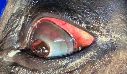 Figure 1
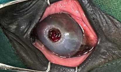 Figure 2
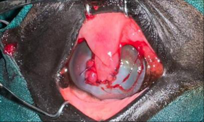 Figure 3
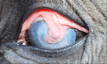 Figure 4
Introduction
In equines, the lateral orientation of the eyeball, the relatively large globe, and pronounced corneal prominence predispose them to a higher incidence of corneal ulcers (Plummer, 2017) [5] as well as periocular and ocular disorders. Corneal injuries are often slow to heal and may progress to complications such as ulceration, descemetocele, and iris prolapse. Superficial corneal damage involves the anterior one-third of the stroma and generally responds to intensive medical therapy (Cutler, 2004) [2] . In contrast, complex deep injuries extend beyond the anterior one-third of the stroma frequently resulting in descemetocele formation and iris prolapse which necessitate immediate surgical intervention (Chmielewski et al., 1997) [1] . In Iris prolapse, full corneal thickness loss results in protusion of a dark brown to black iris tissue through the corneal defect. It may arise secondary to trauma or infections of bacterial, fungal (e.g., Aspergillus, Fusarium), or viral origin (Flores et al., 2020) [3] . While diagnosis can often be made through direct visual inspection of the eye, a comprehensive assessment of the cornea, anterior chamber, lens, and posterior chamber, along with evaluation of vision, is essential to exclude other ocular pathologies. Supportive medical therapy alone is insufficient in iris prolapse. Immediate surgical intervention should be instituted using a conjunctival or corneal graft to cover the defect, protect the globe, and reduce infection risk.
Material and Methods
An eight-year-old mare, weighing 500 kgs was presented with a 14-day history of trauma to the left eye. Clinical examination revealed mucoid ocular discharge accompanied by blepharospasm and photophobia. A central corneal defect measuring approximately 3 mm was identified, through which the brown iris had prolapsed (Fig 1). Opacity was observed around the lesion and along the ventral cornea, whereas neovascularization was evident dorsally. The menace response was intact in both eyes, and episcleral congestion was noted in the affected eye. As per Stoppini & Gilger, (2016) [6] , the eye was evaluated physically by looking for corneal opacity, menace response, and type of neovascularization. To achieve tectonic preservation and restore vision, emergency surgery was carried out under general anesthesia. Anaesthetic induction was performed using intravenous xylazine at 1 mg/kg body weight and ketamine at 5 mg/kg body weight, with maintenance provided by 2.5% isoflurane. The left eyelids were disinfected with povidone-iodine, and the eye was thoroughly irrigated with normal saline. Prior to draping, a few drops of topical gatifloxacin (0.5%) were instilled. The prolapsed iris was carefully debrided with sterile cotton, and the margins of the corneal ulcer were cleared. The remaining viable iris tissue was repositioned into the anterior chamber (Fig 2 ). A 4 × 8 mm section of bulbar conjunctiva was harvested parallel to the dorsal limbus to create a rotational conjunctival graft, which was sutured to the corneal defect using PDS 4-0 in a simple interrupted pattern (Fig 3).
Tarsorrhaphy was then performed with nylon in a horizontal mattress technique, leaving a small medial opening for drug administration. Postoperatively, the mare was administered systemic antibiotics (Ampicillin and Cloxacillin @ 10-50 mg/kg), twice a day for five days and analgesics (meloxicam @ 0.2-0.5 mg/kg) once a day for three days, intravenously. Gatifloxacin was administered topically at 2 drops three times a day for 14 days. Furthermore, topical gatifloxacin with prednisolone (two drops, three times a day) was advised to continue for two more weeks. The tarsorrhaphy sutures were removed after two weeks.
Results and Discussion
Fourteen days after surgery, evaluation of the left eye revealed an intact graft, moderate corneal opacification surrounding the graft with associated neovascularization, and absence of episcleral congestion. At 6 weeks, the conjunctival graft successfully covered and healed the corneal defect, thereby preserving the globe. Severe corneal opacity developed; however, the owner reported partial peripheral vision (Fig 4). Given the chronicity of the injury, delayed treatment could have resulted in phthisis bulbi, endophthalmitis, and eventual globe rupture. While conjunctival pedicle grafts are commonly used in horses to provide structural support and supply blood vessels to an avascular cornea, the remaining graft tissue causes permanent corneal opacity that can impair vision. All biomaterials compromise optical clarity, letting the cornea heal through medical treatment alone when feasible results in significantly less scarring. (Lores et al. 2020) [4] . In this mare, a rotational bulbar pedicle graft was employed since the defect was centrally located and measured 3 mm in diameter. Chmielewski et al. 1997 [1] discussed that fullthickness corneal lacerations exceeding 15 mm that involve or extend past the limbus carry a high risk of poor visual recovery and surgical eye removal (enucleation). So, in present case scenario, there were better chances of partial vision recovery. Several surgical techniques are available for managing iris prolapse, including direct corneal suturing, conjunctival flaps and grafts, and both full-thickness and partial-thickness keratoplasty. Different conjunctival graft types such as rotational bulbar pedicle, hood, island, bridge, 360° complete, and tarsopalpebral grafts each present distinct advantages and limitations. The graft must not only cover the prolapsed area but also adhere to the corneal epithelial cells; otherwise, epithelial migration beneath the graft may delay healing and lead to graft failure. A 360° bulbar conjunctival graft, for instance, impairs vision, complicates ocular response assessment, carries a high risk of failure, causes extensive damage to the bulbar conjunctiva, and reduces drug delivery. Hood and 180° grafts share similar drawbacks but to a lesser extent. Bridge grafts resemble pedicle grafts, except they are anchored at both ends of the eye rather than a single attachment point (Plummer, 2017) [5] . There are advantages and disadvantages of different techniques for correction of staphyloma, so the decision of technique depends on the location of the corneal tear, size of laceration and severity of lesion, most importantly, the surgeon's experience and choice.
References
6 in the original paper- Chmielewski NT, Brooks DE, Smith PJ, Hendrix DV, Whittaker C, Gelatt KN. Visual outcome and ocular survival following iris prolapse in the horse: A review of 32 cases. Equine Veterinary Journal. 1997;29(1):3139.
- Cutler TJ. Corneal epithelial disease. Veterinary Clinics of North America: Equine Practice. 2004;20(2):319326.
- Flores MM, Del Piero F, Habecker PL, Langohr IM. A retrospective histologic study of 140 cases of clinically significant equine ocular disorders. Journal of Veterinary Diagnostic Investigation. 2020;32(3):382388. International Journal of Biology Sciences https://www.biologyjournal.net
- Lores M, Rakestraw P, De Rijck M, Yarbrough T. Use of autologous fascia lata graft to repair a complex corneal ulcer in a mare. Irish Veterinary Journal. 2020;73(1):1-10.
- Plummer CE. Corneal response to injury and infection in the horse. Veterinary Clinics of North America: Equine Practice. 2017;33(3):439-463.
- Stoppini R, Gilger BC. Equine ocular examination: Basic techniques. In: Gilger BC, editor. Equine ophthalmology. 3rd ed. Hoboken (NJ): Wiley Blackwell; 2016. p. 1-39.