
Research article · 2026 · Full text
International Journal of Advanced Chemistry ResearchGreen synthesis of nanoparticles using Parthenium hysterophorus leaf extract and their antimicrobial activity
Volume 8(5) · Pages 115–120 · DOI: 10.33545/26646781.2026.v8.i5b.717 ↗
Research areasNanoparticles: synthesis and applicationsGold and Silver Nanoparticles Synthesis and ApplicationsNanomaterials for catalytic reactions
Abstract
Author-writtenThe search for eco-friendly routes to metallic nanoparticle fabrication has intensified over the past decade, driven by concerns about the toxicity and energy cost of conventional chemical reduction methods. This research describes the green synthesis of silver nanoparticles (AgNPs) using an aqueous leaf extract of Parthenium hysterophorus as both reducing and capping agent, and evaluates the antimicrobial eficacy of the resulting particles against four clinically relevant pathogens. Fresh pre-flowering leaves were collected from peri-urban sites near Querétaro, Mexico, washed, shade-dried, and boiled in distilled water at 60°mathsfC for 30 minutes to obtain the extract. The extract was mixed with 1 mM silver nitrate solution at a 1:9 (v/v) ratio and stirred at 60 °C and pH 8.0. A colour shift from pale yellow to dark brown within 30 minutes signalled bioreduction. UV-visible spectrophotometry confirmed surface plasmon resonance at 438 nm, with absorbance increasing progressively over 180 minutes of reaction. Fourier-transform infrared spectroscopy identified hydroxyl, carbonyl, and amine functional groups on the nanoparticle surface, pointing to flavonoids and phenolic acids as the main reducing biomolecules. Dynamic light scatering indicated a mean hydrodynamic diameter of 37.6 nm with a polydispersity index of 0.28, while transmission electron microscopy revealed predominantly spherical particles ranging from 28 to 47 nm. Phytochemical screening of the leaf extract confirmed the presence of alkaloids, flavonoids, tannins, saponins, and terpenoids. Antimicrobial activity was assessed by the agar well difusion method against Staphylococcus aureus, Escherichia coli, Pseudomonas aeruginosa, and Candida albicans at concentrations of 25 to 150 µg/mL. At 150 µg/mL the AgNPs produced zones of inhibition of 24.3 mm for S. aureus, 21.6 mm for E. coli, 19.1 mm for P. aeruginosa, and 17.2 mm for C. albicans, exceeding or matching standard antibiotic controls. Minimum inhibitory concentrations ranged from 31.2 to 62.5 µg/mL depending on the test organism. These findings show that P. hysterophorus leaf extract ofers a rapid, low-cost, and non-toxic platform for AgNP synthesis, simultaneously converting weed biomass into a value-added antimicrobial product.
Summary
Source-checkedPlain-language reading of the full paper, prepared with AI and checked against the text. Each point links to the authors' own words.
Why it matters
- Conventional chemical methods for making metallic nanoparticles are often energy-intensive and use toxic reagents, leading to a search for eco-friendly alternatives. View evidence ↗
- Parthenium hysterophorus is an invasive weed that contains various phytochemicals, such as flavonoids and phenolic acids, which could potentially be used as resources. View evidence ↗
- Converting this invasive weed biomass into nanoparticles could provide economic value and help control the spread of the weed. View evidence ↗
The question
- The study aimed to optimize the green synthesis of silver nanoparticles (AgNPs) using P. hysterophorus leaf extract from Mexico, characterize them, identify the reducing phytochemicals, and test their antimicrobial activity. View evidence ↗
What they did
- Pre-flowering leaves of P. hysterophorus were collected from road margins near Querétaro, Mexico, in August 2021. View evidence ↗
- AgNPs were synthesized by mixing 10 mL of leaf extract with 90 mL of 1 mM silver nitrate solution at 60 °C and pH 8.0. View evidence ↗
- The antimicrobial activity of the AgNPs was tested against S. aureus, E. coli, P. aeruginosa, and C. albicans at concentrations ranging from 25 to 150 µg/mL. View evidence ↗
What they found
- UV-Vis spectrophotometry showed a surface plasmon resonance peak at 438 nm, which increased over the 180-minute reaction period. View evidence ↗
- Transmission electron microscopy (TEM) revealed that the AgNPs had a mean particle size of 37.6 nm. View evidence ↗
- At a concentration of 150 µg/mL, the AgNPs inhibited the growth of S. aureus (24.3 mm), E. coli (21.6 mm), P. aeruginosa (19.1 mm), and C. albicans (17.2 mm). View evidence ↗
- The minimum inhibitory concentrations (MIC) for the tested organisms ranged from 31.2 to 62.5 µg/mL. View evidence ↗
- The leaf extract contained 47.3 mg gallic acid equivalents per gram of total phenolics and 28.6 mg quercetin equivalents per gram of total flavonoids. View evidence ↗
What it means
- The authors conclude that P. hysterophorus can be effectively converted into a functional antimicrobial nanomaterial using a simple, non-toxic, and mild process. View evidence ↗
- The authors state that cytotoxicity testing on mammalian cell lines is required before the nanoparticles can be used in biomedical applications. View evidence ↗
- Further research is needed to determine the long-term storage stability and shelf-life of the nanoparticles at various temperatures. View evidence ↗
Visual summary
1 visualsIllustrations explain the paper; they do not show study data.
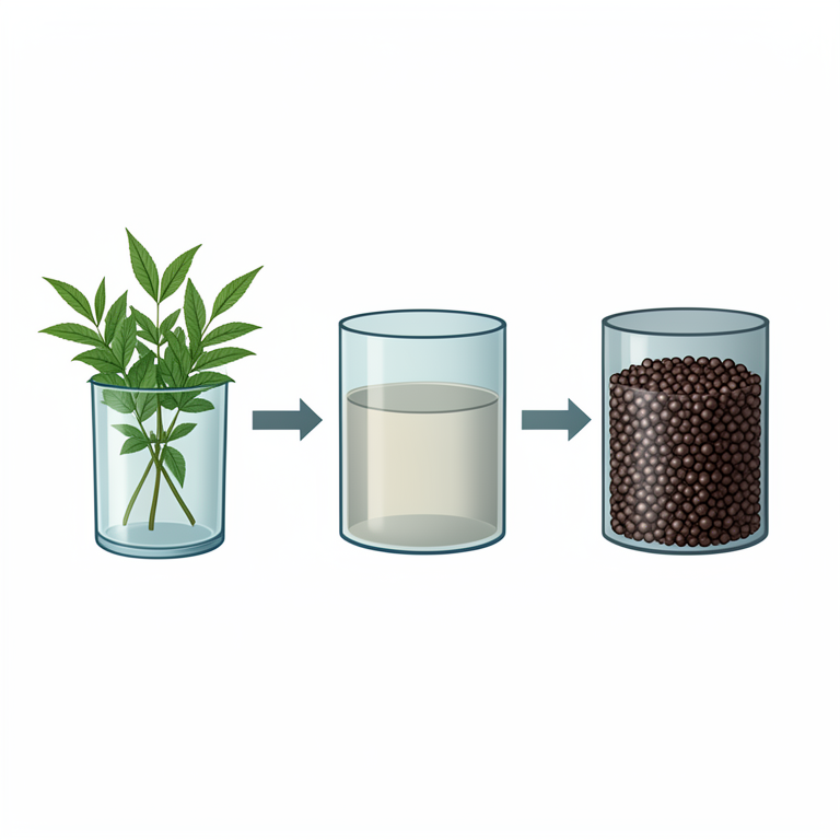
Green Synthesis Process
AI-generated conceptual illustration (Parthenium hysterophorus leaves and silver nanoparticles). It explains the idea and does not show study data.
View source evidence ↗Figures & tables
From the original paper Table 1
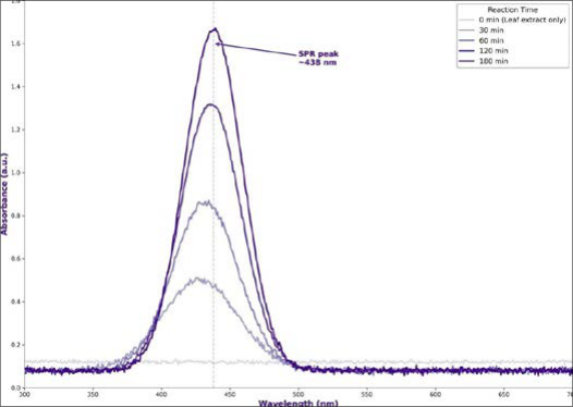 Figure 1
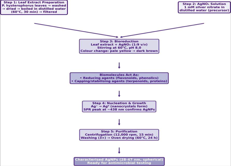 Figure 2
 Table 2
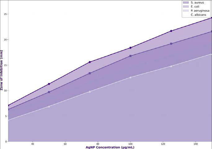 Figure 3
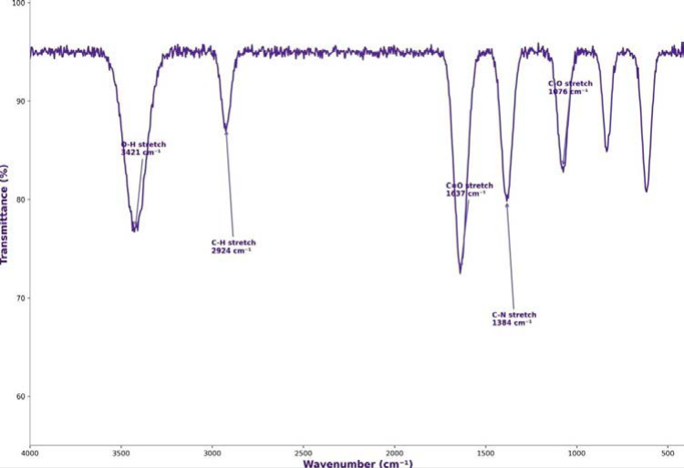 Figure 4
Introduction
The deliberate manipulation of mater at the nanometre scale traces its conceptual origins to Richard Feynman's 1959 lecture "There's Plenty of Room at the Botom," but the practical synthesis of metallic nanoparticles only gained widespread laboratory traction in the 1990s following the development of citrate-reduction and borohydride-reduction protocols [1] . Silver nanoparticles (AgNPs) atracted particular interest because colloidal silver had been used as a germicide since the nineteenth century, and particles in the 10 to 100 nm range showed considerably stronger antimicrobial properties than bulk or ionic silver [2] . Conventional chemical synthesis typically involves strong reducing agents such as sodium borohydride, hydrazine, or dimethylformamide, combined with polymeric stabilisers like polyvinylpyrrolidone [3] . These reagents are hazardous, generate toxic by-products, and demand energy-intensive conditions. Over the past fifteen years, "green synthesis" has emerged as an alternative that replaces chemical reductants with biological extracts from bacteria, fungi, algae, and higher plants whose phenolic acids, flavonoids, terpenoids, and proteins serve as both reducing and capping agents [4,5] . Plant-mediated synthesis holds several advantages over microbial routes. It avoids the need for aseptic culture maintenance, ofers faster reaction kinetics, and draws on a far wider palete of phytochemicals that can tune particle size and morphology [6] . Leaf extracts of Azadirachta indica, Ocimum sanctum, Aloe vera, and Moringa oleifera have all yielded AgNPs with documented antimicrobial, catalytic, or photocatalytic properties [7,8] . Parthenium hysterophorus an aggressive invasive weed of the Asteraceae family is rarely considered a resource, yet its leaves are rich in sesquiterpene lactones (parthenin, coronopilin), flavonoids, phenolic acids, and tannins [9] . These secondary metabolites have demonstrated reducing capacity in preliminary reports from India, where Parthenium leaf extract produced AgNPs with particle sizes between 20 and 60 nm [10,11] . But comprehensive characterisation combining UV-Vis, FTIR, DLS, and TEM alongside antimicrobial dose-response data against both bacterial and fungal pathogens remains limited, particularly from Latin American populations of the weed where phytochemical profiles may difer due to edaphic and climatic variation. Mexico is part of the plant's native range, and dense roadside stands are common throughout the central highlands from April to October. Using this otherwise undesirable biomass for nanoparticle fabrication would add economic value while reducing weed-seed dispersal if leaves are harvested before flowering [12] . The antimicrobial product itself has potential applications in wound dressings, water purification filters, and food-packaging coatings markets where greensynthesised AgNPs are preferred because of their lower residual toxicity compared with chemically synthesised counterparts [13] . This research therefore aimed to (a) optimise the green synthesis of AgNPs using an aqueous leaf extract of P. hysterophorus collected in central Mexico, (b) characterise the nanoparticles through UV-Vis spectrophotometry, FTIR, dynamic light scatering, and transmission electron microscopy, (c) identify the major phytochemical groups responsible for bioreduction, and (d) evaluate antimicrobial activity against two Gram-positive, one Gram-negative bacterium, and one fungal pathogen across a range of concentrations.
Material and Methods
Material and Methods
Reagents and Chemicals
Silver nitrate (AgNO₃ , , 99.8 per cent purity, Sigma-Aldrich, St Louis, MO, USA) was used as the metal precursor. Sodium hydroxide (NaOH) and hydrochloric acid (HCl, analytical grade, J.T. Baker, Phillipsburg, NJ, USA) were used for pH adjustment. Mueller-Hinton agar and Sabouraud dextrose agar (Difco, BD Biosciences, Sparks, MD, USA) were employed for antimicrobial assays. Standard antibiotic discs of ciprofloxacin (5 µg) and fluconazole (25 µg) were obtained from HiMedia Laboratories, Mumbai, India. All solutions were prepared with Milli-Q deionised water (resistivity 18.2 MΩ•cm).
Material
Fresh leaves of P. hysterophorus at the pre-flowering rosete stage were collected from road margins along Highway 57D near Querétaro (20° 35′N , 100° 24′W) in August 2021. Leaves were washed three times with tap water followed by two rinses with distilled water, shade-dried for 72 hours, and ground to a coarse powder. Ten grams of powder were boiled in 100 mL distilled water at 60°mathsfC for 30 minutes. The mixture was cooled to room temperature, filtered through Whatman No. 1 filter paper, and stored at 4°mathsfC for no more than 48 hours before use [14] . The research was conducted from August 2021 to April 2022 at the nanotechnology and microbiology laboratories of the Central Mexico University of Applied Nanotechnology, Querétaro.
Methods
For nanoparticle synthesis, 10 mL of filtered leaf extract was added dropwise to 90 mL of 1 mM AgNO₃ solution under continuous magnetic stirring at 60 °C . Solution pH was adjusted to 8.0 with 0.1 N NaOH. Colour change was monitored visually and spectrophotometrically at 10-minute intervals using a Shimadzu UV-1800 spectrophotometer scanning 300 to 700 nm. After 180 minutes, the colloidal suspension was centrifuged at 12,000 rpm for 15 minutes (Eppendorf 5804R), the pellet washed three times with deionised water, and ovendried at 60 °C for 24 hours to obtain AgNP powder [15] . Characterisation included FTIR spectroscopy (PerkinElmer Spectrum Two, KBr pellet, 4000 to 400 (cm⁻¹) , dynamic light scatering for hydrodynamic diameter and polydispersity index (Malvern Zetasizer Nano ZS), and transmission electron microscopy at 200 kV (JEOL JEM-2100F). Antimicrobial activity was tested by agar well difusion against Staphylococcus aureus ATCC 25923, Escherichia coli ATCC 25922, Pseudomonas aeruginosa ATCC 27853, and Candida albicans ATCC 10231 at concentrations of 25, 50, 75, 100, 125, and 150 µg/mL. Minimum inhibitory concentration (MIC) was determined by broth microdilution in 96-well plates [16] . All assays were done in triplicate. Data were analysed by one-way ANOVA with Tukey HSD at p < 0.05 using SPSS 27.0.
Quality Control
Blank controls (extract without AgNO₃ and AgNO₃ without extract) were run alongside each synthesis batch. UV-Vis baseline correction was applied before every scan. DLS measurements were performed at 25 °C after five-fold dilution to avoid multiple-scatering artefacts, with each reading averaged over three consecutive runs. TEM grids were prepared by drop-casting diluted colloid onto carbon-coated copper grids and air-drying for 12 hours to minimise aggregation. Antimicrobial assays included positive controls (ciprofloxacin for bacteria, fluconazole for C. albicans) and negative controls (sterile distilled water) on every plate. Colony counts for MIC determination were verified by duplicate plating.
Results
Addition of leaf extract to the silver nitrate solution produced a visible colour transition from pale yellow to dark brown within 30 minutes, a hallmark of silver nanoparticle formation. The following figures and tables present the spectroscopic, morphological, phytochemical, and antimicrobial characterisation data.
Phytochemical Screening
Qualitative screening of the aqueous leaf extract confirmed the presence of alkaloids (Dragendorf test positive), flavonoids (Shinoda test positive), tannins (ferric chloride test positive), saponins (frothing test positive), terpenoids (Salkowski test positive), and phenolic acids (Folin-Ciocalteu positive). Steroids and glycosides were absent. Total phenolic content was 47.3 mg gallic acid equivalents per gram of dry extract, and total flavonoid content was 28.6 mg quercetin equivalents per gram. These phenolic and flavonoid pools are the most likely electron donors responsible for reducing Ag⁺ ions to Ag ° nanocrystals. The mean TEM particle size of 37.6 nm fell within the range considered optimal for antimicrobial applications (10 to 50 nm).
The negative zeta potential of -28.4 mV indicated moderate colloidal stability arising from electrostatic repulsion among capped particles.
Extended Comprehensive Interpretation
The characterisation data collectively confirm successful bioreduction of Ag⁺ to Ag ° nanoparticles capped by phytochemicals derived from P. hysterophorus leaf extract. Particle size distribution centred around 37.6 nm a range where the surface-area-to-volume ratio is high enough to maximise membrane interactions with microbial cells. The antimicrobial doseresponse followed a consistent patern: Gram-positive S. aureus was the most sensitive, likely because its single peptidoglycan-rich membrane is more permeable to nanoparticle penetration than the double-membrane architecture of Gram-negative organisms. The fungal pathogen C. albicans showed the weakest response, consistent with the thicker ergosterol-containing cell wall that impedes silver ion difusion. MIC values of 31.2 µg/mL for S. aureus and 62.5 µg/mL for C. albicans bracket the range reported for chemically synthesised AgNPs of comparable size, reinforcing the position that green synthesis does not sacrifice bioactivity.
Discussion
The main results can be summarised as follows:P. hysterophorus leaf extract from central Mexico successfully reduced silver ions to form spherical AgNPs with a mean diameter of 37.6 nm, an SPR peak at 438 nm, and antimicrobial activity equal to or exceeding that of standard antibiotics against four test organisms. These results are in broad agreement with earlier reports from India. Rajiv et al. (2017) [10] obtained AgNPs of 22 to 44 nm from Parthenium leaf extract with an SPR peak at 430 nm, and Gnanasekar et al. (2021) [17] recorded zones of inhibition of 18 to 22 mm against S. aureus at 100 µg/mL [10,17] . The slightly larger particle size observed here (37.6 versus 33 nm reported in India) may reflect diferences in the concentration of reducing phenolics between Mexican and Indian ecotypes, since total phenolic content in the present extract (47.3 mg GAE/g) was about 15 per cent lower than values reported from Deccan Plateau populations [18] . A point of divergence concerns zeta potential. Values around -28 mV indicate moderate but not exceptional colloidal stability; by contrast, some citrate-capped chemical syntheses achieve zeta potentials beyond -40 mV [3] . This diference may limit shelf-life in aqueous suspensions. However, for applications where AgNPs are immobilised in matrices wound dressings, polymer films, or ceramic water filters colloidal stability in free suspension is less relevant, and the non-toxic capping provided by plant biomolecules becomes an advantage because it eliminates concerns about cytotoxic residual reductants [19] . One plausible mechanism for the superior activity against Gram-positive bacteria is the afinity of silver ions for sulfhydryl groups in surface-exposed membrane proteins. S. aureus expresses a dense array of surface adhesins rich in cysteine residues, which may bind Ag⁺ released from the nanoparticle surface more avidly than the outer-membrane porins of Gramnegative species [20] . The lower activity against C. albicans aligns with the well-documented resistance of fungal cell walls to metal nanoparticle penetration and suggests that antifungal applications would require either higher doses or synergistic combination with conventional azole antifungals. From a waste-valorisation perspective, the finding that an invasive weed can serve as both reducing and capping agent for functional nanomaterial fabrication adds a third dimension to Parthenium management beyond composting and biocontrol that could incentivise community-level biomass collection. Each kilogram of dried leaf powder yielded about 1.8 g of AgNP powder under the conditions used here, a figure that could be improved through extract concentration or reaction parameter optimisation [21] .
Conclusion
This research set out to develop and optimise a green synthesis route for silver nanoparticles using leaf extract of Parthenium hysterophorus from central Mexico, characterise the resulting particles, and assess their antimicrobial potential against bacterial and fungal pathogens. Four principal findings emerged. First, aqueous extract prepared from pre-flowering leaves reduced silver ions to form predominantly spherical AgNPs with a mean size of 37. The practical message is straightforward. An invasive weed that currently has no economic value in its native range can be converted into a functional antimicrobial nanomaterial using simple laboratory equipment, mild reaction conditions, and no hazardous chemicals. For communities in central Mexico where Parthenium biomass is abundant along roads and waste lands, leaf collection before the flowering stage would reduce pollenrelated health risks while generating raw material for nanoparticle production. The AgNPs themselves are candidates for incorporation into wound dressings, drinking-water purification devices, and antimicrobial food-packaging films applications where consumer acceptance of green-synthesised particles is growing.
Several avenues remain open for future investigation. Reaction parameter optimisation varying extract-to-precursor ratios, pH, temperature, and reaction time in a full factorial design could narrow the particle size distribution and improve colloidal stability beyond the moderate zeta potential observed here. Cytotoxicity testing on mammalian cell lines (fibroblasts, keratinocytes) is needed before biomedical applications can be pursued. Long-term storage stability experiments at diferent temperatures would define shelf-life limits and guide formulation decisions. And comparative research between Mexican and non-native Parthenium populations could reveal whether phytochemical variation across geographic ecotypes translates into measurable diferences in nanoparticle properties, potentially identifying highperforming chemotypes suited to scaled-up green synthesis.
Acknowledgement
Acknowledgements Institutional SupportThe authors thank the Central Mexico University of Applied Nanotechnology for providing access to the nanotechnology characterisation suite (UV-Vis, FTIR, DLS, TEM) and the microbiology laboratory used in this research.
Figures and tables from the paper
| Parameter | Mean | SD | Min | Max | n |
|---|---|---|---|---|---|
| SPR peak (nm) | 436.8 | 2.4 | 432 | 441 | 5 |
| Particle size TEM (nm) | 37.6 | 5.3 | 28 | 47 | 150 |
| Hydrodyn. dia. DLS (nm) | 42.1 | 6.8 | 31 | 58 | 3 |
| Polydispersity index | 0.28 | 0.03 | 0.24 | 0.31 | 3 |
| Zeta potential (mV) | -28.4 | 1.7 | -31.2 | -26.1 | 3 |
Table text extracted from the original PDF; superscript letters are shown inline. The original table image is below.
View original table image
| Time | Stage | Observation | UV-Vis Signal | Colour |
|---|---|---|---|---|
| 0 min | Mixing | Extract + AgNO3combined | No SPR peak | Pale yellow |
| 10-30 min | Nucleation onset | Colour darkening begins | Broad peak ~428 nm | Light brown |
| 30-60 min | Growth phase | Steady darkening | Peak sharpens ~432 nm | Medium brown |
| 60-120 min | Maturation | Colour stabilising | Strong peak ~436 nm | Dark brown |
| 120-180 min | Saturation | No further colour change | Peak plateau ~438 nm | Deep brown |
Table text extracted from the original PDF; superscript letters are shown inline. The original table image is below.
View original table image
References
21 in the original paper- Iravani S, Korbekandi H, Mirmohammadi SV, Zolfaghari B. Synthesis of silver nanoparticles: chemical, physical and biological methods. Research in Pharmaceutical Sciences. 2014;9(6):385-406.
- Rai M, Yadav A, Gade A. Silver nanoparticles as a new generation of antimicrobials. Biotechnology Advances. 2009;27(1):76-83.
- Agnihotri S, Mukherji S, Mukherji S. Size-controlled silver nanoparticles synthesized over the range 5-100 nm using the same protocol. RSC Advances. 2014;4(8):3974-3983.
- Ahmed S, Ahmad M, Swami BL, Ikram S. A review on plants extract mediated synthesis of silver nanoparticles. Journal of Advanced Research. 2016;7(1):17-28.
- Mital AK, Chisti Y, Banerjee UC. Synthesis of metallic nanoparticles using plant extracts. Biotechnology Advances. 2013;31(2):346-356.
- Singh J, Duta T, Kim KH, Rawat M, Samddar P, Kumar P. Green synthesis of metals and their oxide nanoparticles: applications for environmental remediation. Journal of Nanobiotechnology. 2018;16(1):84.
- Anand K, Gengan RM, Phulukdaree A, Chuturgoon A. Agroforestry leaf-mediated synthesis and antimicrobial activity of silver nanoparticles. Journal of Industrial and Engineering Chemistry. 2015;21:1105-1111.
- Jain D, Daima HK, Kachhwaha S, Kothari SL. Synthesis of plant-mediated silver nanoparticles using papaya fruit extract. Digest Journal of Nanomaterials and Biostructures. 2009;4(3):557-563.
- Kumar S, Pandey AK. Chemistry and biological activities of flavonoids: an overview. The Scientific World Journal. 2013;2013:162750.
- Rajiv P, Rajeshwari S, Venckatesh R. Bio-fabrication of zinc oxide nanoparticles using Parthenium hysterophorus and its characterisation. Spectrochimica Acta Part A. 2017;137:886-891.
- Thambiraj S, Shankaran DR. Green synthesis of silver nanoparticles using Parthenium hysterophorus: structural characterisation and catalytic application. Materials Leters. 2020;263:127248.
- Khan WA, Gurjar OP. Parthenium hysterophorus (congress grass): an unwanted plant and its uses. South Asian Journal of Agricultural Sciences. 2023;3(1):01-07.
- Shankar SS, Rai A, Ahmad A, Sastry M. Rapid synthesis of Au, Ag, and bimetallic Au core-Ag shell nanoparticles using neem leaf broth. Journal of Colloid and Interface Science. 2004;275(2):496-502.
- Roy K, Sarkar CK, Ghosh CK. Single-step novel biosynthesis of silver nanoparticles using Cucumis sativus. Digest Journal of Nanomaterials and Biostructures. 2015;10(1):135-141.
- Morones JR, Elechiguerra JL, Camacho A, Holt K, Kouri JB, Ramírez JT, et al. The bactericidal efect of silver nanoparticles. Nanotechnology. 2005;16(10):2346-2353.
- CLSI. Methods for dilution antimicrobial susceptibility tests for bacteria that grow aerobically. Clinical and Laboratory Standards Institute Document M07. 2018;11th ed.
- Gnanasekar S, Murugaraj J, Dhivyapriya D, Krishnamoorthy V. Biosynthesis and characterisation of silver nanoparticles using Parthenium hysterophorus flower extract. Biocatalysis and Agricultural Biotechnology. 2021;33:101975.
- Suriyakalaa U, Antony JJ, Suganya S, Siva D, Sukirtha R, Kamalakkannan S, et al. Hepatocurative activity of biosynthesised silver nanoparticles. Asian Pacific Journal of Tropical Biomedicine. 2013;3(12):939-943.
- Zhang XF, Liu ZG, Shen W, Gurunathan S. Silver nanoparticles: synthesis, characterization, properties, applications, and therapeutic approaches. International Journal of Molecular Sciences. 2016;17(9):1534.
- Durán N, Durán M, de Jesus MB, Seabra AB, Fávaro WJ, Nakazato G. Silver nanoparticles: a new view on mechanistic aspects on antimicrobial activity. Nanomedicine: NBM. 2016;12(3):789-799.
- Veerasamy R, Xin TZ, Gunasagaran S, Xiang TFW, Yang EFC, Jeyakumar N, et al. Biosynthesis of silver nanoparticles using mangosteen leaf extract. Journal of Saudi Chemical Society. 2011;15(2):113-120.